Chapter 6 The Upper Extremity
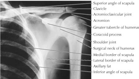
Clavicle fractures occur most frequently at the junction of the middle and lateral thirds of the clavicle (Figure 6-2). The patient characteristically supports the sagging limb with the opposite hand. Subclavian vessels and trunks of the brachial plexus are at risk in fractures of the middle third because they lie behind only the thin subclavius muscle. As the fractured clavicle heals, supraclavicular nerves may be trapped by callous formation, causing chronic neck pain.
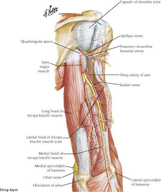
Surgical neck fractures, which are common, may damage the axillary nerve and the posterior humeral circumflex artery as they pass through the quadrangular space (Figure 6-3).
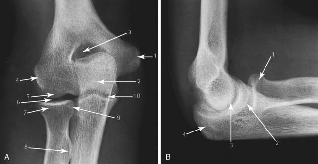
The brachial artery lies just medial to the biceps brachii tendon in the cubital fossa, where the stethoscope is placed when taking blood pressure. The deep branch of the radial nerve may be injured in a fracture of the neck of the radius. See Figure 6-5 for fractures of the radial shaft.

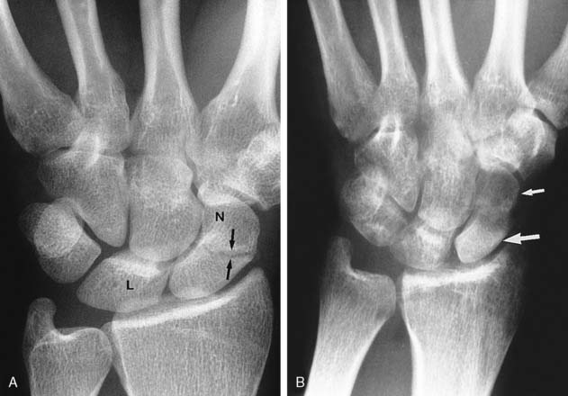
Scaphoid fractures may not show on radiographs for 10 days to 2 weeks, but deep tenderness will be present in the anatomical snuffbox. The proximal fragment may undergo avascular necrosis because the blood supply is interrupted (Figure 6-7), or nonunion of the fracture may occur.
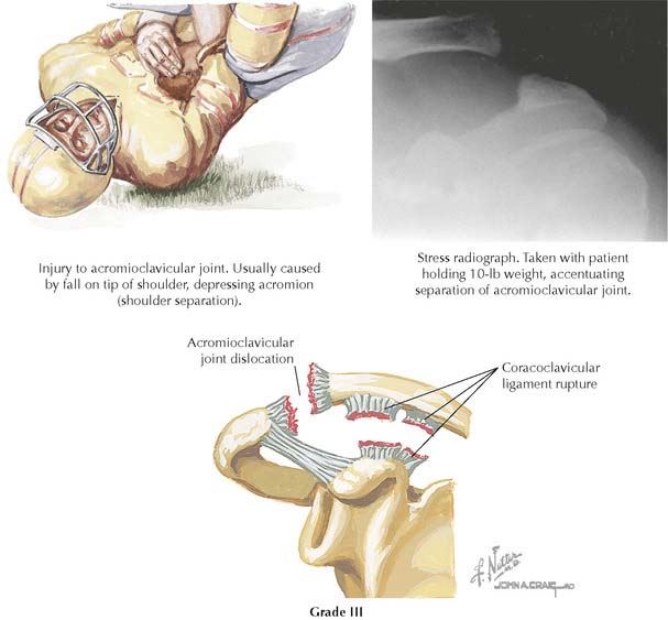
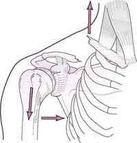
6-8 Shoulder separation (acromioclavicular dislocation) with rupture of coracoclavicular ligament.
(From Greene, W B: Netter’s Orthopaedics. Philadelphia, Saunders, 2006, Figure 14-19.)
Falling on an outstretched hand or a point of the shoulder often dislocates the acromioclavicular joint (shoulder separation). The coracoclavicular ligament tears when the lateral end of the clavicle rides up over the acromion. The weight of the shoulder causes it to drop away, or separate, from the clavicle (see Figure 6-8).
TABLE 6-1 Muscles Producing Movements of Glenohumeral Joint
| Movement | Muscles Involved |
|---|---|
| Flexion | Deltoid (anterior fibers), coracobrachialis, biceps, and pectoralis major |
| Extension | Latissimus dorsi, deltoid (posterior fibers), teres major, and long head of triceps |
| Abduction | Supraspinatus (initiation) and deltoid |
| Adduction | Pectoralis major, latissimus dorsi, and teres major |
| Medial rotation | Subscapularis, pectoralis major, deltoid (anterior fibers), latissimus dorsi, and teres major |
| Lateral rotation | Infraspinatus, teres minor, and deltoid (posterior fibers) |
The glenohumeral joint is the most frequently dislocated large joint (shoulder dislocation). In the usual anterior-inferior dislocation, muscle traction pulls the dislocated humeral head into a subcoracoid position (Figure 6-9). The axillary nerve may be injured. Posterior dislocations comprise only 5% but are seen following convulsions or electric shock. A posterior dislocation may not be apparent on standard AP radiographs.
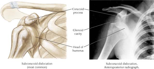
6-9 Dislocation of glenohumeral joint in usual anteroinferior direction (subcoracoid dislocaton).
(From Greene, W B: Netter’s Orthopaedics. Philadelphia, Saunders, 2006, Figure 14-17.)
TABLE 6-2 Muscles Producing Movements of Radiocarpal (Wrist) Joint
| Movement | Muscles Involved |
|---|---|
| Flexion | Flexor carpi ulnaris, flexor carpi radialis, palmaris longus, long finger flexors |
| Extension | Extensor carpi ulnaris, extensor carpi radialis longus and brevis |
| Abduction | Flexor carpi radialis, extensor carpi radialis longus and brevis |
| Adduction | Flexor carpi ulnaris and extensor carpi ulnaris |
Stay updated, free articles. Join our Telegram channel

Full access? Get Clinical Tree


