The Upper Extremity


The Upper Extremity


OBJECTIVES


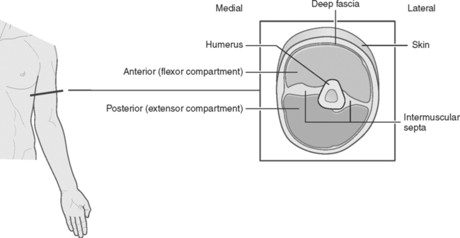
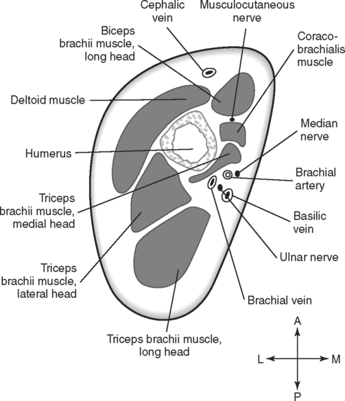
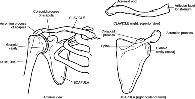
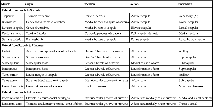
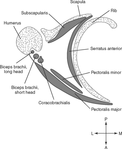
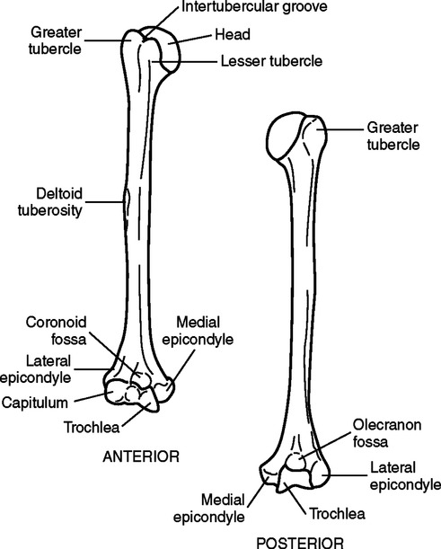
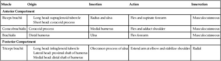
Upon completion of this chapter, the student should be able to do the following:


• Identify the bones that make up the pectoral girdle.


• Describe the location and functions of three groups of muscles associated with the attachment of the pectoral girdle and the upper extremity to the trunk of the body.


• Describe the boundaries and contents of the axilla.


• Identify the skeletal, muscular, vascular, and neural components of the arm.


• Describe the boundaries and contents of the cubital fossa and identify its components in a transverse section.


• Identify the skeletal, muscular, vascular, and neural components of the forearm.


• Name and locate the eight bones in the wrist and discuss the structure and significance of the carpal tunnel.


• Identify the structural components of the arm in transverse sections through the proximal and distal regions.


• Identify the structural components of the forearm in transverse sections through the proximal and distal regions.


• Describe the structure of the shoulder joint and discuss the anatomical relationships of its components.


• Identify structural components of the shoulder joint in transverse sections through the humeral head and the glenoid fossa, and in a coronal section.


• Describe the structure of the elbow joint and discuss the anatomical relationships of its components, including the humeroulnar, humeroradial, and radioulnar articulations.


• Identify the structural components of the elbow joint in sagittal sections through the humerus and the ulna, through the humerus and the radius, and in a transverse section through the radius and the ulna.


Key Terms, Structures, and Features to Be Identified and/or Described


Acromion process of the scapula


Annular ligament


Axilla


Axillary nerve


Basilic vein


Biceps brachii muscle


Brachial artery and vein


Brachial plexus


Brachialis muscle


Brachioradialis muscle


Bursae


Capitate bone


Capitulum of the humerus


Cephalic vein


Clavicle


Coracobrachialis muscle


Coracoid process of the scapula


Coronoid fossa of the humerus


Coronoid process of the ulna


Cubital fossa


Deltoid muscle


Flexor retinaculum


Glenoid fossa


Glenoid labrum


Hamate bone


Humerus


Infraspinatus muscle


Interosseous membranes


Lunate bone


Median cubital vein


Median nerve


Musculocutaneous nerve


Olecranon fossa of the humerus


Olecranon process of the ulna


Pectoral girdle


Pectoralis minor muscle


Pectoralis major muscle


Pisiform bone


Pronator teres muscle


Radial artery


Radial nerve


Radius


Rotator cuff muscles


Scaphoid bone


Scapula


Spine of the scapula


Subscapularis muscle


Supraspinatus muscle


Teres major muscle


Teres minor muscle


Trapezium bone


Trapezoid


Triceps brachii muscle


Triquetral


Trochlea of the humerus


Ulna


Ulnar artery


Ulnar nerve


Anatomical Review of the Upper Extremity


The upper extremity (limb) consists of the arm, forearm, wrist, hand, and fingers. It is attached to the trunk at the shoulder by the pectoral girdle. The arm is the region between the shoulder and the elbow. A single bone, the humerus, makes up the framework of the arm. The forearm extends from the elbow to the wrist. Two bones, the radius on the lateral side and the ulna on the medial side, form the framework of the forearm. The wrist, or carpus, consists of eight small bones, collectively called carpals. The hand has five bones, which are known as the metacarpals, and distal to these are 14 phalanges that form the fingers. These bones are covered with muscle, fascia, and skin. Note that in common use, the terms “upper extremity” and “arm” often are used synonymously. Anatomically, however, this is not correct. “Arm” refers only to the portion of the upper extremity between the shoulder and the elbow.


Attachment of the Upper Extremity to the Trunk


The scapula and the clavicle make up the pectoral girdle, which provides the connection between the upper extremity and the axial skeleton. The bones of the pectoral girdle are shown in Fig. 7-1. Muscles anchor the upper extremity and the pectoral girdle to the trunk of the body. These muscles can be divided into three groups.


One group of muscles extends from the trunk to the scapula. In appropriate combinations, the muscles in this group can move the scapula upward, downward, forward, backward, clockwise, or counterclockwise. These actions assist in movement of the shoulder.


A second group of muscles extends between the scapula and the humerus. These muscles move the arm at the glenohumeral (shoulder) joint. The muscles and tendons that extend over the shoulder strengthen and stabilize the joint.


A third group extends from the trunk to the humerus. This group, which includes the pectoralis major and the latissimus dorsi, adducts the arm. Table 7-1 summarizes the muscles associated with the trunk, the scapula, and the humerus.


TABLE 7-1


Muscles Associated with Trunk, Scapula, and Humerus


Muscle Origin Insertion Action Innervation
Extend from Trunk to Scapula
Trapezius Thoracic vertebrae Spine of scapula Adduct scapula Accessory (XI)
Rhomboids Cervical and thoracic vertebrae Medial border and spine of scapula Adduct scapula Dorsal scapular
Levator scapulae Cervical vertebrae Medial border of scapula Elevate scapula Dorsal scapular
Pectoralis minor Third to fifth ribs Coracoid process of scapula Pull scapula inferiorly Medial pectoral
Serratus anterior First eight ribs Medial border of scapula Rotate scapula Long thoracic nerve
Extend from Scapula to Humerus
Deltoid Acromion and spine of scapula; clavicle Deltoid tuberosity of humerus Abduct arm Axillary
Supraspinatus Supraspinous fossa Greater tubercle of humerus Abduct arm Suprascapular
Subscapularis Subscapular fossa Lesser tubercle of humerus Medial rotation of arm Subscapular
Infraspinatus Infraspinous fossa Greater tubercle of humerus Lateral rotation of arm Suprascapular
Teres minor Lateral margin of scapula Greater tubercle of humerus Lateral rotation of arm Axillary
Teres major Superior lateral margin of scapula Intertubercular groove of humerus Adduct arm Subscapular
Coracobrachialis Coracoid process of scapula Shaft of humerus Adduct arm Musculocutaneous
Extend from Trunk to Humerus
Pectoralis major Clavicle, sternum, costal cartilages Intertubercular groove of humerus Adduct and medially rotate humerus Medial and lateral pectoral
Latissimus dorsi Thoracic and lumbar vertebrae; crest of ilium Intertubercular groove of humerus Adduct and medially rotate humerus Thoracodorsal


image


Axilla


The space at the junction of the arm and the thorax, between the upper limb and the chest wall, is called the axilla The anterior wall of the axilla is formed by the pectoralis major muscle and the pectoralis minor muscle Predominant structures in the posterior wall are the scapula and the subscapularis muscle Medially, the axilla is delineated by the ribs, the intercostal muscles, and the serratus anterior muscle. The narrow lateral wall is formed by the head of the humerus; specifically, it is formed by the intertubercular (bicipital) groove, where the anterior and posterior walls converge. The long head of the biceps brachii muscle is located in the intertubercular groove. The short head of the biceps brachii muscle and the coracobrachialis muscle are closely associated in this same region. The boundaries of the axilla are illustrated in Fig. 7-2.


The axilla functions as a passageway for vessels and nerves that pass between the root of the neck and the arm. The vessels in this region include the axillary artery and vein, together with their branches. The nerves that pass through the axilla are branches of the brachial plexus and innervate the upper extremity. The axilla also contains numerous lymph nodes, which are drained by axillary lymph vessels that pass through this region. The axillary lymph nodes are of particular significance because a large portion of the lymphatic drainage from the breast filters through these nodes, and they are frequently involved with breast cancer.


General Anatomy of the Arm


Osseous Components.


The region from the shoulder to the elbow is the arm, or brachium. The only bone in this region is the humerus, which is the longest bone in the upper extremity. The features of the humerus are shown in Fig. 7-3.


Muscular Components.


The muscles of the arm, or brachium, are arranged in anterior and posterior compartments, which are separated by an intermuscular septum of fascia (Fig. 7-4). The muscles of the arm are summarized in Table 7-2.


The anterior muscle compartment consists of three muscles that act as flexors and are innervated by the musculocutaneous branch of the brachial plexus. The largest of the muscles is the biceps brachii muscle As the name implies, the biceps brachii has two heads of origin. The short head originates on the coracoid process of the scapula, and the long head originates from a tubercle just above the glenoid cavity (supraglenoid tubercle). The two heads merge to form a single muscle belly that inserts on the radial tuberosity and on the ulna by way of an aponeurosis. In addition to being a flexor of the forearm at the elbow, the biceps brachii is a supinator of the forearm. The muscle originates superior to the shoulder and passes over the joint; it therefore helps stabilize and strengthen the shoulder joint. The biceps brachii muscle also acts as a flexor of the arm at the shoulder.


The coracobrachialis is a short muscle on the medial surface of the superior part of the arm. It has a common origin with the short head of the biceps brachii on the coracoid process of the scapula, and it inserts on the medial side of the humerus near the midpoint of the shaft. The coracobrachialis, along with the biceps brachii, acts as a weak flexor and adductor of the shoulder. This muscle is visible only in sections through the upper part of the arm. The coracobrachialis is the only muscle that, although predominantly located in the arm, acts on the shoulder joint.


The third muscle of the anterior compartment is the brachialis muscle This is a deep muscle, underlying the biceps brachii. It has an extensive origin along the anterior surface of the distal half of the humerus, and it terminates on the coronoid process of the ulna. The brachialis is a strong flexor of the forearm at the elbow joint. It is seen only in sections through the lower part of the arm.


The posterior compartment of the arm is occupied by a single, large muscle, the triceps brachii. As the name implies, this muscle has three heads of origin. The long head originates via a tendon from the infraglenoid tubercle. The lateral head attaches to the posterior surface of the proximal shaft of the humerus. The medial head is deep to both the long and the lateral heads. Its origin is on the posterior surface of the shaft of the humerus, distal to the origin of the lateral head. All three heads merge to form a single muscle belly that inserts on the olecranon process of the ulna via a single tendon. An olecranon bursa is located between the tendon and the olecranon process. The triceps brachii muscle is a powerful extensor of the elbow. The long head spans the shoulder joint; consequently, it also helps stabilize that joint. The radial nerve innervates the triceps brachii.


Vascular Components.


The primary arterial blood supply to the arm is the brachial artery and its branches. The brachial artery begins at the inferior border of the teres major muscle, as a continuation of the axillary artery, and ends in the cubital fossa, where it divides into the radial and ulnar arteries. The vessel is superficial throughout its length, and it runs its course in the fascia of the medial intermuscular septum, which divides the muscles into the anterior and posterior compartments. In the septum, it is associated with the basilic vein, the median nerve, and the ulnar nerve Numerous branches supply the muscles of the arm.


One, or possibly two, brachial veins accompany the brachial artery These deep veins ascend through the arm to continue as the axillary vein. In addition to the deep brachial vein, two important superficial veins are in the arm. The cephalic vein is in the superficial fascia, anterolateral to the biceps brachii muscle. As it courses superiorly, it passes between the deltoid and the pectoralis major muscles to empty into the axillary vein. The basilic vein is in the superficial fascia on the medial side of the arm. About one third of the way up the arm from the elbow, the basilic vein passes deep to the superficial fascia and continues upward to merge with the brachial vein to form the axillary vein. Both the superficial cephalic and basilic veins are frequently visible through the skin. Fig. 7-5 shows some of the vascular relationships in the arm.


Nerves in the Arm.


The major nerves traversing the arm are the musculocutaneous, median, ulnar, and radial nerves. A fifth nerve, the axillary nerve, supplies the skin over the upper part of the arm. The nerves are all terminal branches of the brachial plexus. Both the median and ulnar nerves descend the arm without giving off branches. They supply the elbow joint and the forearm. In the uppermost part of the arm, the median nerve may be either lateral or anterior to the brachial artery. About midway down the arm, the nerve crosses over the vessel to the medial side. The ulnar nerve is situated near the brachial artery in the upper half of the arm. It then penetrates the intermuscular septum and descends through the arm just anterior to the medial head of the triceps brachii. The musculocutaneous and radial nerves give off branches to supply the muscles of the arm. The musculocutaneous nerve descends between the biceps brachii and the brachialis muscles. As it descends, its branches innervate those two muscles and the coracobrachialis muscle. The radial nerve enters the arm on the medial side of the humerus, then curves around the bone, in the radial groove, to descend in the intermuscular septum on the lateral side.


Jun 16, 2016 | Posted by in ANATOMY | Comments Off on The Upper Extremity

Full access? Get Clinical Tree

Get Clinical Tree app for offline access