The Thyroid, Parathyroid, and Neck Masses Other Than Lymph Nodes


The Thyroid, Parathyroid, and Neck Masses Other Than Lymph Nodes


Miguel A. Sanchez

Rosalyn E. Stahl


THE THYROID


Biopsy Principles and Techniques

The primary objective of fine-needle aspiration biopsies (FNAs) of the thyroid is to select those patients who require surgery for a neoplastic disorder from those who have a functional or inflammatory abnormality and who can be followed clinically or treated medically (Poller et al, 2000; Werga et al, 2000; Carpi et al, 1996, 1999; Mazzaferri, 1993; Cohen and Choi, 1988; Einhorn and Franzén, 1962; Söderstrøm, 1952). Children and adolescents should not be excluded because they may also harbor malignant tumors (recent summary in Khurana et al, 1999).

The clinical evaluation of a thyroid mass includes radioiodine scanning (scintiscan), ultrasound and biochemical or immunologic thyroid function tests. However, for palpable lesions of the thyroid, this initial work-up is often of limited value. Although most thyroid tumors are “cold” on scintiscan (i.e., do not absorb radioiodine), some are not, but not all “hot nodules” represent functional abnormalities. Eighty per cent of ultrasounds describe the thyroid masses as “partially solid and partially cystic,” which does not contribute to the diagnosis. Thyroid function tests are usually
normal in tumors. Therefore, our approach in evaluating a palpable thyroid mass is to begin with fine-needle aspiration biopsy, which we found to be a safe procedure as have many others (Castro and Gharib, 2000; Werga et al, 2000; Mazzaferri, 1993; Gagneten et al, 1987; Rojeski and Gharib, 1985; Löwhagen and Willems, 1981; Wang et al, 1976; Löwhagen and Sprenger, 1974). Using this approach, we have performed approximately 10,000 fine-needle aspiration biopsies of the thyroid, most guided by palpation and the others under ultrasound guidance.

The use of scintiscanning and ultrasound is not to be dismissed, however, because in individual patients, these tests may complement FNA (Baloch et al, 2000; Tambouret et al, 1999; Capri et al, 1996, 1999; Hagag et al, 1998). For example, scintiscanning may be helpful in differentiating a hyperplastic goiter from a follicular neoplasm (see below), whereas ultrasound provides accurate measurements of the size of nodules that are not considered as suitable for surgery and are followed clinically. Measurement of serum thyroid antibodies, which are not part of standard thyroid function tests, is the most useful blood test to confirm the diagnosis of Hashimoto’s thyroiditis, which may sometimes present as single or multiple thyroid nodules (Wong and Wheeler, 2000) (see below).

We do not recommend routine use of FNA of small, nonpalpable masses found incidentally in patients, especially elderly patients, by ultrasound studies of neck performed for other reasons, for example, carotid artery studies. Nonpalpable malignant lesions are rare, whereas nonpalpable benign thyroid nodules are very common. Even if a small nodule is malignant, it usually is a low-grade papillary tumor of questionable clinical importance (Petez et al, 2001; Kimoto et al, 1999; Tambouret et al, 1999; Carmeci et al, 1998; Hagag et al, 1998; Lin et al, 1997).

In our work, we prefer to use air-dried smears, stained with Diff-Quik. The advantage of this technique is the short preparation and staining time and, despite opinions to the contrary, an excellent quality of nuclear features. With this technique, the colloid stains blue and, because of air drying, the cells are larger than in rapidly fixed smears. Still, rapidly fixed smears stained with Papanicolaou or hematoxylin-eosin are equally valuable and sometimes easier to interpret and to compare with histologic material, as illustrated in this chapter (Koss et al, 1992). Yang et al (1997, 2001) advocated the use of ultrafast Papanicolaou stain as advantageous in recognizing nuclear abnormalities in papillary carcinomas of the thyroid.

It must be stressed that regardless of method used, interpretation of cytologic material from the thyroid is not simple; it requires excellent material and a great deal of experience. Contrary to the belief of many clinicians that aspirating the thyroid is an easy and simple task, they often commit several cardinal mistakes: the aspirate takes too long and the samples are diluted with blood. Other errors include the use of larger caliber needles or “poking around the lesion” with the tip of the needle resulting in hemorrhagic necrosis. Therefore, to ensure the best possible quality of smears, the FNA should be performed and interpreted by an experienced cytopathologist (Dwarakanathan et al, 1989).


Aspiration Biopsy Technique

Aspirations of the thyroid are best performed with 25 or 22 gauge needles by an experienced operator, preferably a cytopathologist. The use of larger caliber needles is not recommended because they cause more trauma and bleeding and result in unreadable bloody aspirates. Similarly, more than one aspiration per nodule does not necessarily yield more diagnostic material but rather causes more trauma to the patient. At the Englewood Hospital Medical Center, the aspiration biopsies are performed by a pathologist. The needle is attached to a 10-cc syringe and a Cameco aspiration gun. The slides are prepared, stained, and microscopically evaluated immediately while the patient waits and, only if the aspirate is not adequate for accurate diagnosis, is the patient re-aspirated. Thus, the number of aspirates can be tailored to the findings in the individual patient and, in most of them, one aspiration is adequate to yield a diagnosis (Aguilar et al, 1998).

The first step in the aspiration is to determine if a neck mass is, in fact, in the thyroid. One should palpate the mass while the patient swallows. If it moves with swallowing, it is in the thyroid. If not, it is probably a lymph node or another adjacent structure.

The aspiration is best performed with the patient supine and neck hyperextended. To avoid inadvertent displacement of the gland during the biopsy, the patient is instructed to refrain from swallowing and speaking, but to continue breathing. After insertion, the needle is rapidly moved in and out to sample different parts of the palpable mass. Because the thyroid is richly vascularized, the actual aspirate must be performed as rapidly as possible, in about 1 to 2 seconds to avoid dilution of samples with blood. If cyst fluid is obtained, the aspiration should continue so long as the fluid keeps flowing.

There are usually no untoward effects from a correctly aspirated thyroid. Potential side effects include pain, swelling, a hematoma, and acute infection, all extremely rare occurrences. Occasionally, an acute infarction of a tumor, most commonly a papillary carcinoma or a Hürthle cell tumor, can occur after aspiration (Kini, 1996, Pinto et al, 1996). This can cause exquisite pain, and may result in difficulty in histologic diagnosis of tumor in the surgically removed specimens (Baloch and LiVolsi, 1999; Ersoz et al, 1997; Vercelli-Retta et al, 1997). Seeding of tumor cells in the needle track was reported in papillary carcinomas (Karwowski et al, 2002; Hales and Hsu, 1990). Two other reports of possible needle track seeding are not well documented (Panunzi et al, 1994; Wang et al, 1976).


Adequacy of Aspiration Biopsy

The issue of what constitutes an adequate thyroid sample has been a subject of debate for several years. Hamburger et al (1989), in a multi-institutional study, declared that six clusters of epithelial cells on two separate smears constitute an acceptable minimum of sampling adequacy. In many instances, however, this criterion is unrealistic. For example,
pure colloid may be consistent with the diagnosis of benign colloid goiter and a pure population of macrophages may be diagnostic of a thyroid cyst or pseudocyst in a patient with past history of hemorrhage. In neither instance is the presence of epithelial cells required for diagnosis. Clearly, the issue depends on the type of lesion (cystic or solid) and the skill of the performer. It has been our experience that the latter is paramount.


Normal Structure of the Thyroid Gland


Anatomy

The thyroid gland is located in the neck, anterior to the trachea, and consists of two conical lobes connected by the isthmus. A third, smaller central lobe, the pyramidal lobe, is infrequently found. The lobes are divided by fibrous septa into lobules, each containing 30 to 40 follicles. The gland is enclosed in a true capsule that may adhere to the trachea and larynx. The thyroid is richly vascularized and has a high rate of blood flow.


Histology

The structural unit of the thyroid gland is the follicle, a closed, approximately spherical space lined with epithelial cells that vary in configuration from flat, to low cuboidal, to high columnar (Fig. 30-1C; see Fig. 30-3D). The height and configuration of the thyroid follicular cells reflect, to some extent, the functional activity. The cells are flat in inactive follicles and cuboidal or even columnar in active thyroid. In hyperthyroidism, the cells are columnar and their cytoplasm may be filled with colloid-containing vacuoles.


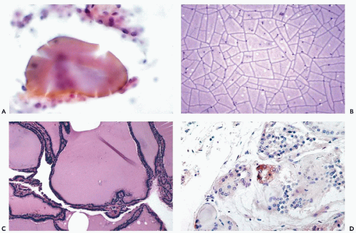
Figure 30-1 Benign thyroid. A. Large deposit of colloid surrounded by a few follicular cells. B. Pure colloid from a colloid nodule. Note the deep purple color and the “cracking artifact” in Diff-Quik stain. C. Histologic section of thyroid removed for colloid goiter corresponding to B. D. “C cells” immunostained for calcitonin by immunoperoxidase stain.

The follicles are filled with colloid, a homogenous eosinophilic substance. Variations in the density and staining properties of the colloid can also be ascribed some functional significance; thin eosinophilic colloid appears to be associated with functional activity, whereas thick, markedly eosinophilic colloid occurs in inactive follicles and in some malignant lesions (Fig. 30-1A,B).

For reasons unknown, the follicular cells may be transformed into large cells with eosinophilic cytoplasm and large, often hyperchromatic nuclei, known as oncocytes (bulky cells) or Hürthle cells (see Fig. 30-8). Such cells were first described by Hürthle in the thyroid of dogs. The role of these cells in the pathology of the thyroid is described below. The cytoplasm of oncocytes is crammed with mitochondria.

The thyroid also contains dispersed calcitonin-producing cells or C cells (Fig. 30-1D). Calcitonin is a hormone governing the metabolism of calcium and, hence, the skeletal system. The C cells may be the source of malignant tumors, known as medullary carcinoma, that may also secrete calcitonin, demonstrable by immunostaining (see below).


Principal Lesions of the Thyroid

The principal lesions of the thyroid gland that may be identified in aspiration cytology are as follows:


  • Cysts


  • Goiters


    • Colloid goiter


  • Thyroiditis


    • Acute


    • Subacute (deQuervain’s)


    • Lymphocytic (Hashimoto’s disease)


    • Riedel’s Struma (fibrosing thyroiditis)


  • Tumors


    • Follicular tumors


    • Follicular adenomas


    • Follicular carcinoma


  • Hürthle cell tumors


    • Hürthle cell adenoma


    • Hürthle cell carcinoma


  • Other carcinomas


    • Papillary and its variants


    • Medullary


    • Anaplastic (large- and small-cell types)


  • Malignant lymphomas


  • Rare malignant tumors


  • Metastatic tumors

It should be emphasized again that, although very specific diagnoses can be made by FNA, the main purpose of the fine needle aspiration is to determine if the patient has a primary thyroid tumor, whether benign or malignant, requiring surgical excision, or has a nonneoplastic condition, such as a goiter or thyroiditis, which can be treated medically. Large benign goiters, obstructing adjacent organs or disfiguring cosmetically, may require surgical intervention, regardless of cytologic findings. Statistical evidence strongly suggests that the use of aspiration biopsy has markedly reduced the total number of thyroidectomies, whereas the proportion of carcinomas in the surgically treated population has increased significantly (Koss et al, 1992).


Clinical Findings

Most patients are referred for FNA because of thyroid enlargement or the presence of a nodule or nodules. For reasons unknown, most patients are females. Age is not important because malignant lesions may occur in the very young and very old. However, it is important to know how long the abnormality has been present and whether its growth was slow, rapid, or sudden. This information may be of diagnostic significance because slowly growing multiple nodules or masses are less likely to be malignant than a more rapidly enlarging solitary nodule. A sudden increase in the size of the nodule suggests a hemorrhage. Still, none of these observations are conclusive and judgmental errors may occur in all situations prior to sampling of the lesion(s).


The Painful Thyroid

Of particular interest is the clinical observation of a thyroid illness presenting with pain as a primary complaint. In our experience, the differential diagnosis of a painful thyroid nodule in the young or middle-aged patient includes:


  • Acute thyroiditis


  • Cyst with acute hemorrhage


  • Subacute or De Quervain’s thyroiditis


  • Infarcted Hürthle cell tumor (rare)


  • Hashimoto’s thyroiditis

In the elderly, a very painful thyroid nodule, or diffuse tenderness, should alert one to the possibility of an anaplastic carcinoma.


Goiters

The terms goiter or struma denote any enlargement of the thyroid gland, which may be diffuse or nodular. The three most common types are the colloid goiter, inflammatory goiter or thyroiditis, and neoplastic goiter caused by benign or malignant tumors.


Colloid Goiter

Synonyms: Adenomatous goiter, diffuse or nodular goiter, endemic goiter, multinodular goiter.


Histology

Colloid goiter is usually caused by hyperplasia of the thyroid gland induced by iodine deficiency. Its histologic appearance varies with the developmental stage of the disease. In the early stages, the changes are bilateral and there is a diffuse enlargement of the gland, made up of small follicles. Later on, some of the follicles may become distended and may coalesce to form nodules, with diameters ranging from less than 1 mm to several centimeters. Degenerative changes, such as hemorrhage, necrosis, cysts (actually pseudocysts) and scar formation, often occur in the nodules. The process may involve the entire gland or it may occur focally and produce a solitary nodule. On the scintiscan, such change is often labeled a “cold nodule,” which is difficult to distinguish from a true neoplasm. For this reason, the colloid goiter is the lesion most often referred for aspiration.


Cytology

The make-up of a needle aspirate from colloid goiter depends on the type of the lesion. The aspirate may be solid, semisolid, or fluid. Solid or semi-solid aspirates often consist of amber colored colloid which, in Papanicolaoustained smears, appears as pink homogeneous material, usually surrounded by a few follicular cells (see Fig. 30-1A) and in Diff-Quik-stained smears, appears bluish-violet. On drying, the colloid may form a typical cracked geometric pattern (see Fig. 30-1B). This type of colloid is derived from distended inactive follicles containing dense central colloid (see Fig. 30-1C). Occasionally, aspirates of colloid goiters yield smears of almost pure colloid with few follicular cells and/or macrophages. Although such smears do not meet the proposed criteria of adequacy because they contain very few follicular cells, they are, in our experience, diagnostic of colloid goiter.


In most cases, however, the smears also contain small cuboidal follicular cells, occurring either singly or in flat sheets wherein the small, spherical nuclei are evenly spaced and honeycomb-forming cytoplasmic borders may sometimes be recognized (Fig. 30-2A). “Naked” follicular cell nuclei are often seen scattered throughout the smear and should not be confused with lymphocytes, which they resemble (Fig. 30-2B). The nuclei of lymphocytes are usually slightly smaller and upon close inspection, usually are surrounded by a very thin rim of blue cytoplasm, at least on one side of the cell. Sometimes entire follicles may be seen in the form of spherical clusters of cells, sometimes with a central deposit of colloid (Fig. 30-3). Swedish observers consider squashed follicles, wherein cell borders cannot be clearly seen as “syncytial balls” and characteristic of colloidal goiter (Fig. 30-3C). In posthemorrhagic nodules or cysts, numerous macrophages, usually containing phagocytized hemosiderin granules, may be observed (Fig. 30-4).

An important source of error is clusters or sheets of spindle-shaped cells of the fibroblastic type. This finding suggests that fibrosis has occurred within the goiter. Sometimes, young, growing fibroblasts from an area of active fibrosis are markedly atypical, with enlarged, irregularly outlined nuclei that may contain large nucleoli and abundant basophilic cytoplasm with multiple cytoplasmic extensions (Fig. 30-5). Mitotic figures may also occur. The atypical fibroblasts may be mistaken for malignant cells and must be interpreted within the context of the clinical situation and other cytologic findings. It is of note that, very rarely, exuberant fibrosis may also occur in papillary thyroid carcinomas (Chan et al, 1991; Us-Krašovec and Golough, 1999).

A fairly high proportion of aspirations in colloid goiter yield variable amounts of fluid which usually indicates degenerative cystic changes (see below). The fluid may be clear, yellow, or brown in color, the latter indicative of a prior hemorrhage. Although the microscopic examination of the cyst fluid is seldom useful, it is mandatory because, occasionally, cystic thyroid carcinomas may masquerade as cystic goiters.

Over time, goiters often become nodular, sometimes in the form of a solitary nodule, usually arising from focal hyperplasia. Such nodules, often of adenomatous nature, usually cannot be reliably differentiated on clinical or ultrasonographic grounds from a “true” adenoma or carcinoma. As a general rule, smears from nonneoplastic nodules contain much colloid and few cells. By contrast, in aspirates from neoplastic lesions, there is little colloid and numerous cells. However, on rare occasions, aspirates of hyperplastic adenomatous nodules are also very cellular and may be very difficult to interpret. Certain features of the smears are sometimes helpful. Thus, the follicular cells tend to form a dispersed pattern in adenomatous goiter, rather than tight clusters or rosettes as in true adenomas. Also, “balls” of follicular cells, representing squashed whole follicles (see Fig. 30-3), are more characteristic of colloid goiter than of follicular adenomas. One has to gauge all these features in order to arrive at a correct interpretation. Although most cases are straightforward, the distinction between a hyperplastic adenomatous nodule and follicular neoplasm can be difficult, as it is in histological sections(Zacks et al, 1998; Busseniers and Oertel, 1993). In such cases, the interpretations and recommendations may include a course of suppression therapy with exogenous thyroid hormones or repeat FNA. If the mass does not regress, a surgical removal must be considered. In such a case, a scintiscan may be helpful. If the mass is “cold,” one would favor surgery rather than medical therapy, since a “cold” nodule would more likely be a true tumor.


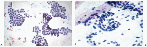
Figure 30-2 Benign follicular cells. A. Follicular cells forming flat sheets with “honeycomb” pattern. B. Clusters of normal follicular cells with many dispersed cells in the background.


Cysts


Histology

Cystic lesions constitute 10% to 30% of all thyroid nodules, depending on the population studied. Nearly all thyroid cysts are pseudocysts that develop in nodular goiters or in adenomas as a consequence of degenerative tissue break-down or a hemorrhage. Pseudocysts are usually formed by fusion of adjacent distended follicles. They are usually lined by flattened or cuboidal cells of thyroid epithelium and may undergo focal squamous metaplasia. The very uncommon true cysts derived from the remnants of the thyroglossal
duct are located in the midline, usually above the thyroid, and are lined by either cylindrical or squamous epithelium (see below). A case of an intrathyroid lymphoepithelial cyst of probable branchial origin was described by Apel et al (1994). It must be stressed that some malignant tumors, particularly papillary carcinomas, are often partly cystic.


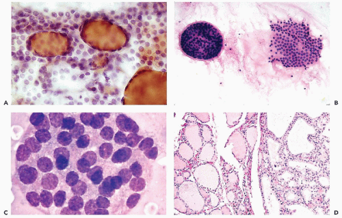
Figure 30-3 Thyroid follicles in benign goiters. A. Several colloid-filled follicles with dispersed follicular cells in the background. B. Left, An entire three-dimensional thyroid follicle. Right, Broken up follicle with a small deposit of colloid in the center. C. Tightly packed, compact thyroid follicle, commonly observed in benign colloid goiters. Note the abundant cytoplasm seen under high magnification with Diff-Quik stain. The term “syncytial balls” has been attached to this formation. D. Histologic section of colloid goiter. Note the variability in the size of the acini. (C: Courtesy of Dr. Sixtén Franzén, Stockholm, Sweden.)


Cytology

Depending on their size, aspirated cysts or pseudocysts of the thyroid yield a few drops to several milliliters of fluid that may be clear, turbid, yellow, brown, or bloody. Preparation of smears may require centrifugation of the fluid. The smears of fluid aspirated from a cyst with a recent hemorrhage contain mainly blood and clusters of well-preserved follicular cells. In cysts aspirated some weeks after the hemorrhage, the few follicular cells may show degenerative nuclear changes in the form of smudging or hyperchromasia and are accompanied by hemosiderin-laden macrophages and by inflammatory cells (see Fig. 30-4). Hemosiderin stains blue or black on Diff-Quik stain and brown in Papanicolaou stain. The sediments of the clear or yellowish fluids obtained from old hemorrhagic cysts usually contain only a few macrophages.

In degenerative cysts, the smears are usually characterized by numerous macrophages with foamy cytoplasm, containing cell debris (see Fig. 30-4A) that may be accompanied by few inflammatory cells. Occasionally, a few clusters of follicular cells may be found in the smears.

If after aspiration of the cyst content, no residual palpable lesion is found, further therapeutic procedures may be avoided. In contrast, if after the evacuation of the fluid, a palpable lesion remains, the aspiration should be repeated in order to rule out the possibility of a tumor masquerading as a cyst. In the sediment of the fluid aspirated from cystic papillary carcinoma, calcified psammoma bodies may be observed as the only abnormality. Thus, the presence of calcified particles in cyst fluid should be interpreted with great caution and mandates further exploration of the lesion. Other unusual cytologic findings, such as the presence of compact clusters of follicular cells, also deserve attention, as they may signal the presence of a thyroid neoplasm.


Thyroiditis

Thyroiditis, or inflammation of the thyroid gland, comprises a large group of diseases that range from acute suppurative
thyroiditis to chronic inflammatory processes. The term also includes a number of transient disorders such as postpartum thyroiditis and drug-induced thyroiditis (recent review in Pearce et al, 2003). Only the common forms of thyroiditis should be sampled by FNA and they are discussed below.


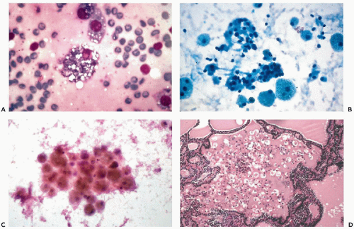
Figure 30-4 Macrophages in colloid goiter. A. A macrophage containing in its cytoplasm, numerous fragments of clear material, most likely fat. B. Numerous large macrophages with faintly vacuolated cytoplasm next to sheets of follicular cells. C. Hemosiderin containing macrophages from a hemorrhagic cyst of the thyroid. D. Follicle filled with colloid containing numerous macrophages. (A,C: High magnification; B: Diff-Quik stain.)

Acute thyroiditis is uncommon. It presents as an erythematous, markedly tender, diffuse process involving the thyroid accompanied by fever and generally does not represent a clinical diagnostic challenge. The aspiration biopsy, very rarely performed, yields follicular cells, neutrophiles, and macrophages. Sodhani (1989) reported the presence of microfilariae in such a case.


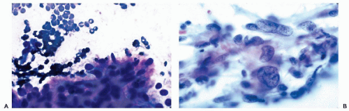
Figure 30-5 Fibrous reaction in colloid goiter. A. Cluster of elongated cells, corresponding to fibroblasts. B. Numerous elongated cells, some with giant nuclei, in reactive fibrosis in a thyroid scar. (A: Diff-Quik stain; B: high magnification.)


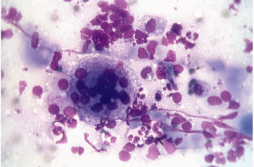
Figure 30-6 Subacute thyroiditis. The smear contains a large multinucleated giant cell surrounded by debris. (Diff-Quik stain.)

With the onset of AIDS, thyroiditis caused by the parasite pneumocystis carinii has been observed (Guttler and Singer, 1988). Several cases of this disease were diagnosed by aspiration cytology of the thyroid (summary in Keyhani-Rofagha and Piquero, 1999). The morphology of the parasite is discussed at length in Chapter 19.

In granulomatous (subacute, de Quervain’s) thyroiditis, the thyroid is slightly to moderately enlarged and tender on digital examination. The patient frequently gives a history of a recent upper respiratory infection or a viral syndrome. The aspirate contains follicular epithelial cells, epithelioid cells, and giant cells of Langhans’ type, together with lymphocytes, plasma cells, and occasionally some granulocytes (Fig. 30-6) (Shabb et al, 1999; Garcia Solano et al, 1997; Löwhagen and Willems, 1981).


Hyperthyroidism

Hyperthyroidism, known in its classical form as Graves’ disease, consists of a goiter, exophthalmos, tremor, and flushed skin. It can be associated with a diffuse goiter or a single hyperplastic thyroid nodule. Aspiration biopsies are practically never performed in diffuse toxic goiter but may be performed on solitary nodules.


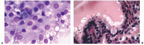
Figure 30-7 Hyperthyroidism (Graves’ disease). A. Follicular cells with eosinophilic cytoplasmic vacuoles in Diff-Quik stain, characteristic of hyperthyroidism. The term “flare cells” has been often attached to this phenomenon. B. Section of thyroid in hyperthyroidism, showing scalloped colloid and tall, vacuolated follicular cells.

Aspirates of untreated hyperplastic thyroid may contain a small number of large follicular cells with slightly enlarged nuclei and abundant cytoplasm with large round vacuoles of variable sizes containing deposits of colloid. In Diff-Quik and other hematologic stains, the cytoplasmic deposits of colloid stain bright red, hence the name “flare cells” (Fig. 30-7). Such cells occur singly or in small clusters, but rarely in large sheets (Myren and Sivertssen, 1962).

Centeno et al (1996) noted scarring of the thyroid and subsequent problems of interpretation of aspirates from patients treated with radioactive iodine for Graves’ disease.


Lymphocytic Thyroiditis or Hashimoto’s Disease


Clinical Data and Histology

This is the most common form of thyroiditis observed in clinical practice. It most often affects middle-aged women and is thought to be an autoimmune disease. Antithyroid antibodies are often elevated in this condition and thyroid function may be reduced, normal, or elevated. Both lobes of the thyroid are usually affected, diffusely enlarged and firm, but asymmetry may occur, with localized nodular enlargement (Wong and Wheeler, 2000). In histologic sections, the thyroid is infiltrated with lymphocytes that may also form lymph follicles with germinal centers (see Fig. 30-8D). The acini are often destroyed or atrophic (see Fig. 30-9D). A major component of the disease is the presence of oncocytes (Hürthle cells). The oncocytes may line glandular structures or form solid sheets of various sizes. The characteristic features of oncocytes, namely, abundant, eosinophilic, granular cytoplasm and large, often pyknotic nuclei of variable sizes, are usually well represented in Hashimoto’s thyroiditis (see Fig. 30-9C,D). Sometimes the Hürthle cells appear as single large cells in otherwise normal follicular epithelium. The term Askanazy cells is sometimes used to describe this feature. Rarely, psammoma bodies may be seen in Hashimoto’s thyroiditis (Dugan et al, 1987). Thyroid carcinomas and malignant lymphomas
may occur in Hashimoto’s thyroiditis. There is a misperception that malignant lymphoma is the most common tumor seen in Hashimoto’s thyroiditis. In fact, from 5% to 10% of patients with this disease will develop papillary carcinoma, and less than 1% develop malignant lymphoma (Carson et al, 1996).

A recently described clinical entity known as subchemical hypothyroidism with antibody-negative chronic thyroiditis, usually occurs in women with symptoms of fatigue, malaise, and hair loss (Wikland, et al, 2003; Wikland et al, 2001). These women often have negative physical examinations and medical work-ups, but are found to have chronic thyroiditis on FNA of their thyroid gland, if the astute clinician considers it in his or her differential diagnosis. The symptoms usually respond dramatically to oral levothyroxine.


Cytology

The aspiration smears are characterized mainly by the presence of a mixture of lymphocytes and oncocytes or Hürthle cells in various proportions. In some cases, the aspirates are dominated by lymphocytes at various stages of maturation, and the smear may resemble an aspirate of a hyperplastic lymph node (Fig. 30-8A). Further search will usually reveal the presence of oncocytes and scattered follicular cells in clusters (Fig. 30-8B). We have observed a case of Hashimoto’s disease in which the pattern was mistaken for a metastatic carcinoma to lymph node (Koss et al, 1992). In other cases, the presence of oncocytes is dominant. In aspirates, these large cells form sheets or gland-like structures. The striking, abundant, eosinophilic and granular cytoplasm and the variability in nuclear sizes are characteristic (Fig. 30-9A,B). Sometimes the nuclei contain fairly large nucleoli, and, very rarely, intranuclear cytoplasmic inclusions (see Fig. 30-8C). Because of nuclear abnormalities, these cells can be mistaken for malignant cells. On further search, lymphocytes and follicular thyroid cells can be observed. On rare occasions, the follicular cells in clusters may mimic a papillary carcinoma (MacDonald and Yazdi, 1999). Still less common is the presence of psammoma bodies (Dugan et al, 1987). These, fortunately very rare findings, may lead to an erroneous diagnosis of papillary carcinoma.


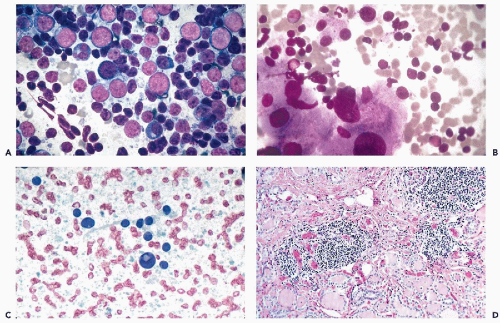
Figure 30-8 Hashimoto’s thyroiditis. A. Smear pattern with lymphocytic predominance. The lymphocytes are in various stages of maturation. This pattern may be misinterpreted as a malignant lymphoma. B. Scattered lymphocytes and large Hürthle cells. C. A Hürthle cell showing an intranuclear cytoplasmic inclusion. D. Histologic pattern of Hashimoto’s thyroiditis showing lymphocytic deposit and follicles lined by eosinophilic Hürthle cells. (A,B: Diff-Quik stain.)

As a general rule, the cytologic features of Hürthle cells in Hashimoto’s thyroiditis can be differentiated from a Hürthle cell tumor. In thyroiditis the oncocytes are large, atypical, and pleomorphic, whereas Hürthle cell tumors usually show monotonous cells, as is the case in histologic sections (see below) (Chen et al, 1998; Ravinsky and Safneck, 1988).
Further, the presence of abundant lymphocytes is very uncommon in Hürthle cell tumors.


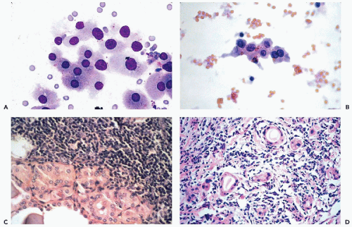
Figure 30-9 Hashimoto’s thyroiditis. A,B. Hürthle cells stained with Diff-Quik (A) and, after fixation, with Papanicolaou stain (B). The difference in size of the cells in the two modes of smear preparation is well shown. Note the variability in the sizes of the nuclei in the benign Hürthle cells. C. Section of the thyroid gland in Hashimoto’s disease showing lymphocytic infiltrate and Hürthle cells lining the adjacent acini. D. Section of the thyroid with follicular atrophy in Hashimoto’s thyroiditis.

The recognition in cytologic samples of either carcinoma or lymphoma, occurring in Hashimoto’s thyroiditis, may be exceedingly difficult. If a carcinoma is suspected, a surgical biopsy should be suggested. If a lymphoma is suspected, flow cytometry studies performed on the aspirate may help in determining a definitive diagnosis without having to resort to surgery.


Riedel’s Struma

Riedel’s struma is a sclerosing inflammatory disorder of the thyroid gland, sometimes associated with a similar tissue reaction in the mediastinum, retroperitoneum, and orbit. On aspiration, the thyroid gland feels very rubbery or firm and the yield from a needle aspiration is nil or very scanty, sometimes containing only a few fibroblast-like cells. This disorder must be distinguished from infiltrating carcinoma, in which a fairly rich cell population would be obtained.


Neoplastic Lesions


Classification

In textbooks and other teaching materials pertaining to the thyroid gland, it is customary to separate benign (adenomas) from malignant lesions (carcinomas). However, because the histologic and cytologic differences between the benign and malignant variant of the follicular cell tumors and Hürthle cell tumors are subtle and the behavior of these lesions is usually difficult to predict either on histologic or on cytologic evidence, these lesions will be discussed in two categories: follicular tumors and Hürthle cell tumors, comprising both the benign and the malignant variants. Other malignant tumors of the thyroid are discussed further on. A brief summary of the key cytologic features of the principal types of thyroid neoplasms is shown in Table 30-1.


Follicular Tumors


Histology

Follicular adenoma is a benign encapsulated tumor composed of follicular cells without invasion of capsule or vessels. Adenomas are, for the most part, composed of smallor medium-sized follicles containing variable amounts of colloid (see Fig. 30-10D), but can be also solid or trabecular. In the latter two forms of adenomas, the follicular cells do not produce colloid. Mixed types of adenomas with foci of varied architecture, including foci of Hürthle cells, also occur. Significant abnormalities of nuclei in the form of enlargement and hyperchromasia may be observed in all types of adenomas.


TABLE 30-1 KEY MORPHOLOGIC FEATURES OF DIFFERENTIAL DIAGNOSIS AMONG COMMON THYROID TUMORS


Follicular cells


Hürthle cells


Clearly malignant cells


Lymhocytes


Colloid


Multinucleated giant cells


Psammoma bodies


Comments and sources of error


Follicular neoplasms


Larger than normal in numerous aggregates; coarse chromatin pattern


Scanty


Uncommon, except in high grade carcinomas


Absent


Scanty


Absent


Rare


Follicular adenoma usually cannot be distinguished from follicular carcinoma


Hürthle cell tumors


Scanty


Dominant, usually monotonous


Uncommon in high grade tumors


Scanty


Scanty


Absent


Exceptional


Hashimoto’s thyroiditis may mimic Hürthle cell tumor


Papillary carcinoma


Clearly enlarged, follicular cells in 3 dimensional papillary clusters; ground-glass nuclei, nuclear grooves and cytoplasmic nuclear inclusions


Rare


In tall cell variant


Scanty, except in Warthin’s type


Condensed droplets


Common


Common


Follicular variant may be confused with follicular neoplasms


Medullary carcinoma


Incidental


Absent


Small, large or spindly-forming amyloid


Rare


Absent


Occasional in large cell type


Absent


Differential diagnosis with metastatic cancer


Undifferentiated carcinoma


Absent


Absent


Small or large


Absent


May be dominant in large cell tumor type


Absent


Differential diagnosis with metastatic cancer and in the presence of multinucleated giant cells with subacute thyroiditis (DeQuervain)


Exceptionally, follicular adenomas may be composed of small follicles lined by epithelial cells with cytoplasm filled with colloid pushing the small, hyperchromatic nuclei to the periphery (signet-ring adenomas). The tumor cells resemble signet ring cells, except for small nuclei (Koss et al, 1992).

Follicular carcinomas are solid tumors of the thyroid that in their well-differentiated form are similar to adenomas and are composed of small- to medium-sized follicles, usually containing some colloid. The cells lining the follicles may be somewhat larger than normal and the individual nuclei hyperchromatic but significant cell abnormalities are rare, and the differences between a follicular adenoma and a well-differentiated carcinoma are often illusory (see Figs. 30-10D and 30-11B). In such cases, only invasion of tumor capsule, adjacent thyroid, or blood vessels constitutes evidence of malignant behavior. Even under those circumstances, the opinion of experts may differ as to whether the tumor is an adenoma or carcinoma (Baloch et al, 2000). DNA ploidy determination is not helpful because follicular carcinoma may be diploid and follicular adenomas aneuploid (Greenebaum et al, 1985). Still, even the very well-differentiated carcinomas may form distant metastases that morphologically resemble normal thyroid, sometimes described in the past as “benign metastasizing struma” (see Fig. 30-13). Tickoo et al (2000) pointed out that, in about one-third of thyroid cancers metastatic to the bones, the metastatic deposit may be better differentiated than the primary tumor.


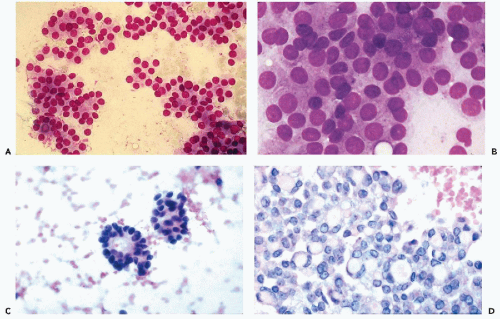
Figure 30-10 Follicular neoplasms. A,B. Flat spherical clusters of follicular cells stained with MGG. Note the absence of colloid and the somewhat enlarged nuclei, when compared with normal acini shown in Figure 30-2. C. One acinar and one solid cluster of follicular cells (Diff-Quik). D. Histologic section of a follicular adenoma, corresponding to C. Note small follicles containing scanty, pale colloid.

Less well, or poorly, differentiated follicular carcinomas are a mixture of follicles lined by distinctly abnormal cells and solid sheets of neoplastic cells with enlarged, atypical nuclei and prominent nucleoli (see Fig. 30-12D). These tumors have a less favorable prognosis than the well-differentiated variety. Clear cell carcinomas of the thyroid are variants of follicular carcinoma.


Cytology

In general, the aspirates of follicular neoplasms are cellular and have an abundant population of cells with little colloid. Smears of follicular adenomas usually contain numerous clusters of follicular cells, usually arranged in flat follicular or papillary structures or flat, tight aggregates. The tumor cells may also form small, rosette-like acinar clusters, containing inspissated colloid (Fig. 30-10). Single follicular cells and nuclei stripped of cytoplasm (“naked nuclei”) are scattered throughout the smear. Anisonucleosis may be present. The nuclear chromatin is uniformly distributed and the nucleoli are small and barely
visible. Small, indistinct intranuclear clear inclusions may occur and do not necessarily indicate a malignant tumor (see Fig. 30-10B). The papillary configuration of some of the colloid-free clusters may be reminiscent of papillary carcinoma. However, the clusters in papillary cancer are often multilayered and complex and show specific nuclear abnormalities that are not evident in follicular tumors (see below). Aspiration smears from follicular carcinomas are also highly cellular with little or no colloid. The cells occur primarily in clusters and are often arranged in follicle-like structures. Thus, they may closely resemble the cytologic presentation of follicular adenoma. In well-differentiated follicular carcinoma, cellular atypia may be minimal, and the general impression from the smear may suggest a benign lesion, rather than a carcinoma (Fig. 30-11). Therefore, in such cases, the cytologic diagnosis should be “follicular neoplasm or tumor,” clearly indicating that surgical excision and histologic examination is mandatory for a reliable differential diagnosis between follicular adenoma and a well-differentiated carcinoma.

In less well-differentiated forms of follicular carcinoma, nuclear atypia is present but it rarely reaches high levels of abnormality. The nuclei may vary in size and show some hyperchromasia (Figs. 30-12A and 30-13A). Rarely, nuclear pallor and small intracytoplasmic nuclear inclusions (nuclear holes) may be noted (Fig. 30-13A). Particularly valuable is the presence of prominent, large nucleoli within the follicular cells (see Fig. 30-12B). In such cases, the cytologic diagnosis of follicular carcinoma may be justified but we still prefer to sign out these cases as “follicular neoplasm-excision recommended” because some follicular neoplasms with severe atypia are encapsulated and their malignant nature is uncertain. In such cases, the histologic classification may vary between “atypical follicular adenoma” and “follicular carcinoma without invasion,” depending on the preference of the pathologist. Still, in some of these cases, recurrent or even metastatic tumor may be observed, sometimes many years after the removal of the primary (Fig 30-13B).


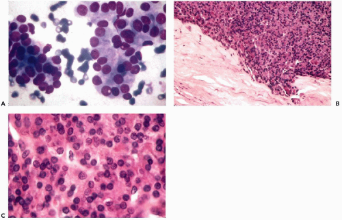
Figure 30-11 Follicular carcinoma. A. Aggregates of follicular cells with nuclear enlargement and small intranuclear cytoplasmic inclusions. The colloid is scanty. B. Periphery of the follicular tumor showing invasion of capsule. C. Histologic section of the same tumor composed of sheets of follicular cells with intranuclear cytoplasmic inclusions. (A: Diff-Quik stain.)


Hürthle Cell Tumors (Oxyphilic or Oncocytic Adenoma and Carcinoma)


Histology

These usually encapsulated tumors are composed of sheets and follicles composed of large, eosinophilic Hürthle cells with granular cytoplasm. Intracytoplasmic lumens, containing thyroglobulin, have been observed in these cells (Gonzales-Campora et al, 1986). The follicles contain little or no colloid (Fig. 30-14D). Nuclear abnormalities in the form of large, irregular, hyperchromatic nuclei are common. The configuration of nuclei has no prognostic significance because most such tumors are encapsulated
and do not recur or metastasize after careful surgical removal. A papillary variant of Hürthle cell tumor and a variant of papillary carcinoma composed of Hürthle cells with lymphoid stroma (Warthin’s-like tumor) are discussed below with papillary carcinomas. Fewer than 10% of the Hürthle cell tumors invade the capsule, blood vessels, and adjacent organs and, therefore, must be considered malignant (Fig. 30-15D). Because of their unpredictable behavior, surgical removal of these tumors is the treatment of choice (Nguyen et al, 1999). Infarction of these tumors is an infrequent, but known, complication of FNA (Kini, 1996; Pinto and Mandreker, 1996).


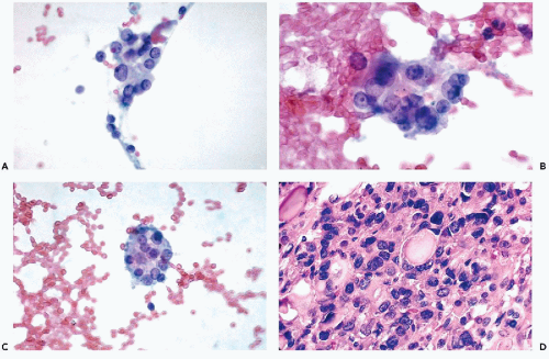
Figure 30-12 Follicular carcinoma with rapid progression in a 38-year-old man. A. Cluster of follicular cells showing variability in the nuclear sizes. B. High-power view of the tumor cells showing prominent nucleoli within the enlarged nuclei. C. Follicular structure of tumor cells. Note the presence of nucleoli. D. Histologic section of the primary tumor showing marked nuclear abnormalities.


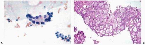
Figure 30-13 Follicular carcinoma. A. Thyroid aspirate. A cluster of acinar cells with central colloid. Nuclear pallor and small intranuclear cytoplasmic inclusions may be observed in some of the cells. This cluster could not be identified as malignant. B. Biopsy of bone, same patient. Bone metastasis of well differentiated thyroid carcinoma. This is an example of the so-called “benign metastasizing struma.”


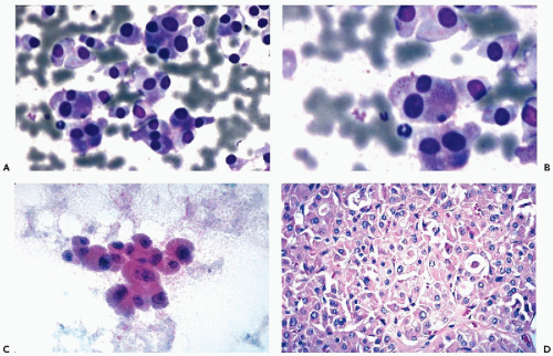
Figure 30-14 Malignant Hürthle cell tumor. A-C. Several examples of Hürthle cells in the tumor shown in D. Note the large Hürthle cells and some variability in cell and nuclear sizes. D. Section of the Hürthle cell tumor corresponding to A, B, and C. (A,B: Diff-Quik stain; B: high magnification.)


Cytology

In contrast to Hashimoto’s thyroiditis, where the Hürthle cells are dispersed or form small clusters and show considerable nuclear variability, in Hürthle cell neoplasms, the cells are usually of uniform size and shape and form tight aggregates or clusters. Small, basophilic cytoplasmic granules are better seen in air-dried smears stained with hematologic stains. The nuclei are large, vary in size, and may contain visible nucleoli (see Figs. 30-14 and 30-15). Intracytoplasmic nuclear inclusions may be observed (Koss et al, 1992). Galera-Davidson (1997) and Yang and Khurana (2001) observed that the presence of intracytoplasmic lumens containing thyroglobulins and capillary vessels (named transgressing vessels) in aspirates is more likely to occur in the malignant variant of a Hürthle cell lesion than in a benign tumor or Hashimoto’s thyroiditis. The most important point of differential diagnosis of Hürthle cell tumor is Hashimoto’s thyroiditis with little or no lymphocytic component.


Papillary Carcinoma and Its Variants


Clinical Data and Histology

Papillary carcinomas are the most common malignant tumors of the thyroid, usually presenting as a palpable thyroid nodule that does not absorb radioactive iodine and, therefore, is “cold” on scintiscan (see Fig. 13-17A). The tumors occur mainly in women but have been observed in men and in children, the latter either spontaneous or after exposure to radioactive elements, such as the recent Chernobyl disaster (Tronko et al, 1999).

The histologic structure of these tumors is fairly characteristic: anastomosing, papillary branches of the tumor are lined by cells with opaque or clear homogeneous nuclei, sometimes showing visible nucleoli. Clear, sharply demarcated, intranuclear cytoplasmic inclusions and psammoma bodies are commonly observed. The connective tissue core of the tumor branches is richly vascularized (see Figs. 30-16D and 30-17D).

This group of tumors may have unusual clinical presentation. Although, in most instances, a nodule is observed in the thyroid, the first clinical manifestation of the tumor may be a metastasis to a lymph node of the neck from a small, occult primary tumor. Such metastases may be cystic. The faulty term “lateral aberrant thyroid” has sometimes been used in the past to describe these situations (recent reviews in Coleman et al, 2000; Verge et al, 1999).

The behavior of papillary carcinomas is unpredictable. Most of the tumors are indolent and do not recur or metastasize after removal, even in the presence of metastases to neck lymph nodes. In some cases, however, widespread metastases to lung, the skeleton, central nervous system and, occasionally, other organs may be observed.

Only gold members can continue reading. Log In or Register to continue

Stay updated, free articles. Join our Telegram channel

Jun 8, 2016 | Posted by in PATHOLOGY & LABORATORY MEDICINE | Comments Off on The Thyroid, Parathyroid, and Neck Masses Other Than Lymph Nodes

Full access? Get Clinical Tree

Get Clinical Tree app for offline access