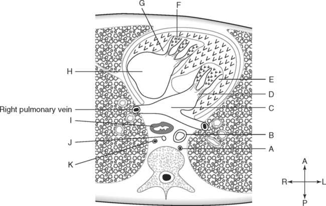
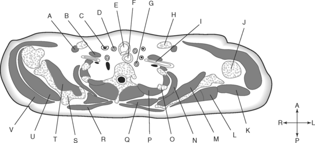
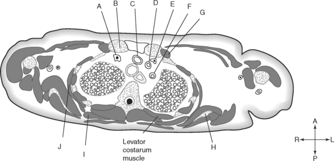
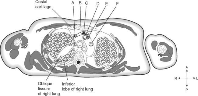
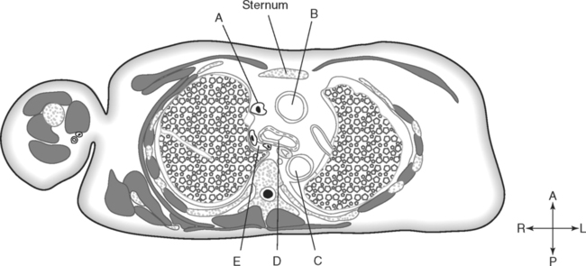
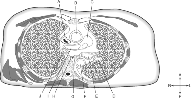
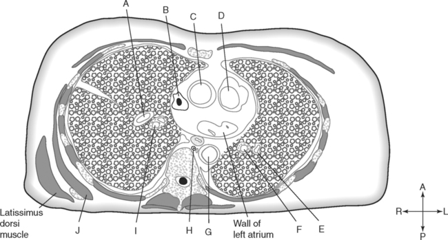
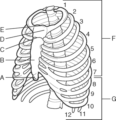
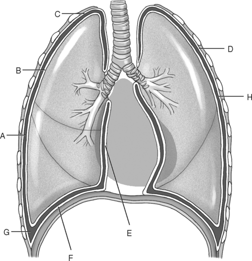
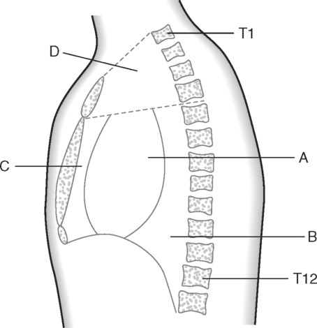
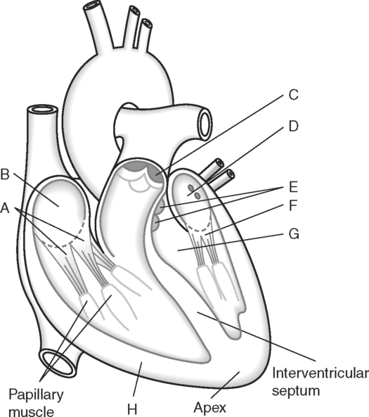
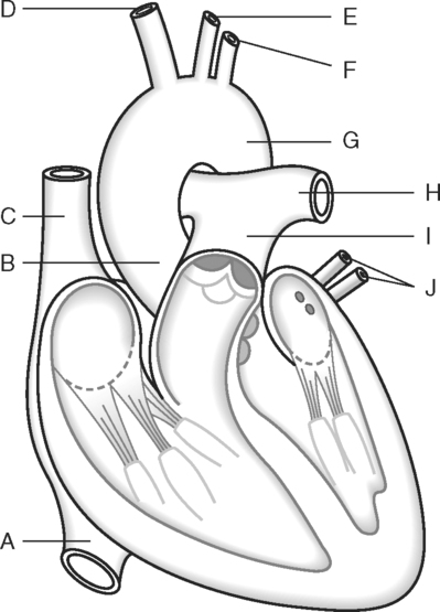
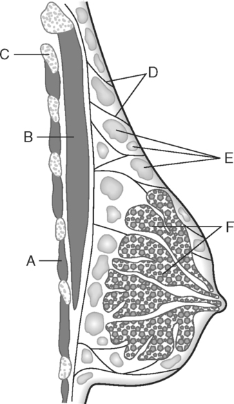
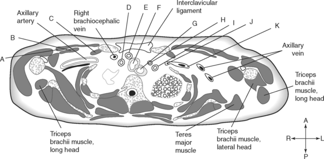
Identify the Features Indicated in Fig. 2-1: Identify the Features Indicated in Fig. 2-2: 1. Which layer of the pleura is closely attached to the lung? 2. What type of fluid is produced by the pleural membrane? 3. What specific portion of the parietal pleura is closest to the pericardial sac? Identify the Structures Indicated in Fig. 2-3: 1. What specific portion of the mediastinum contains the heart? 2. What is the boundary between the superior mediastinum and the inferior mediastinum? 3. Name three structures and/or tissues that are found in the anterior mediastinum. 4. Name four vessels and/or structures that are found in the posterior mediastinum. Identify the Structures Indicated in Fig. 2-4: 1. The valve between the right atrium and the right ventricle is called the ______________________ valve. 2. The chordae tendineae are anchored to projections of myocardium called ______________________. 3. The valve located between the most posterior chamber of the heart and the chamber with the thickest walls is the ______________________ valve. 4. Valves located in the outflow vessels from the heart are called ______________________ valves. 5. Which valve is located at the exit from the right ventricle? Identify the Structures Indicated in Fig. 2-5: 1. Which chamber of the heart is associated with the pulmonary veins? 2. On which side of the heart do the venae cavae enter? 3. What vessel exits the heart from the right ventricle? 4. Which vessel is the middle branch from the arch of the aorta? Identify the Structures Indicated in Fig. 2-6: 1. Which chamber of the heart is most anterior? 2. The lymphatic vessel posterior to the esophagus is called the ______________________. 3. Which chamber of the heart is most directly related to the esophagus? Identify the Structures Indicated in Fig. 2-7: 1. The skeletal muscle most closely related to the breast is the ______________________ muscle. 2. Fibrous connective bands that help to support the breast are called _____________________ or ligaments. 3. The parenchyma of the breast is arranged in ______________________. Identify the Structures Indicated in Fig. 2-8: 1. What blood vessel is prominent posterior to the anterior scalene muscle? 2. What is the small muscle along the posterior surface of the clavicle? 3. Which is more posterior, the right subclavian artery or the right common carotid artery? 4. Name the four muscles of the rotator cuff. 5. The muscle that appears to enclose the ribs and intercostal muscles like parentheses is the ______________________. 6. Which is more posterior, the brachiocephalic vein or the subclavian artery? Identify the Structures Indicated in Fig. 2-9: 1. What muscle extends from the ribs to the vertebral transverse process in this section? 2. What blood vessel is closest to the apex of the left lung? 3. What two blood vessels join to form the brachiocephalic vein? 4. Lateral to the clavicle, what name designates the continuation of the subclavian artery and vein? In Fig. 2-10 Color or Outline: Identify the Structures Indicated in Fig. 2-10: 1. What bones are anterior to the brachiocephalic veins? 2. What two muscles form the anterior wall of the pectoral region? 3. What is the name of the specific serous membrane that is closely applied to the lung surface? 4. What single vessel branches from the aorta to supply the right side of the head and the right upper extremity? 5. What is the name of the ligament between the two clavicles? In Fig. 2-11 Color or Outline: Identify the Structures Indicated in Fig. 2-11: 1. Which is longer, the right brachiocephalic vein or the left brachiocephalic vein? 2. Which is more anterior, the left brachiocephalic vein or the brachiocephalic artery? 3. What is the name of the muscle that, in transverse sections, appears to enclose the rib cage like a set of parentheses? 4. Which is more to the left, the trachea or the esophagus? 5. Which of the two fissures in the right lung is more superior? In Fig. 2-12 Color or Outline: Identify the Structures Indicated in Fig. 2-12: 1. What vessel curves over the root of the right lung to drain into the superior vena cava? 2. Which is most to the right side, the ascending aorta, the descending aorta, or the superior vena cava? 3. What lymphatic vessel is to the right of the descending aorta? 4. Which is more anterior, the descending aorta or the esophagus? 5. Which is more to the right, the trachea or the descending aorta? 6. Which is most posterior, the left main bronchus, the thoracic duct, or the esophagus? In Fig. 2-13 Color or Outline: Identify the Structures Indicated in Fig. 2-13: 1. Which is most to the left side, the superior vena cava, the pulmonary trunk, or the ascending aorta? 2. Which is most posterior, the superior vena cava, the right main bronchus, or the right pulmonary artery? 3. What vessel is posterior to the superior vena cava but anterior to the right bronchus? 4. Which is most to the right side, the azygos vein, the descending aorta, or the esophagus? 5. Which is most anterior, the superior vena cava, the esophagus, or the right main bronchus? In Fig. 2-14 Color or Outline: Identify the Structures Indicated in Fig. 2-14:
The Thorax
Basicmedical Key
Fastest Basicmedical Insight Engine

