Chapter 3 The Skin
“The power of making a correct diagnosis is the key to all success in the treatment of skin diseases; without this faculty, the physician can never be a thorough dermatologist, and therapeutics at once cease to hold their proper position, and become empirical.”
Basic Terminology and Diagnostic Techniques
1 How many skin diseases exist? What are the two main categories of skin lesions?
There are more than 1400 skin diseases. Yet, only 30 are important, common, and worth knowing. The first step toward their recognition is the separation of primary from secondary lesions (Table 3-1).
Primary lesions result only from disease and have not been changed by additional events (such as trauma, scratching, or medical treatment; see Table 3-1). To better identify primary lesions, pay attention to their colors, shape, arrangement, and distribution.
Secondary lesions instead have been altered by outside manipulation, medical treatment, or their own natural course.
Table 3-1 Dermatologic Lesions
skin lesions | ||
---|---|---|
Primary | Secondary | Special |
Solid (Nonpalpable) | Crusts | Purpurae |
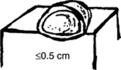
• Macules (≤0.5 cm) | Scales | Petechiae |
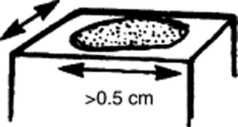
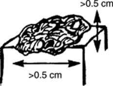
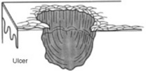
• Patches (>0.5 cm) | Ulcers | Ecchymoses |
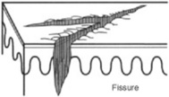
Fissures | Teleangiectasias | |
Solid (Palpable) | Excorations | Comedones |
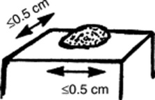
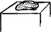
• Papules (≤0.5 cm) | Scars | Burrows |
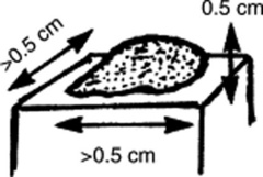
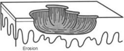
• Plaques (>0.5 cm) | Erosions | Target lesions |
• Nodules (deeper plaques) | Lichenification | |
• Wheals (pruritic plaques) | Atrophy | |
• Tumors (larger nodules) | Scars | |
Sinuses | ||
Fluid-Lesions | ||
• Vesicles (fluid-filled papules) | ||
• Pustules (pus-filled papules) | ||
• Bullae (fluid-filled plaques) | ||
• Cysts (fluid-filled nodules) |
2 What are the major primary lesions?
Macules: Flat, nonpalpable, circumscribed areas of discoloration ≤0.5 cm in diameter. Typical macules are the familiar freckles.
Patches: Flat, nonpalpable areas of skin discoloration >0.5 cm in diameter (i.e., a large macule). A typical patch is the one of vitiligo.
Papules: Raised and palpable lesions ≤0.5 cm in diameter. They may or may not have a different color from the surrounding skin. A typical papule is a raised nevus.
Plaques: Raised and palpable lesions >0.5 cm in diameter (i.e., a large papule). Usually confined to the superficial dermis, they may result from the confluence of papules. A typical plaque is that of psoriasis.
Nodules: Raised, palpable, and elevated lesions >0.5 cm in diameter, which, unlike plaques, go deeper into the dermis. Since they are below the surface of the skin, the overlying cutis is usually mobile. Typical nodules are those of erythema nodosum.
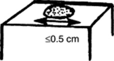
Tumors: Nodules that are either >2 cm in diameter or poorly demarcated. Usually neoplastic.
Wheals (hives): Raised, circumscribed, edematous, and typically pruritic plaques that are pink or pale but typically transient. Classic wheals are the lesions of urticaria, or of a mosquito bite.
Vesicles (blisters): Fluid-filled, circumscribed, and raised lesions that contain clear serous fluid and are ≤0.5 cm in diameter. Typical vesicles are those of herpes simplex.
Bullae: Vesicles >0.5 cm in diameter. Commonly seen in patients with second-degree burns. Presence of a bulla is so important that it usually trumps all other concomitant primary lesions.
Cysts: Raised and encapsulated lesions that contain fluid or semi-solid material. Typical are the cysts of acne.
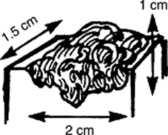
Pustules: Pus-filled papules. Typically seen in patients with impetigo or acne.
Purpura: Skin extravasation of red cells, which, based on size, may present as petechiae or ecchymoses. Palpable purpura is never normal and argues for an antigen-antibody complex (vasculitis). Often localized to the lower extremities, the lesions of Henoch-Schönlein are typical examples of a palpable purpura. Internal organs (kidneys, GI tract) are often involved too.
Petechiae: Reddish-to-purple discolorations, caused by a microscopic hemorrhage. These are <0.5 cm in diameter and usually in clusters. With the exception of color, they resemble papules or macules (depending on whether they are palpable or not). Typical petechiae are those of typhus. The lesions of thrombocytopenic thrombotic purpura (TTP) are typical petechiae too.
Ecchymoses (bruises): Reddish-to-purple discolorations larger than petechiae. Except for color, they resemble plaques and patches (depending on whether they are palpable or not). Typically located below an intact epithelial surface.
Spider angiomas: These are arterial teleangiectasias, i.e., vascular arterial lesions that resemble the legs of a spider. They fill from the center and blanch whenever this is compressed.
Venous spiders: These are venousteleangiectasias, i.e., vascular venous lesions that also resemble the legs of a spider. Hence, they fill from the periphery, not the center. They empty with pressure.
(Figures adapted from Willms JL, Schneiderman H, Algranati PS: Physical Diagnosis. Baltimore, Williams & Wilkins, 1994, with permission.)
3 What are the major secondary lesions?
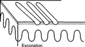
Excoriations: Linear erosions produced by scratching. Often raised, scratch marks may also present as crust on top of a primary lesion that has been partially scratched off. They are almost exclusively confined to the eczematous diseases.
Lichenification: A typical skin thickening seen in chronic pruritus with recurrent scratching. Resembles the callus formation of palms and soles after recurrent trauma. Lichenified skin is hardened, leather-like, with prominent markings and some scaling. Like excoriation, lichenification is typical of eczematous diseases. In fact, it is considered pathognomonic of atopic dermatitis.
Scales: Raised lesions presenting as flaking of the upper skin surface. In fact, they represent thickening of the stratum corneum, the uppermost layer of the epidermis. Scales may be white, gray, or tan. They may also be small or rather large. They provide the squamous component to papulosquamous diseases. They are extremely common in the scalp, where they suggest either banal processes (dandruff) or more serious conditions (seborrheic dermatitis, psoriasis, and tinea capitis).
Crusts: Raised lesions produced by dried serum and blood cell remnants. Usually preceded by fluid-filled primary lesions (i.e., vesicles, pustules, or bullae). The most familiar crust is the “scab” of impetigo.
Erosions: Depressed lesions produced whenever the epidermis is either removed or sloughed. They are moist, usually red, and well circumscribed. Classic erosions are those of chickenpox following rupture of a vesicle.
Ulcers: Depressed lesions produced whenever not only the epidermis but also part (or all) of the dermis is gone. Ulcers are concave, often moist, and at times inflamed or even hemorrhagic. They heal with scarring. A classic ulcer is that of the syphilitic chancre.
Fissures: Depressed lesions presenting as narrow, linear, and vertical cracks that penetrate through the epidermis, reaching at least part of the dermis. Classic fissures are those of the athlete’s foot.
Atrophy: Usually the nonspecific end-product of various skin disorders. It is characterized by a pale and shiny area, with loss of cutaneous markings and full skin thickness.
Sinuses: Connective channels between the surface of the skin and deeper components.
(Figures from Fitzpatrick JE, Aeling JL: Dermatology Secrets. Philadelphia, Hanley & Belfus, 1996, with permission.)
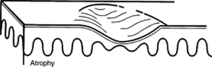
4 Are there other ways to classify skin lesions?
Many ways. One divides lesions into four groups based on the relationship with the surrounding skin:
5 What is the pattern of distribution?
Beside the distribution in the body (i.e., generalized versus localized), this descriptor refers to the relationship of lesions to one another:
6 What is the configuration of a skin lesion?
It is the outline of the lesion as observed from above. The most common configurations are:
Annular: Doughnut-shaped lesions. Fungal infections present as red rings with the scaly surface.
Linear: Lesions arranged in a line. For example, streaks of small vesicles on an erythematous base. The most common linear lesion is the rash of poison ivy, also called rhus dermatitis (rhus is the Greek word for sumac, which describes various shrubs or small trees). Some species of sumac, or rhus, include poison ivy and poison oak—both cause an acute itching rash on contact.
Reticular: Lesions organized in a net-like cluster
Gyrate: Lesions with a serpiginous (or polycyclic) configuration—as in gyrate erythema
7 How should an initial cutaneous exam be done?
From head to toe. Patients should fully disrobe, so that the entire body can be inspected—including palms, soles, scalp, and mouth. If a total skin exam is not feasible, a targeted exam of lesions, as guided by history, is also appropriate. Either way, always attempt to complete at least an upper body exam, since the trunk represents a large but easily examinable surface area.
8 How should a specific lesion be examined in order to better classify it?
Not only by looking at it, but also by touching it, which is key for determining its characteristics and relationship to the surrounding skin: papular, sclerotic, soft, mobile, rough, or smooth. Note that, in contrast to other fields of medicine, a thorough physical examination is key for dermatologic diagnoses. In fact, it is much more key than history.
9 And so, what are the required components of a dermatologic diagnosis?
Based on the aforementioned criteria, a dermatologic diagnosis requires first of all the recognition of both the primary and the secondary lesions. Once that is done, lesions must be assessed based on:
Morphology: Color, shape, dimensions (width and height, if necessary), elevation/depression, and palpable features (smoothness, induration, tenderness, scaling, and crusting)
Distribution (body location): Generalized versus localized
Distribution (arrangement to one another): Clustered, confluent, dermatomal
10 What are the tools necessary for a dermatologic diagnosis?
Wood’s lamp: A fluorescent and long-wave ultraviolet light that has been narrowed to 360 nm. Developed by Robert Wood (1868–1955), it is commonly relied on for detecting fungal lesions, areas of hypopigmentation, and porphyrin compounds. In a darkened room, it reveals fungal infections (like tinea capitis) as sharply marginated patches of bright blue-green. Since melanin absorption is at 360 nm, it also identifies areas of vitiligo or tinea versicolor (hypopigmented patches as pale-white, and depigmented areas as bright-white). Finally, it makes porphyrin compounds stand out as coral red fluorescence, like in erythrasma, a bacterial infection of intertriginous areas (axillae).
11 How does one prepare a potassium hydroxide (KOH) stain for fungi?
Scrape a few scales off the skin, and place them onto a glass slide. Then, pour potassium hydroxide (10%) onto the slide, and apply a coverslip. After gently warming up the slide (usually over a match), examine the preparation under a microscope for hyphae and spores.
12 What is a Tzanck test?
A test commonly used for herpes virus infection. Pioneered by the Russian dermatologist Arnault Tzanck (1886–1954), it is carried out by unroofing a vesicle with a scalpel, scraping its base, applying the material to a microscope slide, fixing it with 95% alcohol, and preparing it with a standard Wright or Giemsa stain. If positive, the test reveals multinucleated giants cells, confirming that the cause of the lesion is either herpes simplex or varicella zoster.
13 What skin appendages should be part of a thorough dermatologic exam?
Definitely hair (including eyebrows, eyelashes, and scalp), but also fingernails and toenails. As opposed to hair, these are rarely of concern to the patient, yet they may narrow the differential diagnosis, guide work-up, and provide important clues for the identification of systemic illnesses. Hence, it is the physician’s responsibility to examine them thoroughly.
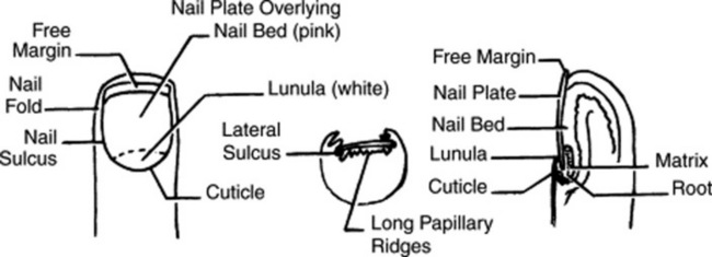
14 How should fingernails and toenails be assessed?
If covered by polish, clean them first with a solvent like acetone. Then pay attention to color and shape but also to anatomic details (Fig. 3-25):
Lunula: The white half-moon at the proximal edge of the nail bed
Cuticle: The thin skin adherent to the nail at its proximal portion
Perionychium: The epidermis forming the ungual wall at the sides and back of the nail
Although fingernails tend to be more informative than toenails (since they grow more rapidly and suffer fewer traumas), always examine them both. Inspect them first without applying any pressure. Then, blanch the fingertip to see if a pigmented lesion changes color (which would argue for discoloration of the vascular bed rather than the nail plate). Finally, place a penlight against the finger pulp and shine it through the nail. If upon illumination a discoloration disappears, it is also more likely to be in the vascular bed than in the soft tissue or matrix. When indicated, scrape the nail plate surface, and do a potassium hydroxide preparation to rule out fungal disease. Note that nail changes due to systemic disease (as opposed to trauma) often occur in the matrix, so that the leading edge of the abnormality (for example, a pigmentation change) is usually shaped like the distal portion of the matrix. To estimate the time of initial insult, measure the distance from the proximal nail fold (cuticle) to the leading edge of the pigmentation change, remembering that nails grow 0.1–0.15 mm/day.
15 What systemic conditions are associated with changes in nail shape or growth?
Clubbing: Inflammatory bowel disease, pulmonary malignancy, asbestosis, chronic bronchitis, chronic obstructive pulmonary disease, cirrhosis, congenital heart disease, endocarditis, atrioventricular malformation and fistulas (see also question 19)
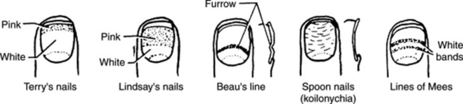
Koilonychia (spoon nails) (Fig. 3-26): Iron deficiency anemia, hemochromatosis, Raynaud’s disease, systemic lupus disease, trauma, nail-patella syndrome
Onycholysis: Psoriasis, infection, hyperthyroidism, sarcoidosis, trauma, amyloidosis, connective tissue disorders
Pitting: Psoriasis, Reiter’s syndrome, incontinentia pigmenti, alopecia areata
Beau’s lines (see Fig. 3-26): Any severe systemic illness that disrupts nail growth, Raynaud’s disease, pemphigus, trauma
Yellow nail: Lymphedema, pleural effusion, immunodeficiency, bronchiectasis, sinusitis, rheumatoid arthritis, nephrotic syndrome, thyroiditis, tuberculosis, Raynaud’s disease
16 What systemic conditions are associated with changes in nail color?
Terry’s nails (see Fig. 3-26): Hepatic failure, cirrhosis, diabetes, congestive heart failure, hyperthyroidism, malnutrition
Azure lunula: Wilson’s disease, silver poisoning, quinacrine
Lindsay’s nails (half-and-half nails) (see Fig. 3-26): Specific for renal failure
Muehrcke’s lines: Specific for hypoalbuminemia
Lines of Mees’ (see Fig. 3-26): Arsenic poisoning, Hodgkin’s disease, congestive heart failure, leprosy, malaria, chemotherapy, carbon monoxide poisoning, other systemic insults
Dark longitudinal streaks: Melanoma, benign nevus, chemical staining, normal variant in darkly pigmented people
Longitudinal striations: Alopecia areata, vitiligo, atopic dermatitis, psoriasis
Splinter hemorrhage: Subacute bacterial endocarditis, systemic lupus erythematosus, rheumatoid arthritis, antiphospholipid syndrome, peptic ulcer disease, malignancies, oral contraceptive use, pregnancy, psoriasis, trauma
Telangiectasia: Rheumatoid arthritis, systemic lupus erythematosus, dermatomyositis, scleroderma
Nails
19 What is clubbing?
A condition that can be (1) idiopathic; (2) congenital (dominant trait); or (3) a clue to serious underlying pathology, including cardiovascular, hepatobiliary, mediastinal, endocrine, gastrointestinal, neoplastic, infectious, and, especially, pulmonary (see Chapter 13, questions 101–116).
20 What is onycholysis?
“Loosening of the nail” in Greek, it consists of separation of the nail plate from the nail bed. This begins at the free edge and progresses proximally, causing a traumatic uplifting of the distal plate. Usually incomplete, it results in white discoloration of the affected area, often complicated by secondary microbial colonization. Typically traumatic, it may also result from any local problem that separates the plate from the nail bed, such as periungual warts or onychomycosis. It also may originate from eczematous or drug disorders, reactions to acrylic nails or nail-hardeners, and psoriasis. The latter may at times occur with complete separation of the nail, which is either mechanically lifted off the bed or separated by traumatic bleeding. Absent local explanations, thyrotoxicosis must be ruled out (“Plummer’s nails”). This may also cause brown nail discoloration.
21 What is nail pitting?
An early but nonspecific sign of psoriasis that can also occur in alopecia areata and chronic dermatitis. Nails are pocked by multiple tiny depressions, which may eventually cause the nail to crumble. Pitting is due to loss of parakeratotic cells from the surface of the plate, starting in the proximal nail matrix and migrating distally.
22 What are the nail findings of psoriasis?
Many, affecting 10–55% of patients and <5% of those with no other cutaneous manifestation. If untreated, they can lead to functional and social impairment. They are especially common in psoriatic arthritis (53–86% of patients), even though only 10–20% of all psoriatic patients have the arthritis.
Pitting (previously discussed)
Oil spot or salmon patch/nail bed: The most diagnostic nail sign; it consists of a translucent, yellow-red discolored patch in the nail bed, resembling a drop of oil beneath the plate.
Beau’s lines in the proximal nail matrix (see later)
Leukonychia: Areas of white nail plate; due to parakeratotic foci in the mid-matrix
Subungual hyperkeratosis: Excessive proliferation of the nail bed that can lead to onycholysis
Nail plate crumbling: Weakened nail plate, bed, and matrix from diseased underlying structures
Splinter hemorrhages: Longitudinal black lines due to tiny capillary hemorrhages between the nail bed and plate; analogous to Auspitz sign (pinpoint bleeding under the psoriatic plaque)
Dilated tortuous capillaries in the dermal papillae
Spotted lunula: Distal matrix involvement characterized by erythema of the lunula
23 What is koilonychia (spooning)?
A “spoon-shaped” deformity of the nail, usually large enough to hold a drop of liquid. Normal in infants (but resolving after the first few years of life), it also may result from trauma, recurrent exposure to petroleum-based solvents, or nail-patella syndrome. It is also seen in Raynaud’s disease or lupus erythematosus, but rarely in isolation. Finally, it can occur with iron deficiency (with or without anemia) or, paradoxically, hemochromatosis (check iron, hemoglobin).
24 What is yellow nail syndrome?
An autosomal-dominant disease characterized by absent/hypoplastic and easily dislocated patellae, skeletal abnormalities, nephropathy, glaucoma, and fingernail dysplasia. This consists of slow nail growth, with a “heaped-up” or thickened appearance, excessive transverse curvature, loss of the lunula, and a yellowish/greenish discoloration. Eventually, the nail thickens and may even detach. Yellow/green nails may result from any condition that slows nail growth (such as infection, like Pseudomonas) or impedes lymphatic circulation. Hence, they have been associated with lymphedema of the extremities or face. They also have been linked to respiratory involvement (including chronic bronchiectasis or sinusitis), pleural effusions, internal malignancies, immunodeficiency syndromes, and rheumatoid arthritis (usually as a result of thiol drugs, such as bucillamine and gold sodium thiomalate). Microvascular hyperpermeability with protein leakage would explain the link of yellow nail syndrome with infection, hypoalbuminemia, pleural effusion, and lymphedema.
25 What are brittle nails (onychorrhexis)?
Nails whose distal plate splits into layers, with irregular, frayed, and torn borders—almost resembling the scaling of dry skin. Present in 20% of adults (and even more in older subjects), brittle nails may be hereditary or simply aging-related. They also may be due to strong solvents or repeated immersion in water (e.g., dishwashers). Dysmetabolic states (such as hyperthyroidism, malnutrition, and iron or calcium deficiency) must always be ruled out.
26 What is longitudinal ridging (Reedy nails)?
A normal variant of patients older than 50, but one that also can occur in younger subjects. It may even represent a brittle nail variation. Ridges typically extend from the proximal nail fold to the distal plate, with some being very prominent, especially in older women. They are usually multiple, but at times may be single—like in patients with lichen planus (see questions 38 and 210–216).
27 What is nail beading?
A condition due to faster matrix turnover. It is quite common in rheumatoid arthritis (RA). In a study of 119 RA patients and an equal number of controls, “global” beading (i.e., involving >50% of the nail) on at least six fingernails (or four toenails) was highly suggestive of underlying disease, with a positive predictive value of 95%. Beading, however, is uncommon in early RA, thus limiting its diagnostic value. Beading can occur with itraconazole too, since this also increases nail growth.
28 What is onychogryphosis?
A thickened nail plate, often due to trauma plus mycotic infection. Eventually, the nail curves inward, resembling a claw (gryphon is dragon in Greek) and “pinching” the nail bed (pincer nail).
30 What is leukonychia?
A condition characterized by white spots, patches, or streaks between the nail and nail bed. It is due to tiny bubbles of subungual air trapped in the plate layers after trauma. Leukonychia (white nail in Greek) may involve the entire nail (total), or present instead as lines (striate) or dots (punctate). Total leukonychia is a congenital dominant disorder, whereas striate or punctate forms are instead traumatic. The latter resolve with time, since spots eventually grow out with the nail plate
31 What are Beau’s lines?
Transverse grooves on the fingernails of patients recovering from a serious illness (like a myocardial infarction). This produces a transient intermittent inflammation of the matrix, resulting in arrested growth. Grooves will progress distally with the nail, eventually coming into view and finally moving out as the nail elongates. First described by the French Joseph H.S. Beau (1806–1865).
32 What are bitten nails lesions?
Lesions that may resemble Beau’s lines, insofar as they, too, are transversal indentations (no pun intended), although, more commonly, they present as absence of the free nail edge, produced by nervous biting and chewing. They are an important clue to underlying psychosocial pathology.
33 What are Muehrcke’s lines (ML)?
Two arcuate, narrow, and transverse white lines—parallel to the lunula and separated by an otherwise normal nail. Named after the American nephrologist who first described them in 1956, ML usually involve the second, third, and fourth fingers. They reflect a vascular abnormality in the nail bed (typically, subungual edema), and not in the nail plate. Hence, they do not progress distally with nail growth. Common in hypoalbuminemia (<2.2 gm/100 mL), they disappear with its resolution. In his original study, Muehrcke found paired, transverse, white bands in 23/31 (74%) patients with nephrotic syndrome and 8/9 with hypoalbuminemia (<2.3 gm/100 mL) from other causes. Lines were instead absent in all healthy subjects, and in those with albumin >2.2 gm/100 mL. Bands were more prominent after albumin had been <1.8 gm/100 mL for at least 4 months. In another study by Conn and Smith, Muehrcke lines were seen in 10/44 (23%) patients with hypoalbuminemia from various debilitating illnesses, but absent in those with normal serum levels. Hence, ML occurs in hypoalbuminemia from many reasons, including nephrotic syndrome, but also liver disease and malnutrition. Additionally, they can occur in pellagra, Hodgkin’s disease, sickle cell anemia, or nail damage from paraquat and chemotherapeutic agents. Although transverse white bands in the nail plate are often due to trauma to the matrix at the proximal nail fold (leukonychia), Muehrcke’s (and Mees’) lines are instead associated with a systemic disease. They typically span the entire breadth of the nail bed/plate, tend to be more homogenous, have a contour similar to the distal lunula (with a rounded distal edge and smoother borders), occur on several nails at once, and typically follow a generalized insult. Trauma-induced transverse white bands tend to be more linear, do not spread across the entire breadth of the nail plate, resemble the contour of the proximal nail fold (where trauma occurred), and have a history of localized trauma to the cuticle.
34 What are Mees’ lines?
They are transverse white lines distal to the cuticle, and typically laid down during a generalized illness or after a poisoning. In contrast to Muehrcke’s lines, Mees’ lines (also called Reynolds’ or Aldrich’s) are in the nail plate. Hence, they move distally with it. Differential diagnosis includes arsenic or thallium (2–3 weeks after an acute poisoning, or following chronic exposure), cancer chemotherapy, Hodgkin’s lymphoma, and other systemic disorders, such as severe cardiac or renal disease. They were first described by the Dutch physician R.A. Mees.
35 What are the traumatic changes of the nail?
Subungual hematomas are posttraumatic hematogenous extravasation in the nail bed. When partially digested and oxidized, a subungual hematoma may turn into a brownish and transversal dark band, often associated with indentation or damage of the nail plate.
37 What is longitudinal melanonychia?
A condition characterized by brownish/blackish longitudinal streaks or bands. Very common in dark-skinned individuals, it reflects melanin deposition from a normal increase of melanocytes in the nail bed. Yet, it also may indicate a much more ominous subungual melanoma. Hence, it presents a clinical challenge. When in doubt, always biopsy the nail matrix and bed.
38 What is lichen planus of the nail?
A condition present in 10% of patients with lichen planus. The most common finding is thinning of the nail plate, leading to longitudinal grooving and ridging (see also question 26). Hyperpigmentation, subungual hyperkeratosis, onycholysis, and longitudinal melanonychia can also be present.
40 What are splinter hemorrhages?
Tiny and linear red hemorrhages, extending from the free margin of the nail bed toward the proximal end. Traditionally linked to subacute bacterial endocarditis or trichinosis, they are more commonly due to trauma. According to conventional teaching, traumatic “splinters” extend all the way into the edge of the nail, whereas embolic splinters (like those of endocarditis) are fully contained within the nail bed. There is, however, little evidence to support this differentiation.
41 What are azure half-moons in nail beds?
The nails of Wilson’s disease (hepatolenticular degeneration). Lunulae are not white, but bluish.
42 What are Lindsay’s nails?
Half-and-half nails, with a white proximal half, and a dark distal half (usually brownish, but also reddish or pink). Named after the American physician who first described them in 1967, Lindsay’s nails have a sharp linear demarcation between the two halves, parallel to the nail edge. Of the 25 patients first described by Dr. Lindsay, 21 had chronic renal failure and two had minor nephropathy.
43 What are Terry’s nails?
Nails characterized by whitening of the proximal 80% of the nail, leaving a small rim of peripheral reddening, usually as sharply demarcated as in Lindsay’ nails. Named after the British physician who described them in 1954, Terry’s nails are seen in older subjects and patients with heart failure, cirrhosis, or non–insulin-dependent diabetes. They probably are due to subungual edema.
44 What are red half-moons in nail beds?
A variety of Terry’s nails, also described by him in the same 1954 Lancet paper. They are characterized by a lunula that is not white, but red. They also are called the nails of cardiac failure. In fact, all these half-and-half nails (Lindsay’s, Terry’s, and the red half-moons in nail beds) reflect conditions causing subungual edema, such as cardiac, hepatic, and renal disease.
45 How does vasculitis of the nail present?
With typical nail fold changes, like cuticular hypertrophy (with small, hemorrhagic infarcts) and periungual telangiectasias. These are common in scleroderma or dermatomyositis.
47 What is tinea unguium?
A dermatophytic infection of the nail, characterized by onychauxis (hypertrophy of the nail plate caused by keratin pileup); onycholysis (nail plate separation from the nail bed); and dystrophic, thickened, broken, and discolored nails (white, yellow, brown, black). Browning of the plate (with peeling and splitting of the edge) also occurs, together with ridging and white spots. Note that, although dystrophic nails are often onychomycotic, half of them are not (and instead due to trauma, psoriasis, or lichen planus). Hence, always do scraping and KOH staining to rule out fungal infection.
48 What is paronychia?
An acute or chronic inflammation of the perionychium, with redness, swelling, and tenderness.
49 How does pseudomonal infection of the nail present?
With a greenish nail, due to iron compounds produced by pseudomonas organisms invading the nail between the nail plate and nail bed. The darker the discoloration, the deeper the bacterial progression into the plate layers. This eventually causes it to lift altogether from the nail bed.
Hair
51 How is the hair assessed?
Region by region, first the scalp and then the body—including axillae, extremities, and pubic region. Evaluate quantity and distribution of hair, plus thickness and texture. Since alopecia is often caused by inflammation of the scalp, look for scaling, redness, crusts, and papules. Note that of the three scaling scalp lesions (seborrheic dermatitis, psoriasis, and tinea capitis), only tinea is associated with alopecia.
52 What is folliculitis?
An infection of hair follicles, most commonly due to staphylococci, but also gram-negative organisms and even fungi. In fact, Pityrosporum, the organism responsible for tinea versicolor, can overgrow in the follicles of patients with HIV and cause folliculitis.
53 What is eosinophilic folliculitis?
A disorder of unknown etiology, characterized by recurrent crops of sterile pustules and papules, often intensely pruritic, and presenting in multiple cycles of remissions and exacerbations. Although labeled as a folliculitis, it is actually a “sterile eosinophilic pustulosis” since lesions are not restricted to the hair follicle. It is probably an autoimmune response against sebocytes or some other components of sebum that usually occurs in the third to fifth decade of life. It can affect all races, but predominates in Asians. It presents as an area of erythematous papules and pustules, facial in 85% of patients, but also on the back and extensor surface of upper extremities. Papules eventually become confluent, creating indurate plaques with a healing center and a spreading periphery. These ultimately fade away, leaving residual hyperpigmentation and scaling. In HIV-infected patients, eosinophilic folliculitis (EF) presents with widespread urticarial lesions or large erythematous plaques with excoriations, often severe and persistent. Also seen in lymphoma, leukemia, myelodysplastic syndrome, atopy, and bone marrow transplantation.
Fluid-Filled Lesions: PUS (Pustules)—Table 3-2
Acne
54 What does acne look like?
The hallmark of acne (from the Greek acme, blooming) is the variety of its lesions, both inflammatory and noninflammatory, blossoming on the face and trunk of patients. Noninflammatory lesions include closed comedones (whiteheads) and open comedones (blackheads). Comedo is the Latin word for glutton, since this is indeed a juicy clump of sebum and keratin around a hair follicle. Closed comedones are dome-shaped papules 1–2 mm in size. When open, their content is exposed to air and thus blackened through oxidation. Hence, open comedones are 1–2-mm papules with a black keratinous plug closing the orifice of a sebaceous follicle. Inflammatory lesions are instead papules and pustules, as well as cysts (suppurative nodular lesions). Cystic lesions are more likely to scar if left untreated. Scarring may be pitted, hypertrophic, or papular.
Table 3-2 Fluid-Filled lesions
Fluid-Filled Lesions | ||
---|---|---|
Pus-Filled | Clear Fluid | |
Pustular | Vesiculo-Bullous | Bullous |
Acne vulgaris | H. simplex | Pemphygus vulgaris |
Acne rosacea | H. zoster/varicella | Pemphygoid |
Steroid acne | Dermatophytoses | Drug reactions |
• Erythema multiforme | ||
• Stevens-Johnson | ||
• TEN | ||
Folliculitis (bacterial/fungal) | Insect bites | Poison ivy/contact dermatitis |
Intertriginous candidiasis | Dermatitis herpetiformis | Bullous impetigo |
Porphyria cutanea tarda | ||
Lupus erythematosus |
55 Who develops acne?
Usually puberal teens, although girls may develop it one or more years before menarche. Men tend to have more severe acne, but this usually resolves by the second decade. In women, it often lingers well into the 40s and 50s, often with perimenstrual flares.
56 What are the other clinical presentations of acne?
The two most common are steroid acne and acne rosacea, although the latter is more than a variant of acne, and probably a completely different process of middle-aged and older people.
57 What is acne rosacea?
An acne-like rash that affects the central and flush/blush areas of the face (forehead, nose, cheeks, chin). Vascular lability is so typical that many patients have a history of facial flushing in childhood or early teens, often triggered by dietary and environmental factors (like hot drinks, alcohol, spicy foods, but also temperature changes and even emotions). Ocular rosacea may be accompanied by conjunctival injection, and rarely, chalazion and episcleritis. Although rare, there may be extrafacial involvement of the neck and upper part of the chest. Overall, symptoms of rosacea are intermittent, but can progressively lead to permanent flushing of the skin, with telangiectasias of cheeks and nose, lymphedema, and, ultimately, skin coarseness with acne-like papulopustular eruption. Yet, unlike acne, there is neither seborrhea (i.e., greasiness of the skin) nor comedones. In fact, rosacea may present with drying and peeling. Hyperplasia of sebaceous glands may eventually lead to a thickened and disfigured nose (rhinophyma). Still, this can be an isolated finding, and in fact, some authors even consider it a separate entity. Note that although rosacea is much more frequent in subjects of Celtic ancestry, it has also been described in dark-skinned individuals, such as African-Americans.
58 What is steroid acne?
An acne-like rash that may begin as early as 2 weeks after administration of systemic corticosteroids. In contrast to plain acne, lesions are monomorphous (generally pustules and dome-shaped papules) and covering the trunk, shoulders, and upper arms. A rosacea-like syndrome (including perioral dermatitis) also can result from the indiscriminate use of potent corticosteroids on the face.
Fluid-Filled Lesions: Clear Fluid (Vesiculobullous Diseases)
Herpes Simplex
60 How does herpes simplex present?
With pain or tingling. This precedes the onset of small erythematous papules/plaques of uniform size and shape, which eventually develop into grouped umbilicated vesicles on an erythematous basis, progress to pustules, and finally ulcerate, erode (especially in the genital areas), and crust. The most common locations are labial (vermilion border of the lip) and genital, although other areas may be affected, including the eye and presacrum.
61 Who develops herpes simplex?
Any age group, even though certain involvements are age-specific, such as stomatitis in children, labialis in adults, genitalis in sexually active subjects, and lumbosacral in patients older than 40.
62 What is the typical clinical course of herpes simplex?
Primary (initial) outbreaks: Usually more severe, with pain, edema, and a prolonged course. Still, the majority of primary infections go unnoticed because symptoms are either absent or so minimal that only the recurrent episodes are recognized.
Secondary (recurrent) disease: Less severe and shorter in duration. In fact, the hallmark of mucocutaneous herpes simplex is its ability to remain dormant in ganglia, eventually recurring in areas of primary infection. Frequency of recurrences varies, but for genital and labial herpes, the average is four episodes per year. Approximately 50% of patients with genital herpes have one or more episodes, often from local trauma, menses, and even stress.
63 What are the other clinical presentations of herpes simplex?
Herpetic gingivostomatitis: Presents in children and young adults with fever, malaise, sore throat, painful vesicles, and erosions of tongue, palate, gingiva, buccal mucosa, and lips
Herpetic whitlow (middle English for white flaw; also referred to as a felon): This is an occupational hazard of medical and dental professionals caused by exposure to the virus in a patient’s mouth. It is characterized by vesicles and edema of a digit, sometimes associated with erythema, lymphangitis, and lymphadenopathy of the arm. It may last for several weeks.
Herpes simplex in immunosuppressed patients: Frequently produces more severe and persistent ulceration, as well as disseminated cutaneous and systemic lesions
64 How is herpes simplex diagnosed?
Usually by clinical appearance (grouped umbilicated vesicles on an erythematous base). Still, a Tzanck preparation is diagnostic. Direct immunofluorescent antibody staining of infected cells is also an effective and rapid method of diagnosis. Viral cultures grow herpes simplex virus in several days. Biopsy of lesions demonstrates reticular and ballooning degeneration of the epidermis, multinucleated giant cells, and intranuclear inclusions.
Varicella
65 What are the features of varicella?
The classic lesion is a 2–3-mm elliptical vesicle surrounded by erythema, commonly described as a “dew drop on a rose petal.” This quickly converts into a pustule, then umbilicates and crusts. The crust falls off in 1–3 weeks, leaving a shallow pink depression that may result in scarring. New vesicles appear in successive crops, resulting in nonclustered lesions in all stages of development. These first appear on the face and scalp, then the trunk, and 2–3 days later on the arms and legs in a centrifugal fashion. Mucous membranes are also involved, especially the mouth.
66 Who develops varicella?
Mostly children younger than 10 years. Only 5% of cases occur in subjects older than 15.
67 What is the typical clinical course of varicella?
Incubation is 2 weeks, with patients being highly contagious for 2–4 weeks (from 1–2 days before exanthema until crusting of all lesions).
68 How are the other clinical presentations of varicella?
More severe in adults than in children, with higher fever, constitutional symptoms, and pneumonia (4% of cases). During pregnancy, varicella can be transmitted to the fetus, producing serious developmental abnormalities. In immunocompromised patients, it causes a more extensive and persistent rash, with higher morbidity and mortality, and even hemorrhagic complications.
69 How is varicella diagnosed?
By the classic appearance of the rash, often supported by a history of recent exposure. Conversely, to distinguish disseminated herpes simplex from varicella, a culture is often needed.
Herpes Zoster
70 What are the clinical features of herpes zoster?
The hallmark of zoster (girdle in Greek) is its clustered pattern, as opposed to the nonclustered pattern of varicella. The rash is usually preceded by paresthesia and pain in the involved dermatome (Saint Anthony’s fire). This is often mistaken as musculoskeletal, due to its intensity and deep location. Hence, zoster should always be considered in patients with deep dermatomal pain with no historical or physical explanations. The prodromal discomfort is then followed by the appearance of erythematous plaques, which, in sequence, develop into (1) grouped vesicles, (2) pustules, (3) umbilicated pustules, and (4) crusts. The rash is unilateral, does not cross the midline, and generally appears in only one dermatome, with trigeminal and T3 to L2 being the most common. It is not unusual to see a few vesicles just outside the involved dermatome.
71 Who develops herpes zoster?
Older subjects, with more than two thirds of cases occurring in patients older than 50 and less than 10% of cases in those younger than 20. It is also more common in immunosuppressed patients. Zoster in infants is usually associated with a history of maternal varicella infection during gestation.
72 What is the typical clinical course?
The rash erupts and progresses to complete crusting over 1 week, then resolves over several weeks. Postherpetic neuralgia (pain persisting after all crusts have fallen off) occurs in 15% of patients and is more common in the older population.
73 What are the other presentations of herpes zoster?
Involvement of the eye (ophthalmic branch): Heraldic lesions appear on the tip of the nose, due to infection of the nasociliary nerve. Get immediate ophthalmologic consultation.
Ramsay Hunt syndrome: Due to involvement of the geniculate ganglion with facial paralysis. Lesions occur over the external auditory canal or tympanic membrane, with or without vertigo, tinnitus, deafness/hyperacusis, unilateral loss of taste, and decrease in tear formation and salivation. Described by the Philadelphia neurologist James Ramsay Hunt (1874–1937).
Zoster in immunocompromised patients: Seen in patients with AIDS and malignancies (especially lymphocytic leukemia or Hodgkin’s) and those undergoing immunosuppressive therapies. Usually more severe and often disseminated. Chronic eruptions can be the first sign of HIV infection.
Scabies (see questions 255–257)
Dermatitis herpetiformis
74 What is dermatitis herpetiformis (DH)?
An immune-mediated blistering disease. First described by Duhring in 1884, DH is uniformly linked to gluten-sensitive enteropathy—usually asymptomatic. Hence, it can be controlled by a gluten-free diet. Typical of 20- to 40-year-old Northern Europeans. May degenerate into cancer.
75 What are the skin lesions of DH?
They are clusters of flesh-colored to erythematous herpetiform papules and vesicles. These are inflammatory, pruritic, and mostly located on the extensor surfaces (elbows, knees, buttocks, shoulders, and the posterior/nuchal scalp). Lesions may often present not as vesicles, but as erosions and crusts. Palms and soles usually are spared, and so is the buccal mucosa.
76 What is the course of DH?
A lifelong one, with remissions and exacerbations. Symptoms can be controlled with dapsone in 24–48 hours. A gluten-free diet can also provide control without medications.
Pemphigus and Pemphigoid
77 What is Pemphigus?
A group of autoimmune and potentially life-threatening mucocutaneous diseases characterized by intradermal blisters and circulating IgG against keratinocytes. Binding of autoantibodies results in acantholysis (i.e., loss of cell–cell adhesion), and antibody titers correlate with disease activity. Pemphigus (from the Greek pemphix, blister) comprises three subsets: pemphigus vulgaris (70% of all cases), pemphigus foliaceus, and paraneoplastic pemphigus.
78 What are the clinical features of pemphigus vulgaris (PV)?
It can affect all races, although more commonly Jewish and other Mediterranean groups. Its peak of onset is during the fifth and sixth decades. The primary lesion is a flaccid blister filled with clear fluid, and originating from a normal or erythematous skin base. Since blisters are fragile, they rupture easily, producing large, shallow, and painful erosions that heal slowly. Hence, erosions are the most common skin manifestation of the disease. Still, mucosal lesions (usually oral) are the most common presenting manifestation, affecting 50–70% of patients and often preceding cutaneous involvement by several months. Since intact bullae are rare in the mouth, most patients have irregularly shaped erosions of gingival, buccal, or palatine mucosa. These are painful, slow to heal, and interfere with eating, drinking, or swallowing. Other involved mucosae include conjunctiva, esophagus, larynx, labia, vagina, cervix, penis, urethra, and anus.
79 Are there any other causes of PV?
A form of PV (but also BP, see question 80) can be drug induced, resulting from penicillamine, captopril, thiol-containing compounds, and rifampin. Emotional stress can also trigger it. Finally, PV may occur in other autoimmune diseases, including myasthenia gravis and thymoma.
80 What is bullous pemphigoid (BP)?
Another autoimmune and blistering skin disease, except that in contrast to PV it rarely involves mucous membranes, it rarely erodes (lesions are subepidermal and thus the bullae have thicker roofs), and it is typically pruritic. Caused by the binding of circulating IgG autoantibodies to the skin basement membrane, BP is more common than PV, affects older patients (average age 65), and runs a chronic course marked by remissions and exacerbations. Onset may be subacute or acute, with widespread, tense, and often pruritic blisters. These may at times arise from persistent urticarial lesions, but also from chronic nonbullous inflammatory diseases, such as lichen planus and psoriasis. Rupture of the blisters leaves painful and disabling erosions, especially when involving palms and soles. Still, the primary lesion of BP is the bulla: tense, arising from normal-appearing as well as erythematous skin, and affecting any part of the body (even though flexural areas are usually a preferential site). Involvement of ocular and oral mucosae is rare (only 10–25% of patients), but when it occurs, oral intake may be limited because of dysphagia.
81 What is Nikolsky’s sign? What is its significance?
It is a sign described in 1896 by Pyotr W. Nikolsky, a Russian dermatologist who taught at Warsaw and Rostov. It consists in the superficial separation of normal-appearing epidermis into an erosion, as a result of the shearing stress produced by sliding a finger on it. It is due to poor adhesion of the epidermal cells (acantholysis). Hence, it occurs in bullous diseases like pemphigus, but not pemphigoid, where the epidermal split is much deeper. Still, it is not entirely specific for PV, since it can occur in other active blistering diseases, such as scalded skin syndrome.
82 What is Asboe-Hansen sign?
Another sign of PV; lateral pressure on the edge of a blister may spread it into unaffected skin.
Drug Reactions
83 What are the most important cutaneous manifestations of drug reactions?
Erythema multiforme (EM), toxic epidermal necrolysis (TEN), and Stevens-Johnson syndrome (SJS). Even though EM may be entirely distinct, SJS and TEN probably represent different manifestations of a single disease. A recent classification based on percentage of epidermal detachment defines SJS as denuding <10% of body surface area (BSA), overlapping SJS/TEN as denuding 10–30%, and classic TEN >30%. Bullous EM, previously grouped with SJS, causes epidermal detachment in <10% of BSA, but has acral target lesions or raised atypical targets.
84 What is erythema multiforme (EM)?
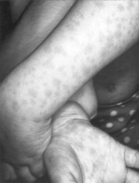
A relatively benign process characterized by target or targetoid lesions, with or without blisters, in a symmetric and acral distribution. In fact, the rash favors palms and soles, dorsum of hands, face, and extensor surfaces of extremities (Fig. 3-27). It is often associated with oral lesions, but rarely involves more than one mucosal surface. Although it can be caused by drugs, it is most commonly a sequela of herpes virus infection. It has low morbidity, no mortality, but frequent recurrences. It may be associated with epidermal detachment, yet denudation always involves <10% of BSA.
85 What is Stevens-Johnson syndrome (SJS)?
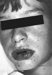
A potential dermatologic emergency. First described in 1922 by the American pediatricians Albert Stevens and Frank Johnson, SJS is characterized by widespread purpuric macules and targetoid lesions, usually more common on face and torso, and with concomitant mucosal involvement of more than one site (usually the eyes, mouth, and genitalia; Fig. 3-28). Lesions may undergo full-thickness epidermal necrosis, although this is limited by definition to <10% of cutaneous surface. Hence, mortality is much less than in TEN (only 5%).
86 What is toxic epidermal necrolysis (TEN)?
Also known as Lyell’s syndrome, this is a true dermatologic emergency characterized by widespread skin and mucosal denudation. Skin lesions are erythematous and target-like macules associated with full-thickness epidermal necrosis and detachment of >30% BSA (Fig. 3-29). It is fatal in 50% of the cases, usually because of sepsis and respiratory distress. Mortality is related to BSA involvement: 11% for BSA (which is actually more of a SJS-TEN transitional form), and 35% for BSA >30%.
87 Describe the presentation of SJS/TEN
Constitutional symptoms (fever, cough, or sore throat) precede the cutaneous lesions by 1–3 days. Then photophobia appears, followed by a diffuse “cayenne pepper” eruption that is extremely tender to touch and symmetrically distributed on the face and upper torso. This consists of poorly defined erythematous macules with darker purpuric centers (targetoid lesions) that coalesce, blister, and ulcerate. Sheets of skin may then lift off as in a severe thermal burn, especially in TEN.
88 What is the difference between “targetoid” lesions of SJS/TEN and “targets” of EM?
The SJS/TEN lesions have only two zones of color: a central dusky purpura (or central bulla), surrounded by a macular erythema. Conversely, a classic target lesion of EM has three zones of color: a central dusky purpura (or central bulla), a surrounding edematous pale zone, and a surrounding macular erythema. Except for the central bulla, the initial lesion of SJS/TEN is also typically flat. Conversely, lesions of EM are more likely palpable.
89 What are the skin manifestations of SJS/TEN?
Lesions begin symmetrically on face and upper torso, and then extend rapidly. Maximal extension occurs in 2–3 days, but occasionally takes only a few hours. Individual macules are usually present around large areas of confluence. Lesions may predominate in sun-exposed areas, but may also affect the entire epidermis, including nail beds. The hairy scalp, however, usually remains intact. Palms and soles develop a painful edematous erythema. Flaccid blisters are typically present, together with full-thickness epidermal necrosis. Nondenuded areas have a wrinkled paper appearance. A Nikolsky sign is easily demonstrated by applying lateral pressure to the bullae. Full skin detachment usually occurs in areas subjected to pressure, such as shoulders, sacrum, or buttocks. Areas of denuded epidermis are dark red with oozing surfaces.
90 Is there any mucosal involvement in SJS/TEN?
Yes. Mucosae are involved in almost all patients with SJS/TEN—not only those with extensive cutaneous manifestations. In fact, mucosal lesions may precede skin lesions. Preferential sites include the oropharynx and esophagus, tracheobronchial tree, GI tract, genitalia, and anus. Painful oral erosions cause severe crusting of the lips, increased salivation, and impaired alimentation. Intact expectorated cylindrical casts of bronchial epithelium may also occur, and involvement of genitalia can lead to painful micturition. Patients with GI manifestations often develop a profuse protein-rich diarrhea. Of all mucosal lesions, the most problematic are the ocular ones, since they often leave sequelae. Initially, the conjunctivae are erythematous and painful, with lids often stuck together. Efforts to loosen them may result in tearing of the epidermis. Eventually, pseudomembranous conjunctival erosions form synechiae between the eyelids and conjunctivae, with inverted eyelashes, keratitis, corneal erosions, and corneal/conjunctival neovascularization.
91 What are the sequelae of SJS/TEN?
Variable. Some patients rapidly lose large areas of the skin in just a few days, whereas others begin re-epithelialization almost as quickly. Predicting the course at initial presentation is not possible. Overall, re-epithelialization is usually complete within 3 weeks, but pressure and mucosal areas may remain eroded and crusted for 2 weeks or longer. Long-term sequelae of SJS/TEN vary based on site:
Disabling eye lesions may cause blindness in 40% of TEN survivors. Persistent watery eyes or a sicca-like syndrome can also occur.
Cutaneous lesions may resolve with a mottling of hyperpigmentation and hypopigmentation.
Genitourinary lesions may result in phimosis or vaginal synechiae.

Stay updated, free articles. Join our Telegram channel

Full access? Get Clinical Tree

