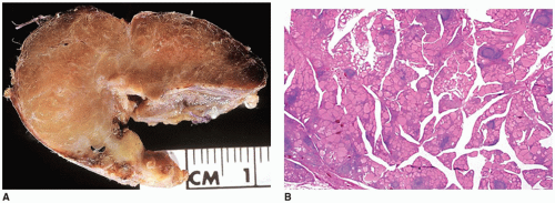
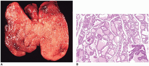
The Pineal, Pituitary, Parathyroid, Thyroid, and Adrenal Glands


The Pineal, Pituitary, Parathyroid, Thyroid, and Adrenal Glands


M. John Hicks, M.D., D.D.S., M.S., Ph.D.

Nicole Cipriani, M.D.

Peter Pytel, M.D.

Hiroyuki Shimada, M.D., Ph.D., F.R.C.P.A.


PINEAL GLAND


Anatomy and Physiology

The pineal gland is a small, cone-shaped midline structure that is attached to the superior-posterior border of the third ventricle. This 50- to 150-mg tan-brown structure sits just rostral to the quadrigeminal plate. It develops at approximately 7 weeks’ gestation from an evagination of the ependymal lining covering the caudal portion of the roof of the third ventricle (1). Based on magnetic resonance (MR) imaging studies, the pineal gland increases in size from birth through 2 years of age, at which time it remains constant in size through adolescence. No size difference has been noted between male and female children. In children older than 2 years of age, the average pineal gland measures 6.5 × 4.8 × 4 mm. At approximately 5 years of age, calcifications, in the form of corpora arenacea, develop. These calcifications increase with age, giving the pineal gland a hyperdense appearance on computed tomography (CT) imaging starting around at puberty. Pineal calcifications are observed in 8% of children by age 10, 20% at puberty, and 40% by age 20.

Histologically, the pineal gland is composed of nests of cells in lobular profiles, with a resemblance to the “zellballens” of paraganglia, surrounded by connective tissue septa containing blood vessels and nerve fibers (2,3,4). The pinealocytes, or chief cells, have basophilic cytoplasm with large irregular nuclei and prominent nucleoli and are arranged in cords or follicles within the lobules. Randomly distributed throughout the pineal gland in perivascular areas and between pinealocytes are astrocytes as the second main cell population of the pineal gland. In the late third-trimester fetus and neonate, two populations of pineal parenchymal cells are identified with the small cell population disappearing with advancing age. The pinealocytes are immunoreactive for synaptophysin (SYN), chromogranin (CHR), and neurofilament protein (NFP), and the interstitial astrocytes are immunoreactive for S100 and glial fibrillary acidic protein (GFAP) (1).

In lower vertebrates, the pineal gland is located more superficially and directly photosensitive. In higher vertebrates, the pineal receives afferent connections from the retina (5). The major hormone produced by the pineal gland is the indoleamine, melatonin, which plays a role in circadian rhythm regulation and gonadal steroidogenesis. Other physiologic functions attributed to the pineal gland include a role in modulating the hypothalamic-pituitary-gonadal axis, hormonal rhythms, sleep cycle, and body temperature. Destruction of the pineal gland by a benign cyst or tumor has led to precocious puberty. Interference with the inhibitory effect of melatonin on gonadal steroidogenesis represents one mechanism. Melatonin levels have been reported elevated in some children with primary pineal tumors (6). Melatonin levels may be useful in determining the adequacy of pineal tumor resection when the level was increased before surgery. Other aspects of the anatomy and function of the pineal gland are discussed in more detail by Reiter (1).


Developmental Disorders

Pineal agenesis has been reported as a component of other midline central nervous system developmental syndromes with absence of the corpus callosum, such as in Aicardi syndrome (7). The contrasting abnormality, pineal gland hyperplasia, has been reported in children with genital enlargement (7).

Pineal cysts (glial cyst) are a relatively common radiologic finding on MR and an incidental finding in 25% to 40% of autopsies. There is a female predilection (eFigure 21-1) (2,4,6). On CT and MRI imaging, the content of pineal cysts exhibits similar properties as CSF. A pineal cyst larger than 1 cm in diameter may cause headache, vertigo, and visual disturbances (1). Symptomatic cysts have been treated by surgical excision (8,9). Possible mechanisms for pineal cyst development include persistence of the ependymal-lined pineal diverticulum, secondary cavitation within the pineal gland, or development secondary to prior hemorrhage in the
gland (1). An ependymal lining accompanied by reactiveappearing astrocytes can be seen (eFigure 21-2). Typically cases show a wall that exhibits features of piloid gliosis. Approximately 5% of children with hereditary retinoblastomas have pineal cyst (10). Cyst formation is also seen in pineal neoplasms (6,11).


Acquired Disorders

Neoplasms of the pineal gland region account for 2% or less of all primary CNS tumors in children and are discussed in more detail by Burger and Scheithauer (2) and in Chapter 10. They may present with features including hydrocephalus from aqueductal obstruction, Parinaud syndrome, ataxia, or diplopia. Classically, there are three histogenetic categories: tumors of pineal parenchyma, gliomas, and germ-cell tumors (12). Overall gliomas account for about 17% of pineal neoplasms and include pilocytic astrocytomas, diffuse astrocytomas, and anaplastic astrocytomas (13). In children, they are proportionally less common than in adults. Germ-cell tumors account for about 27% of cases (eFigure 21-3) overall. They are typically said to be more common in East Asian populations, but one recent study did not confirm this finding (14,15). They include the same morphologic spectrum found in gonadal germ-cell tumors (eFigures 21-4 to 21-6) (16,17). A recent study found frequent mutations leading to KIT/RAS pathway activation (15). The mentioned calcifications that form with normal aging are typically not seen in children under age 10 to 12 years on CT imaging studies. In younger children, calcification can be a feature worrisome for a germ-cell tumor because pineal germ-cell neoplasms often appear on CT as solid masses with dense calcifications (eFigure 21-7A to C). On MR, the solid portion is isodense to brain on T1-weighted images and hyperintense to brain on T2-weighted images, while calcifications are hypointense on both pulse sequences.

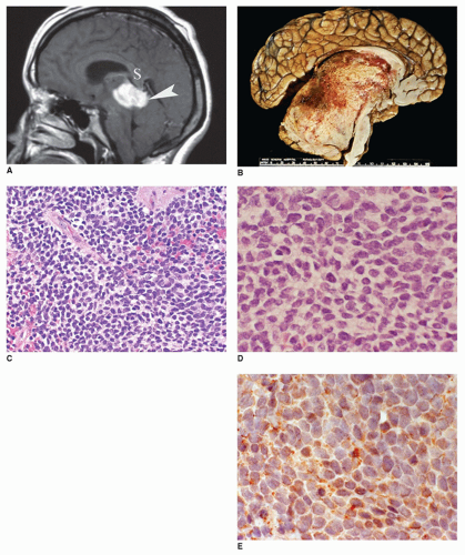
Pineal parenchymal tumors (PPTs) also account for some 27% of pineal tumors (12). These include pineocytoma, pineoblastoma, and PPT of intermediate differentiation (2,18,19,20). Pineoblastomas, like germ-cell tumors, preferentially occur in the first decade of life in contrast to pineocytomas, which are seen in the second decade and into adulthood. In the pediatric population, almost 60% of PPTs are pineoblastomas (mean age, 2 to 3 years) and another 10% are pineocytomas (mean age 10 to 12 years) (16,21). The M:F gender ratio for pineoblastomas varies among series from 5:1 to 1:2 for children 16 years of age or younger (16,19). Pineal tumors may compress the tectum and aqueduct of Sylvius causing findings of hydrocephalus (eFigure 21-7). On imaging studies, pineocytomas are hypo- to isointense to brain on T1-weighted images and hyperintense to brain on T2-weighted images (eFigure 21-8). Pineoblastomas are variable in their MR appearance. They may be large and lobulated and have areas of necrosis (Figure 21-1A) causing a heterogeneous appearance. PPTs generally enhance markedly after intravenous gadolinium contrast administration. These tumors are assigned to the following grades: pineocytoma (WHO grade I), pineoblastoma (WHO grade IV), PPT of intermediate differentiation (WHO grade II or III) (18-21). Prognosis of PPTs is dependent on stage, tumor volume, histologic type, and NFP immunostaining (19,21). Pineocytoma has a favorable survival (85% to 90%, 5 years), whereas the 5-year survival in pineoblastoma is less than 25% (18,19,21). Among pineoblastomas, tumors with mutated Rb1 gene are more aggressive with decreased survival when compared to sporadic pineoblastomas without RB1 mutation.

Pineoblastoma is a high-grade primitive embryonal tumor akin to other central primitive neuroectodermal tumors (cPNET). Grossly, they are tan-gray, soft, infiltrative tumors that may exhibit hemorrhage, necrosis, and extension into the leptomeninges (2) (Figure 21-1B). Sheets of primitive round to slightly ovoid cells with irregular, hyperchromatic nuclei and scant cytoplasm are observed on histologic examination. Mitotic figures and apoptotic bodies are readily identified (Figure 21-1C, D, eFigures 21-9 and 21-10). Focal necrosis and Homer Wright rosettes are present in some cases, but pineocytomatous rosettes found in pineocytomas are lacking. Infrequently, photoreceptor differentiation is indicated by the presence of Flexner-Wintersteiner-like rosettes. Tumor cells are immunoreactive for SYN (Figure 21-1E, eFigure 21-11), to CHR and NFP to a lesser degree, and to retinal S antigen in about 50% of cases (2,22). Trilateral retinoblastoma syndrome is defined by the development of a pineoblastoma as a midline intracranial malignancy in the setting of hereditary retinoblastoma (2,10).

Pineocytoma, unlike pineoblastoma, has a lobular appearance like other examples of endocrine or neuroendocrine neoplasms, is well circumscribed, and displaces surrounding structures. The tumor cells are uniform with small central nuclei and conspicuous eosinophilic cytoplasm with an absence of pleomorphism, necrosis, and mitotic figures. Homer Wright and Flexner-Wintersteiner rosettes and large GFAP-positive fibrillary areas, referred to as pineocytomatous rosettes, are observed in these tumors (eFigures 21-12A, B and 21-13). Like pineoblastoma, tumor cells are immunoreactive for SYN, CHR, NFP, and neuron-specific enolase (NSE), in addition to retinal S antigen in approximately 30% of cases (eFigure 21-14) (22). Neurosecretory granules are identified ultrastructurally in contrast to their usual absence in pineoblastomas. PPT of intermediate differentiation shows histologic features of both pineoblastoma and pineocytoma. Some atypia and mitotic activity may be seen, but their prominence does not reach that seen in pineoblastomas.

Other rare tumors described in this location include papillary tumor of the pineal region (PPTR) (23), atypical teratoid/rhabdoid tumor (AT/RT) (24), pleomorphic xanthoastrocytoma (PXA) (25), Langerhans cell histiocytosis (LCH), lipoma, and meningioma. Pineal involvement with acute lymphocytic leukemia has been reported. Infections, vascular malformations, epidermoid cyst, hemorrhage, and apoplexy are nonneoplastic lesions of the pineal gland in children.


FIGURE 21-1 • Pineoblastoma in a 3-year-old girl. A: Sagittal postgadolinium T1-weighted image shows a markedly enhancing, lobulated mass (arrowhead) in the pineal region below the splenium of the corpus callosum (S). B: This large, tan-gray, infiltrative pineal tumor has a heterogeneous appearance with hemorrhage, necrosis, and leptomeningeal extension. (Used with permission, Dr. David Louis, Department of Pathology, Massachusetts General Hospital, Boston, Massachusetts.) C: This pineal tumor is composed of sheets of primitive round to slightly ovoid cells (H&E stain, original magnification 200×). (Courtesy of Dr. Joe Parisi, Mayo Clinic, Rochester, Minnesota.) D: The tumor cells have irregular, hyperchromatic nuclei and scant cytoplasm. Mitotic figures were also present (H&E stain, original magnification 400×). (Courtesy of Dr. Joe Parisi, Mayo Clinic, Rochester, Minnesota.) E: The tumor cells demonstrate immunoreactivity with SYN (immunostain for SYN, original magnification 400×). (Courtesy of Dr. Joe Parisi, Mayo Clinic, Rochester, Minnesota.)


PITUITARY GLAND


Anatomy and Physiology

The pituitary regulates key physiologic processes including growth, metabolism, reproduction, and homeostasis. The pituitary gland extends by a narrow stalk from the hypothalamus into the sella turcica within the sphenoid bone (26,27,28). The pituitary gland is a small ovoid structure that is divided into a red-brown anterior lobe (adenohypophysis), a gray-white posterior lobe (neurohypophysis), and an indistinct intermediate lobe. The pituitary gland weighs approximately 100 mg at birth and increases in weight
during adolescence to its adult weight of 500 to 600 mg. The adenohypophysis accounts for 80% of the gland (26,27). The neonatal pituitary gland is especially prominent, owing to its stimulation by maternal hormones, but it undergoes some involution in the postnatal period, followed by increased growth through the age of 3 years. A notable increase in the size of the gland occurs with menarche and pregnancy. Generally, the pituitary gland in women after puberty weighs more than the gland in men. Suprasellar extension of the pituitary gland during puberty has been reported as a normal variant.

The pituitary gland receives its vascular supply from two hypophyseal arteries that branch from the internal carotid arteries and give rise to two anastomosing networks of capillaries that surround the stalk and adenohypophysis. The hypophyseal portal circulation, which arises from the second capillary plexus, supplies the adenohypophysis (26,27). A thin diaphragm, arising from the dura, covers the opening to the sella turcica, but in the center of the diaphragm, the pituitary stalk passes through an aperture. The pituitary gland is not covered by meninges. The periosteal dura lines the sella turcica.

The adenohypophysis is composed of three cell types on histologic examination: the chromophobes, acidophils, and basophils, accounting for 50%, 40%, and 10% of adenohypophyseal cells, respectively (eFigure 21-15). Five main hormonally active cell types are identifiable in the adult gland, which express six different hormones. The cell types and their respective hormones are the somatotrophs (growth hormone), lactotrophs (prolactin), corticotrophs (ACTH), gonadotrophs (FSH/LH), and thyrotrophs (TSH), accounting for 40% to 50%, 10% to 30%, 10% to 20%, 5% to 10%, and 5% of the adenohypophyseal cells, respectively (27,28). Stimulating and inhibitory hypothalamic factors released into the hypophy-seal-portal circulation regulate the release of these hormones from the adenohypophysis (eFigure 21-16).

The folliculostellate cells are agranular; immunostain for S100, GFAP, and vimentin (VIM); and extend between the other adenohypophyseal cells. These cells are thought to have a paracrine regulatory function on the hormone-producing cells (27). Calcified concretions are an incidental finding in the anterior pituitary of ostensibly normal fetuses and neonates.

The posterior pituitary (neurohypophysis) contains the axonal processes of neurosecretory neurons that originate in the supraoptic and paraventricular nuclei of the hypothalamus to secrete vasopressin and oxytocin, respectively. Vasopressin and oxytocin are stored in secretory granules (Herring bodies) in the nerve endings (27). The intermediate lobe contains melanotrophs. These produce proopiomelanocortin (POMC), a precursor for endorphins as well as melanocyte-stimulating factor (MSH).

The development of the pituitary is complex and depends on a series of signals that are expressed in distinct spatial and temporal patterns. Defects in these signals can lead to developmental defects as outlined below. The anterior and intermediate lobes develop from oral ectoderm, while the posterior lobe arises from neuroectoderm through an evagination of tissue from the base of the developing diencephalon. During the 4th week of gestation, an outpouching of ectoderm from the roof of the stomatodeum (primitive oral cavity) grows dorsally toward the diencephalon as Rathke pouch. Along this route of migration, progenitor cells of the future adenohypophysis may lag behind as potential sources of ectopic anterior pituitary. Constriction and disappearance of Rathke pouch during the 5th to 6th gestational week separate the adenohypophysis from the stomatodeum. Concurrently, the elongating Rathke pouch passes between the developing presphenoid and basisphenoid bones of the skull and joins with the infundibulum, a diverticulum arising from the diencephalon, as the future neurohypophysis. The first vestiges of the hypothalamic-hypophyseal portal circulation are seen at 7 weeks’ gestation, with the process being complete at 18 to 20 weeks’ gestation.

Somatotrophs and corticotrophs are identified immunohistochemically in the adenohypophysis between the 5th and 12th gestational week. By 12 to 13 weeks’ gestation, thyrotrophs and gonadotrophs are seen. At 13 to 16 weeks’ gestation, lactotrophs first appear. During the sixth gestational month, innervation of the neurohypophysis with axonal processes from the supraoptic and paraventricular nuclei takes place.

The differentiation of the oral ectoderm into the terminal anterior pituitary cell types with expression of hormones and receptors is under the control of a large complement of genes and transcription factors (eFigure 21-17). Several excellent reviews discuss the role of these factors in pituitary organogenesis in more detail (29,30,31,32). The physiology of the different cell types, their specific hormones, and mechanisms of action of these hormones are beyond the scope of this chapter, but is detailed by others (26,27,30,32,33).


Imaging

Due to its small size and location within the bony sella, the pituitary is best evaluated with dedicated MR imaging. The adenohypophysis is isointense to gray matter and has a flat superior margin until puberty, when the margin becomes slightly convex, especially in females. The neurohypophysis is hyperintense compared to brain on T1-weighted images, producing the posterior pituitary “bright spot.” The pituitary stalk (infundibulum) is normally midline and no larger than the basilar artery on axial images. Developmental lesions may be detected on imaging. Posterior pituitary ectopia is seen as an abnormal location of the posterior pituitary as a bright spot along the infundibulum or near the infundibular recess of the third ventricle (eFigure 21-18). Rathke cleft cysts are well circumscribed, round or lobulated, and isodense to CSF on CT. The signal intensity of the cyst content is variable on MR, depending on the protein content of the fluid. The cyst contents are generally isointense to slightly hyperintense to CSF on T1-weighted images and isointense to slightly hypointense to CSF on T2-weighted images (eFigure 21-19).


Inflammatory or infiltrative disorders are optimally demonstrated on MR images. Lymphocytic and granulomatous hypophysitis and LCH appear similar on imaging studies. With these conditions, the hypothalamus and infundibulum appear enlarged, with usually uniform enhancement following intravenous administration of gadolinium (eFigures 21-20 to 21-23). Primary pituitary tumors are best evaluated by MRI studies with and without contrast. Microadenomas do not distort the gland, but are hypointense to the normal gland on T1-weighted images and enhance less than the normal gland on early dynamic postgadolinium imaging (eFigure 21-24). Macroadenomas distort the gland and the infundibulum and enhance uniformly and intensely.


Developmental Disorders

Anomalies in pituitary gland development are outlined in Table 21-1 (34). Agenesis, complete absence, of the pituitary gland is rare as an isolated finding. Isolated agenesis of the pituitary has been noted in infants of diabetic mothers, as a presumed form of diabetic embryopathy. Pituitary dysfunction in neural tube defects is well documented. Agenesis can be associated with other midline and craniofacial abnormalities (27). In most cases of human holoprosencephaly, a hypoplastic gland can still be found in contrast to findings in animal studies (35). In the presence of pituitary agenesis, the thyroid gland, the adrenal glands, and gonads are expectedly diminutive. The posterior pituitary or neurohypophysis may be present.

Hypopituitarism, defined as diminution or absence of one or more anterior pituitary hormones, is estimated to occur in 1:4,000 to 10,000 live births. In general, isolated hormone deficiencies and combined pituitary hormone deficiencies (CPHDs) can be distinguished. Hypopituitarism can be the result of developmental defects or a rare secondary effect of traumatic brain injury, treatment for childhood cancer, or meningitis (5,36,37,38). Hypopituitarism can also be the result of neoplastic and inflammatory processes discussed in detail below. The complex development of the pituitary with contribution of two separate embryonic tissues is guided by a number of cellular signals (Table 21-2). Disruption in signals that regulate the early steps of this development results in syndromic defects with variably associated CNS, eye, and peripheral manifestations. This is in contrast to defects in signals guiding later steps of development, differentiation, and function that may cause selective hormone deficiencies.


TABLE 21-1 CONGENITAL AND DEVELOPMENTAL ANOMALIES OF THE PITUITARY GLAND


Agenesis


Hypoplasia


Ectopic pituitary


Duplication


Rathke cleft cysts


Pars intermedia cyst


Dermoid cyst


Empty sella syndrome


Hamartoma


Teratoma


Isolated growth hormone deficiency


Combined pituitary hormone deficiency (CPHD)


Cranial vault abnormalities involving sella turcica


Transsphenoidal encephalocele


Persistent craniopharyngeal canal


Modified from Parks JS, Felner EI. Hypopituitarism. In: Kliegman RM, Behrman RE, Jenson HB, Stanton BF, eds. Nelson textbook of pediatrics, 18th ed. Philadelphia, PA: Elsevier, 2007; Chapter 558.


Early signals include Lhx3, Lhx4, and the sonic hedgehog (SHH) signaling pathway (Table 21-2). Mutations in Lhx3 and Lhx4 result in aplasia or hypoplasia of the anterior pituitary and the intermediate lobe as well as other manifestations. These may include a short rigid spine and hearing loss (Lhx3) or cerebellar abnormalities and Chiari malformation (Lhx4). Holoprosencephaly can be the result of many different mutations (Chapter 10—Nervous System). Disruption of SHH signaling is a common cause. Mutations in GLI2, a factor in the SHH pathway, have been shown to cause congenital hypopituitarism in isolation or in association with other craniofacial anomalies and polydactyly (39,40). The importance of SHH mutations as the cause of hypopituitarism in humans is less clear than it appears to be in animal studies (40).

Mutations in HSEX1, SOX2, and SOX3 are linked to septooptic dysplasia with pituitary hypoplasia, midline forebrain defects, and optic nerve hypoplasia. OTX2 mutations can cause anophthalmia and pituitary defects (41). The transcription factors PROP1 and POU1F1 are expressed during pituitary development. Mutations in either of these result in CPHD. Some mutations disrupt isolated pituitary hormones. Isolated GH (growth hormone) deficiency has been linked to mutations in the GH1 gene encoding growth hormone or the gene encoding the GHrH receptor, but also SOX3 and HESX1. Congenital deficiency of ACTH has been linked to TBX19 mutations. Kallmann syndrome results from FEZH mutations causing failure of GnRH neurons to develop (42).

Other syndromes with hypopituitarism include MELAS syndrome, Rieger syndrome, trisomy 18, trisomy 13, Pallister-Hall syndrome, neurofibromatosis, Fanconi anemia, and ataxia-telangiectasia.

Anencephaly is characterized by the presence of anterior pituitary tissue within the mass of cerebrovascular tissue (eFigure 21-25A, B). The presence of somatotrophs, lactotrophs, and gonadotrophs is demonstrated by immunohistochemistry. Corticotrophs and thyrotrophs present in the pituitary in the second trimester disappear owing to lack of hypothalamic stimulation during the third trimester. A distinct neurohypophysis is absent. The adrenal glands are hypoplastic at birth (eFigure 21-25C, D).

Ectopia of anterior pituitary type tissue is common and invariably an incidental finding typically in the roof of the nasopharynx or as a pharyngeal pituitary. Persistence of
Rathke pouch in the roof of the oronasopharynx, the source of the pharyngeal pituitary gland, has been reported in a number of conditions, including the anencephalic fetus, spina bifida, trisomy 18, and Meckel syndrome. Ectopia of the posterior pituitary has been associated with mutations in genes responsible for pituitary organogenesis (eFigure 21-18) (43). Ectopic pituitary adenomas (PAs) are documented in the suprasellar region, clivus, nasopharynx, and paranasal sinuses mainly in adults, but also in children.


TABLE 21-2 MANIFESTATIONS ASSOCIATED WITH MUTATIONS IN SELECTED GENES INVOLVED IN PITUITARY DEVELOPMENT


Gene


Function


Manifestations


LHX3, LHX4


Development and maintenance of adenohypophysis


CPHD, IPHD, pituitary hypoplasia, ectopia of neurohypophysis, Arnold-Chiari I malformation, cerebellar abnormalities, short rigid spine, hearing loss


HESX1 SOX2 SOX3


Early development of pituitary gland


CPHD, IPHD, pituitary hypoplasia, ectopia of neurohypophysis, septo-optic dysplasia


POU1F1(PIT 1)


Differentiation of the somatotrophs, lactotrophs, and thyrotrophs


Growth hormone, prolactin and TSH deficiency


PROP 1


Differentiation of the somatotrophs, lactotrophs, thyrotrophs, and gonadotrophs


30%-50% of cases of familial CPHD


Tpit


Differentiation of corticotrophs


ACTH deficiency


Gli 2, Gli3


Holoprosencephaly and panhypopituitarism, Hall-Pallister syndrome


PTX2


Rieger syndrome


CPHD, combined pituitary hormone deficiency; IPHD, isolated pituitary hormone deficiency; ACTH, adrenocorticotropic hormone; TSH, thyroid-stimulating hormone.


Based on data from Lap-Yin Pang A, Martin MM, Martin ALA, et al. Molecular basis of diseases of the endocrine system. In: Coleman WB, Tsongalis GJ, eds. Molecular Pathology: The Molecular Basis of Human Disease. Amsterdam, The Netherlands: Elsevier, 2009:435-463.


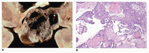
Rathke cleft cysts may become symptomatic because of compression of intrasellar or suprasellar structures. Rare cases present with growth retardation in children (the socalled pituitary dwarfism) (eFigure 21-19) or central precocious puberty. Morphologically, these exhibit similar morphologic features as those found with colloid cysts: the cyst is filled with thickened mucoid secretions or dark fluid (eFigure 21-26) and lined by ciliated columnar or low cuboidal epithelium. Microscopic incidental cystic rests with similar morphologic features are a common incidental finding in the pars intermedia in 2% to 26% of autopsies. Other cystic lesions in the region of the pituitary include craniopharyngioma (CRP) and intrasellar arachnoid cyst. A distinguishing feature of the CRP is mixed cystic and solid areas with the palisading and squamoid-type epithelium (Figure 21-2A, B). Because CRP and Rathke cleft cyst have a shared histogenesis, ciliated columnar epithelium may be seen on occasion in a CRP. Abscess formation and hypophysitis are rare complications in Rathke cleft cysts.

Pituitary duplication is a rare disorder that is ascribed to a duplication of the prechordal plate and anterior aspect of the notochord. Two distinct pituitaries, each with its own stalk, are the typical presentation. This anomaly has been seen with partial twinning, median cleft facial syndrome, precocious puberty, and fetal exposure to meclizine (teratogenic effect). A midline hypothalamic mass of disorganized neurons is accompanied by other midline developmental anomalies, including a duplicated sella, cleft palate, hypertelorism, agenesis of corpus callosum, and vertebral anomalies. Nasopharyngeal teratomas have been reported in association with pituitary duplication in infancy.

Empty sella syndrome (ESS) is usually an incidental finding in young children in contrast to adults. The primary form of ESS results from a defect in the diaphragm covering the opening to the sella turcica, with arachnoid tissue extending through the diaphragmatic defect. Increased CSF pressure leads to enlargement of the sella turcica and compression of the pituitary gland along the floor of the sella turcica, giving the appearance of an empty sella turcica (eFigures 21-27 and 21-28). Pituitary infarction, pituitary atrophy from a tumor, other mass lesion, and prior hypophysectomy account for secondary ESS.


Acquired Disorders

Inflammatory and infiltrative disorders are known to involve the pituitary gland including infections, noninfectious inflammatory conditions, and infiltrative processes. Examples of these diseases are congenital syphilis, mycobacteriosis, lymphocytic-granulomatous hypophysitis, LCH, sarcoidosis (44), Wegener granulomatosis (45), iron overload, storage disorder, Rosai-Dorfman disease (RDD), and Hurler syndrome.


FIGURE 21-2 • Craniopharyngioma. A: This gross brain image shows a suprasellar cystic lesion filled with a dark brown fluid containing cholesterol debris. B: This adamantinomatous variant consists of ribbons of epithelial cells with pseudopalisaded nuclei at the periphery of the lobules surrounding cystic spaces. The inner cells in the more solid areas have a loose, stellate appearance. The so-called wet keratin is seen as intermixed stacks of necrobiotic squames. This image is from a 7-year-old girl, who presented with headaches and decreased visual acuity and was found to have a suprasellar mass (H&E stain).

Lymphocytic hypophysitis was first described as a disease of pregnant woman presenting with hypopituitarism. This condition is now recognized as occurring in other settings, including children as young as 9 years of age, but is generally uncommon in children. It is regarded as an autoimmune condition, because of its association with Hashimoto or chronic lymphocytic thyroiditis (CLT). The adenohypophysis (lymphocytic adenohypophysitis) and neurohypophysis (lymphocytic infundibuloneurohypophysitis) may be involved. In general, the designation of lymphocytic hypophysitis is used to describe both conditions. The pituitary is enlarged with a firm consistency and contains an inflammatory infiltrate of small lymphocytes intermixed with plasma cells (eFigure 21-29A to C). Eosinophils and some macrophages are also seen. Fibrosis is common, but may not be apparent in a small biopsy. Hypopituitarism, diabetes insipidus, and mass lesion symptoms are the usual clinical manifestations in both children and adults. Because the pituitary and sella are enlarged, a PA is often the clinical impression.

Granulomatous hypophysitis with epithelioid or caseous granulomas has the differential diagnosis of infection (tuberculosis), sarcoidosis, rupture of Rathke cleft cyst, LCH, and idiopathic granulomatous hypophysitis. Granulomas are not a feature of lymphocytic hypophysitis, although a nosologic and etiologic relationship may exist between these idiopathic inflammatory disorders.

Xanthogranulomatous inflammation (cholesterol granuloma) of the sellar region is an inflammatory reaction characterized by cholesterol clefts, lymphoplasmacytic infiltrates, hemosiderin deposits, fibrosis, foreign body giant cells, histiocytes, and eosinophilic necrotic debris. Although xanthogranulomatous inflammation may be associated with an adamantinomatous craniopharyngioma (CRP), this pattern has also been observed in idiopathic cases primarily in adolescents and young adults lacking a CRP component.

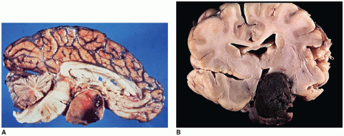
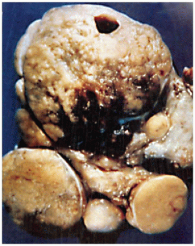
Vascular lesions with hypopituitarism are uncommon in children, but hemorrhagic infarction of a pituitary macroadenoma, referred to as pituitary apoplexy or pituitary tumor apoplexy, is one such example (Figure 21-3A, B) (46). Pediatric cases of pituitary apoplexy may be milder than in adults, having a more indolent course and associated with a more favorable outcome (47). Sheehan syndrome is the result of severe maternal intrapartum hypotension leading to pituitary infarction in the postpartum period. Presumed ischemia of the pituitary in sickle cell crisis is associated with decreased growth hormone secretion and impaired growth in affected children. Some cases of septo-optic dysplasia, classified as a developmental anomaly, are thought to represent a vascular disruption of the anterior cerebral artery. Vascular lesions due to stalk transection may occur secondary to trauma.

Nonneoplastic cysts identified in children on radiologic studies are not clinically evident unless the sella turcica is expanded, which leads to hypopituitarism and diabetes insipidus. Cystic dilatation of Rathke pouch remnants is common; however, these cysts are usually less than 5 mm in diameter (eFigures 21-19 and 21-26). Rathke cleft cysts arise from the squamous epithelium of the Rathke cleft and infrequently become enlarged with symptoms resembling a CRP. Arachnoid and dermoid cysts are also regarded by some as congenital defects. An intrasellar arachnoid cyst must also be distinguished from a CRP.

Pituitary hyperplasia is a nonneoplastic proliferation of one of the functional adenohypophyseal cell types. It is a polyclonal proliferation leading to pituitary enlargement and may produce a suprasellar mass. In children, somatotroph hyperplasia is reported in the McCune-Albright syndrome (MAS) and gigantism. Pituitary hyperplasia has also been reported in primary hypothyroidism. During pregnancy, the pituitary gland doubles in size due to the proliferation
of the lactotrophs (responsible for prolactin secretion) and decreases in size postpartum (27).


FIGURE 21-3 • Pituitary apoplexy. A: Sagittal section of brain showing hemorrhage within a pituitary macroadenoma. B: Coronal section showing hemorrhagic infarction of a 2-cm diameter well-circumscribed pituitary macroadenoma.

Pituitary adenoma (PA) is a monoclonal neoplasm of the adenohypophysis. Up to 10% of all PAs present in the first two decades of life. Over 90% of these are diagnosed in the second decade, and less than 10% before 10 years of age. Between 15 and 19 years of age, PAs are the most common CNS tumor, being twice as common in girls as boys (26,46,48,49). Reports of adenomas occurring in children less than 4 years of age are uncommon. The youngest example of an ACTH-secreting PA occurred in a 7-month-old infant with Cushing disease. Most PAs are sporadic, but a number of cases are linked to familial mutations (Table 21-3). About 20% of PAs in the pediatric populations are the result of AIP mutations (50,51,52). Most of the AIP-associated PAs express growth hormone with or without prolactin. Other cases are linked to germline mutations in MEN1, VHL, SDHB, SDHC, SDHD, and PRKAR1A (Carney complex) (50,53,54,55). Cases with SDH subunit mutations typically exhibit prominent cytoplasmic vacuolation (55).

In terms of function, the majority of PAs in children are prolactinomas (53%). The remaining tumors are ACTH-secreting tumors (31%), growth hormone-secreting tumors (9%), and endocrine-inactive (null cell tumors) (3%) (48,56,57). ACTH-secreting adenomas are more common before puberty, in contrast to prolactinomas and growth hormone-secreting tumors, which are more common after puberty.

The clinical manifestations of PAs in children are variable. Headaches and visual field defects are the most common clinical manifestations of mass effect. Prolactinomas may result in amenorrhea and galactorrhea in girls and gynecomastia and hypogonadism in boys. Children with ACTH-secreting adenomas present with Cushing disease. Children with somatotropin or growth hormone-secreting adenomas present with gigantism.

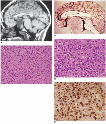
Most prolactinomas, growth hormone-secreting tumors, and endocrine-inactive PAs are macroadenomas (>10-mm tumors in diameter) (Figure 21-4A, B). In contrast, ACTH-secreting adenomas are more often microadenomas (<10-mm tumor in diameter) (eFigures 21-30 and 21-31) (58). Macroadenomas are more common than microadenomas in children, consistent with the fact that prolactinomas are more common than ACTH-secreting tumors. PAs are classified on the basis of five-tiered features: endocrine activity, imaging studies and operative findings, histology, immunohistochemistry, and ultrastructure (59). PAs are soft and gray-red and measure less than or equal to 2 cm. On the basis of imaging criteria, four grades of tumors are recognized: grade I (<l cm in diameter), grade II (intrasellar lesion >1 cm in diameter or with suprasellar expansion without invasion), grade III (small or large locally invasive tumor with bony invasion of the sella turcica), and grade IV (large invasive tumor involving the bone, hypothalamus, or cavernous sinus) (26). Larger aggressive tumors are more likely to be cystic, hemorrhagic, and necrotic (Figure 21-3A, B). One or more concurrent histologic patterns (diffuse, trabecular, papillary) may be evident. The degree of cellularity is variable from highly cellular to more scantily cellular tumors with hyalinized or amyloid-like stroma. The tinctorial quality of the cytoplasm has given rise to the characterization of PAs as basophilic, acidophilic, or chromophobic with some limitations. The tumor cells are generally rounded with some occasional spindling with the rounded central or eccentric nuclei. The tumor cells may have plasmacytoid qualities in the presence of an eccentric nucleus and prominent basophilic, acidophilic, or amphophilic cytoplasm. Prolactinomas are typically composed of chromophobes or slightly acidophilic cells with a solid or papillary pattern and hyalinized stroma with or without microcalcifications (Figure 21-4C, D, eFigure 21-32). PAs tend to lack a capsule (eFigure 21-30). Electron microscopy


and immunohistochemistry are adjuncts to characterize these tumors (Figure 21-4E, eFigure 21-33). In addition to specific hormonal immunostaining, PAs are positive for SYN, CHR, and NSE (30).


TABLE 21-3 ENDOCRINE ORGANS INVOLVED IN SELECTED FAMILIAL TUMOR SYNDROMES


Syndrome


Inheritance


Gene


Pituitary


Parathyroid


Thyroid


Adrenal


Other Endocrine Organ Manifestations


Beckwith-Wiedemann


paternal uniparental disomy


CDKN1C/NSD1


ACN


Pancreatoblastoma, adrenal cytomegaly, neuroblastoma


Carney complex (LAMB and NAME syndromes)


AD


PRKAR1A


Adenoma


PTC


ACN


Cowden


AD


PTEN


FTC/PTC


Familial adenomatous polyposis coli


Gardner’s syndrome


AR/AD


APC


PTC, cribriform-morula variant


Familial medullary thyroid carcinoma


AD


RET Exons 10 and 11


Codons 618, 620,209,611


MTC


Familial paraganglioma-pheochromocytoma


SDHD (PLG1), SDHAF2 (PLG2), SDHC (PLG3), SDHB (PLG4)


Non-MTC in SDHB, SDHD


PHEO


Paraganglioma


Hyperparathyroidism-jaw tumor


AD


HRPT2


Adenoma/carcinoma


Li-Fraumeni


AD


TP53, CHEK2


can


McCune-Albright


GNAS


Adenoma


Hyperplasia


Hyperplasia/adenoma


MEN 1 (Wermer syndrome)


AD


MEN1 (Menin, 11q13)


Adenoma


Adenoma/hyperplasia


ACN


Islet cell neoplasia


MEN 2A (Sipple syndrome


AD


RET, Exons 10 and 11, Codons 609, 611, 618, 620, 634


Adenoma/hyperplasia


MTC


PHEO


Paraganglioma, enteric ganglia


MEN 2B (Wagenmann-Froboese syndrome


AD


RET Exon 16, Codon 918


MTC


PHEO


Mucosal neuromas, enteric ganglioneuromatosis


Neurofibromatosis type 1


AD


NF1


PHEO


Paraganglioma


Von Hippel-Lindau


AD


VHL1, VHL2A, VHL2B, VHL2C


PHEO


Islet cell neoplasia Paraganglioma


Peutz-Jeghers


AD


STK11/LKB1


Non-MTC


Werner


AR


WRN


Non-MTC


Papillary renal neoplasia


PTCPRN


PTC


Familial papillary thyroid carcinoma with clear cell renal carcinoma


t(2;3;8)


PTC


Familial papillary carcinoma with or without oxyphilia


TCO


PTC


Familial papillary thyroid carcinoma


NMTC1


PTC


Familial multinodular goiter with papillary thyroid carcinoma


MNG1, MNG3


PTC


Familial papillary thyroid carcinoma


PTC1


PTC


Familial follicular thyroid carcinoma


NMCT1


PTC


ACN, adrenocortical neoplasm; AD, autosomal dominant; FTC, follicular thyroid carcinoma; MTC, medullary thyroid carcinoma; non-MTC, nonmedullary thyroid carcinoma; PHEO, pheochromocytoma; PTC, papillary thyroid carcinoma.


Modified from Table 5.01. Eng C. Inherited tumor syndromes. Introduction. In: DeLellis RA, Lloyd RV, Heiz PU, eds. World Health Organization Classification of Tumors: Pathology and Genetics. Tumors of Endocrine Organs. Lyon, France: IARC, 2004;210.


FIGURE 21-4 • Pituitary adenoma. A: A PA is shown in this sagittal T1W MR image of an 11-year-old boy with a cystic expansile mass (macroadenoma) arising within the sella turcica and extending upward (Courtesy of James Smirniotopoulos, M.D., Bethesda, Maryland). B: Sagittal section of brain showing a pituitary macroadenoma, prolactinoma, with a homogeneous cut surface. C: The normal architecture of the pituitary gland is replaced by a diffuse growth pattern of cells. The normal histologic pattern of acidophils, basophils, and chromophobes arranged in a cord-like pattern is replaced by a single population of cells with acidophilic cytoplasm (H&E stain, original magnification 200×). D: The tumor cells are large with irregular nuclei and acidophilic cytoplasm (H&E stain, original magnification 400×). E: Tumor cells are immunoreactive for prolactin in this pituitary macroadenoma (Immunostain for prolactin, original magnification 400×). (Images C-E, courtesy of Dr. Joe Parisi, Mayo Clinic, Rochester, Minnesota.)

In terms of clinical behavior, macroadenomas are more likely to be invasive in contrast to the smaller expansile microadenomas. It is debatable whether PAs in children are more aggressive than their adult counterparts. Invasive adenomas are characterized by extension into dura, bone, and cavernous sinus. These features are generally not well documented in the pathologic examination. There is a certain degree of correlation between proliferative activity, as determined by MIB-1 (Ki67) nuclear immunostaining, and observed invasiveness of the tumor.


Pituitary blastoma is a rare pediatric lesion linked to germline mutations in DICER1, composed of various cell types, including primitive Rathke-type epithelium, and folliculostellate and secretory adenohypophyseal cells (60,61). Pituicytoma is a rare neoplasm arising from specialized glial cells, pituicytes, in the neurohypophysis, presenting as a lowgrade spindle cell lesion with glial differentiation (62).

Craniopharyngiomas (CRPs) are thought to be derived from remnants of the Rathke pouch and account for 3% to 4% of primary CNS tumors in children (63). Generally, the tumor is found in the suprasellar region (Figure 21-2A, eFigures 21-34 to 21-36). Rare cases may be parasellar or ectopic in the pineal gland region. The differential diagnosis includes PA, infection, inflammatory processes, vascular malformations, and Rathke cleft cyst. Imaging is helpful in distinguishing among CRPs, PAs, and Rathke cleft cysts.

CRPs in children are most commonly diagnosed between 5 and 14 years of age with no gender predilection (63). Tumors occurring during infancy are uncommon. Compressive symptoms, including pituitary dysfunction with retarded growth, are the principal clinical manifestations (64). Diabetes insipidus due to posterior pituitary involvement is infrequent. A calcified cystic suprasellar mass is characteristic on CT and MR scans (eFigure 21-36). Surgical resection may be followed by a recurrence (2). The gross specimen consists of cyst fragments with a yellow to dark brown appearance. Fluid contents have a dark oily appearance (crankcase) with cholesterol crystals and fragments of keratinous debris. In children, the histologic features are typically adamantinomatous or ameloblastic. A papillary squamous pattern is seen more often in adults (26,29). Beta-catenin mutations are seen in the adamantinomatous pattern (26,29), while the papillary squamous pattern is linked to BRAF mutations (65). In adamantinomatous CRPs, epithelial lobules are arranged in a cloverleaf-like pattern. Palisading of the cells adjacent to the randomly distributed fluid-filled cystlike spaces is a characteristic feature. Aggregates of necrotic, keratinized cells, or “wet” keratin accompanied by dystrophic calcification, are other features (Figure 21-2B, eFigure 21-37). Fibrosis, chronic inflammation, and cholesterol clefts are observed in the solid areas. A xanthogranulomatous reaction is prominent in some cases, especially with a ruptured cyst. Although CRPs are regarded as clinically benign, adherence to the hypothalamus and extension into the surrounding brain parenchyma occur in some cases. Cytokeratin expression has been used to distinguish CRP from Rathke cleft cyst. An uncommon CRP variant is one with adamantinomatous features together with elements of a PA, a so-called collision tumor. In many of these “collision tumors,” the adenoma is nonfunctional; however, immunohistochemistry displays gonadotropin, prolactin, ACTH, and TSH staining.

Other tumefactive lesions of the pituitary and sellar region include the ganglion cell tumor (so-called gangliocytoma), LCH, granular cell tumor, RDD, and salivary gland hamartoma. Gangliocytomas are regarded as neoplasm by most observers, but hamartoma by others. These may be found in association with PAs, pituitary hyperplasia, or as a distinct mass. These lesions have been classified as either mixed adenoma-gangliocytoma or pure gangliocytoma.

In addition to PA and CRP, the midline region of the sella and suprasellar area is another common anatomic site besides the pineal region for intracranial germ-cell tumors. 60% to 70% of all primary intracranial germ-cell tumors arise in the pineal region and 30% to 40% in the sellar or suprasellar area. Cases associated with prominent inflammatory infiltrates may be mistaken for an inflammatory disease process if the neoplastic cells are not appreciated. Visual field defects, diabetes insipidus, and panhypopituitarism are the principal clinical manifestations of suprasellar germ-cell tumors.

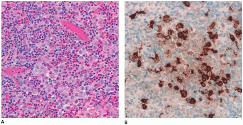
Langerhans cell histiocytosis (LCH) is well documented in the CNS with involvement of brain parenchyma and/or the hypothalamic-pituitary axis, causing central diabetes insipidus (66). The pituitary stalk is thickened on imaging studies (eFigures 21-21 to 21-23). Almost 15% of those with multisystem LCH have hypothalamic-pituitary involvement. There is limited documentation of the pathology in such cases because the diagnosis is usually established on the basis of a biopsy from a more accessible site. A mixture of Langerhans cell histiocytes, characterized by large, convoluted, and indented nuclei, that are CD207 (langerin, more specific) and CD1a (less specific) positive, mixed with lymphocytes, plasma cells, and eosinophils, is diagnostic for LCH (Figure 21-5A, B, eFigure 21-38A to C).

RDD has craniospinal manifestations in a minority of cases, including the sellar-suprasellar region. In 50% of cases, RDD is limited to this site.

Salivary gland rest or heterotopia is an incidental microscopic finding on the surface of the posterior pituitary. Other neoplasms of presumed salivary gland type, granular cell tumor of the pituitary and pituitary stalk, leukemia, lymphoma, and metastatic involvement of the pituitary are restricted to adults in most cases. Both primary and metastatic germ-cell neoplasms also occur in the pituitary.


PARATHYROID GLANDS


Anatomy/Physiology

The parathyroid glands, usually four in number, are pink-red, oval 4 to 6 mm in diameter glands located in proximity (usually posterior) to the thyroid gland or even embedded within the thyroid (intrathyroidal). The inferior and superior parathyroid glands arise as endodermal outpouchings from the dorsal bulbar portion of the third and fourth pharyngeal pouches, respectively, during the fifth gestational week in the 8- to 9-mm-stage embryo as bilateral cellular clusters. Concurrently, the thymus arises from the ventral aspect of the third pharyngeal pouches. Both the thymus and inferior parathyroid glands initially migrate together caudally with the heart. During the descent, the thymus and inferior
parathyroid glands separate and the inferior parathyroid glands localize to the inferior aspect of the thyroid gland. Formation of the parathyroid glands is associated with the genes EOLVO and GCM2 on chromosome 6 (6p24.2). Other genes associated with parathyroid development and migration include HOX3a (12q13), PAX1 (7q36), EYA1 (8q13.3, branchiootorenal syndrome 1), SIX1 (14q23, branchiootorenal syndrome), and TBX1 (22q11.2, DiGeorge syndrome). Dysregulation or mutation in these genes results in absence, hypoplasia, or ectopic parathyroid glands.


FIGURE 21-5 • Langerhans cell histiocytosis. A: Biopsy from the pituitary stalk in an adolescent with diabetes insipidus. The infiltrate is composed of foamy histiocytes that were immunoreactive with CD1a, in a background of lymphocytes, eosinophils, and plasma cells (H&E stain, original magnification, 400×). (Courtesy of Dr. Joe Parisi, Mayo Clinic, Rochester, Minnesota.) B: CD1a positivity in histiocytic cells in a patient with LCH (immunostain for CD1a, original magnification 400×). (Courtesy of Dr. Joe Parisi, Mayo Clinic, Rochester, Minnesota.)

In children, the combined weight for all four parathyroid glands increases with age from a mean weight of 5 to 10 mg each in the neonatal period to a combined weight of 120 mg for adult males and 140 mg for adult females by age 30 years. In children less than 10 years of age, the mean weight of all four glands is less than 60 mg. Between the ages of 11 and 20 years, the mean weight of all four glands has been recorded as less than 100 mg. A more recent study of parathyroid gland weight in children 9 to 19 years of age indicated individual gland weights can range between 10 and 80 mg.

The parathyroid glands in children tend to be solid and cellular with minimal fat. A connective tissue capsule encloses the gland. Chief cells are the predominant cell type. Blood vessels are intermixed among the parenchymal cells, and small delicate capillaries are present between the cells. The polyhedral chief cells have a small central nucleus and pale to clear cytoplasm. Oxyphil (oncocytic) cells are not observed generally until puberty, if at all. Adipocytes within the gland initially appear around puberty with fatty change gradually accounting for 25% to 30% of the total gland weight after age 18 years (67).

Calcium homeostasis is regulated by the interaction of parathyroid hormone (PTH), calcitonin, and vitamin D (eFigure 21-39) (68,69). In response to hypocalcemia, PTH is released from the chief cells, which is accompanied by an increase in PTH mRNA within hours of the onset of hypocalcemia. Hyperplasia of chief cells occurs within weeks. In contrast, hypercalcemia inhibits the release of PTH by activation of the chief cell calcium receptor (Table 21-4). Serum phosphate levels, independent of vitamin D3, also affect PTH release (68,69). The anatomy and physiology of the parathyroid glands is discussed in more detail elsewhere (70).


Imaging

The parathyroid glands are small and difficult to appreciate on imaging studies when not enlarged. Patients with hyperparathyroidism (HPT) are best evaluated with ultrasonography (US) and/or radionuclide scintigraphy. In children, high-resolution US should be the first-line imaging modality. Enlarged parathyroid glands in the neck are typically identified posterior to the thyroid gland. As parathyroid glands are best identified based on proximity to the thyroid gland, ectopic glands pose a diagnostic challenge. Radionuclide scintigraphy is more accurate than US (87% versus 80%), particularly for ectopic glands. The combination of nuclear
scintigraphic studies and US provides the highest accuracy for preoperative localization of hyperfunctioning glands.


TABLE 21-4 PRIMARY HYPERPARATHYROIDISM ETIOLOGY IN CHILDHOOD


Parathyroid adenoma


Sporadic (nonsyndromic)


HPT-jaw tumor syndrome


Parathyroid hyperplasia


Sporadic (nonsyndromic)


Neonatal HPT


Familial isolated HPT


MEN1


MEN2a


Parathyroid carcinoma


Differential Diagnosis of Hypercalcemia in Children


Elevated parathyroid hormone


Hyperparathyroidism, primary, secondary, or tertiary


Parathyroid hyperplasia


Parathyroid adenoma


Parathyroid carcinoma


Ectopic parathyroid hormone production


Hypervitaminosis D


Sarcoidosis


Subcutaneous fat necrosis of newborn


Familial hypocalciuric hypercalcemia


Idiopathic hypercalcemia (Williams syndrome)


Thyrotoxicosis


Hypervitaminosis A


Hypophosphatasia


Prolonged immobilization


Thiazide diuretics


MEN, multiple endocrine neoplasia.


Based on data from DeLellis RA, Mazzaglia P, Mangray S. Primary hyperparathyroidism: a current perspective. Arch Pathol Lab Med 2008;132:1251-1262.


Nuclear medicine studies utilize 99mTc sestamibi, which localizes to hyperfunctioning parathyroid glands, as well as the thyroid gland and salivary glands. Sestamibi scans can be performed in several ways. In dual-isotope single-phase imaging, the patient is administered and labeled with sestamibi and 123I and 99mTc pertechnetate, which are taken up by the thyroid gland. The images are subtracted to show the activity only in the hyperfunctioning parathyroid glands (eFigure 21-40). Alternatively, a single-isotope dual-phase technique may be employed. Sestamibi washes out of the thyroid and salivary glands faster than the parathyroid glands, so delayed images show relatively greater uptake in the hyperfunctioning parathyroid gland. Single-photon emission computed tomography (SPECT) imaging in addition to planar imaging helps to localize the abnormality in the anterior-posterior plane. Fusion of SPECT imaging to x-ray-based CT adds additional anatomic information that aids in precise localization of the parathyroid gland, which may be particularly useful in tumor recurrence after surgery.

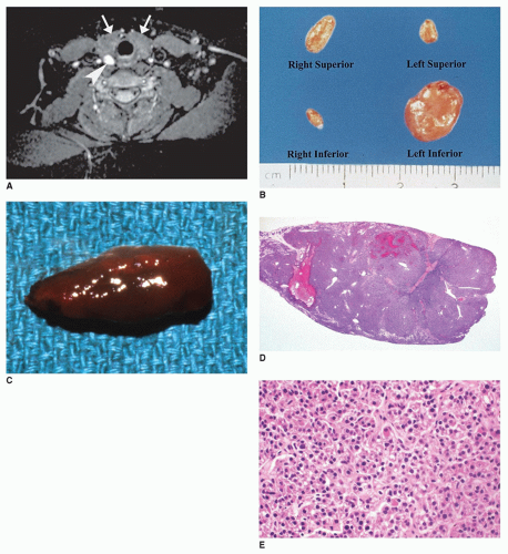
Parathyroid adenomas may also be demonstrated on CT and MR, but the accuracy of these studies for preoperative localization is no greater than for US. Parathyroid adenomas may be present at the typical gland location or located anywhere from the mandible to cervical thymus to mediastinal thymus or within the substance of the thyroid gland (intrathyroidal). On CT, adenomas are usually well defined and enhance intensely following intravenous contrast administration (eFigure 21-41). On MR, adenomas are generally of intermediate signal on T1-weighted images and of high signal intensity on T2-weighted images and enhance intensely following intravenous administration of gadolinium chelate (Figure 21-6A). Prior to the advent of laboratory screening, patients with undiagnosed, prolonged HPT developed characteristic findings on bone radiographs as well as nephrocalcinosis and nephrolithiasis. These findings are now rarely encountered (eFigures 21-42 and 21-43).


Developmental Disorders

Supernumerary parathyroid glands are found in up to 16% of the population, with an additional single gland being the most common presentation. Supernumerary glands, with as many as 12 glands, may be of normal size or rudimentary. Parathyroid adenomas and carcinoma have been reported in ectopic parathyroid glands in children (eFigure 21-41) (71).

Ectopic parathyroid tissue or a normally formed gland is relatively common within the thyroid or thymus. Ectopic parathyroid tissue has also been observed as scattered small nests in the soft tissues of the neck and mediastinum owing to aberrant migration or premature separation of parathyroid primordia during fetal development. Not surprisingly, nests of parathyroid tissue may be accompanied by equally diminutive nests of thymic tissue. Aberrant parathyroid and thymus are known to present as a recurrent lateral neck mass in children. Heterotopic parathyroid tissue has also been observed at remote sites, including the vagina.

Agenesis-hypoplasia of the parathyroids, due to a defect in pharyngeal pouch development or defective neural crest migration, is uncommon as an isolated finding with associated congenital hypoparathyroidism. Agenesis-aplasia is more frequently associated with other syndromes, including 22q11.2 deletion syndrome (DiGeorge anomaly, DiGeorge syndrome), Smith-Lemli-Opitz syndrome type II, X-linked recessive hypoparathyroidism, Kenny syndrome, Kearns-Sayre syndrome, and trisomy 18. The parathyroid glands may be absent in up to 50% of patients with 22q11.2 deletion syndrome. Anomalies of the aortic arch, thymus, thyroid, and C-cells in addition to abnormal facial development are also observed. Parathyroid hemorrhage is reported in osteogenesis imperfecta and refractory hypocalcemia (see Chapter 3).


FIGURE 21-6 • Parathyroid adenoma. A: Axial T2-weighted MR image shows the hyperintense parathyroid adenoma (arrowhead) posterior to the thyroid gland (arrows) in a 14-year-old girl. B: Parathyroid adenoma in a child with primary HPT is seen as a solitary enlargement of the left inferior gland. (Courtesy of Robert Dufour, M.D., Washington, DC.) C: Parathyroid adenoma in a 16-year-old girl who presented with flank pain due to nephrolithiasis, elevated serum calcium, decreased phosphate, and increased parathormone levels. The parathyroid gland was enlarged and red-brown on gross examination. D: The enlarged parathyroid gland shows a hypercellular parenchyma composed of chief cells without intraglandular fat on low power. Necrosis was absent (H&E stain). E: The parathyroid gland is composed of a monotonous population of chief cells with no intraglandular fat. Mitotic figures were absent (H&E stain).

Cyst(s) of the parathyroid are rare in children and usually asymptomatic. These cysts may represent cystic degeneration of an adenoma or result from a presumed aberration in development. Other cysts in the neck may contain both parathyroid and thymic tissues, as developmental cysts derived from the third pharyngeal pouch. Parathyroid cysts are classified as functional or nonfunctional, based upon whether elevated PTH levels or symptoms of HPT are identified. These lesions account for as many as 10% of HPT cases and account for about 1% of all thyroid and parathyroid masses. These cysts are often mistaken for thyroid cysts, because there is no radiologic or ultrasound method that differentiates parathyroid cysts from thyroid cysts. These cysts may be located anywhere from the mandible to the mediastinum. The cysts typically contain clear colorless fluid when aspirated, compared with the cloudy, gelatinous to bloody aspirate fluid from thyroid cysts. The aspirate material tends to be acellular or paucicellular with rare histiocytes or parathyroid cells. Resection of the cysts shows a smooth, glistening semitransparent to fibrous wall, which maybe loosely
attached to adjacent thyroid tissue and surrounding soft tissue. Microscopic examination shows a fibrous to fibromembranous cyst wall with parathyroid tissue embedded within the cyst wall. Only rarely have parathyroid cysts been associated with MEN syndromes.


Acquired Disorders

Hypercalcemia in childhood may be a manifestation of (a) increased PTH secretion by an adenoma or hyperplasia (primary HPT); (b) PTH-related peptide-induced hypercalcemia of malignancy; (c) mutations involving the calcium-sensing receptor gene (CASR) (familial hypocalciuric hypercalcemia [FHH], neonatal HPT) or PTH receptor; (d) conditions associated with vitamin D excess (sarcoidosis, tuberculosis, granulomatous disorders); (e) medications; (f) immobilization; or (g) other endocrine disorders (69,72,73). Anorexia, fatigue, constipation, weight loss, weakness, and mental status changes are some of the clinical manifestations. Metastatic calcifications in various organs may result in organ damage if the hypercalcemia is not recognized (73).

Primary HPT is uncommon in children with a prevalence of two to five cases per 100,000 (Table 21-4) (74). Most children are greater than 10 years of age at diagnosis, with a male predilection in contrast to a female predilection in adults (Figure 21-6B) (74,75). A solitary adenoma is the etiology in 80% to 90% of cases. The serum calcium level is usually elevated greater than 12 mg/dL. Hereditary syndromes contribute to about 25% of cases with parathyroid hyperplasia.

In neonatal HPT and MEN syndromes, four gland hyperplasia is a common finding (eFigure 21-44). Other heredofamilial settings with HPT are HPT with or without fibroosseous tumor of the jaws, MEN1, and MEN2a (Chapter 28).

MEN1, an autosomal dominant disorder, is characterized by parathyroid gland hyperplasia, pituitary adenoma, pancreatic endocrine tumors, extrapancreatic neuroendocrine tumors, adrenocortical neoplasms (ACNs), angiofibromas, and lipomas (Table 21-2) (76). The MEN1 gene, a tumor-suppressor gene, has been mapped to chromosome 11q13 and encodes the protein menin, which is involved in transcriptional regulation, genome stability, and cell division (43). In addition to parathyroid gland hyperplasia, medullary thyroid carcinoma (MTC) and pheochromocytoma (PHEO) are the other associated tumors in MEN2a, an autosomal dominant disorder, with RET gene (10q11.2) mutation, which encodes a tyrosine kinase receptor. HPT-jaw tumor (HPT-JT) syndrome, an autosomal dominant disorder, is associated with inactivating mutations in the tumor-suppressor gene HRPT2 (1q25-32), which encodes parafibromin. Solitary or multiple enlarged parathyroid glands are accompanied by fibroosseous lesions of the mandible or maxilla (43). Parathyroid carcinoma is reportedly more common in this syndrome. Renal cysts, hamartomas, renal cell carcinoma, and Wilms tumor (WT) are other associated lesions and tumors. Isolated familial HPT, distinct from HPT-JT syndrome, is a rare disorder without other associated endocrinopathies, but with germline mutations involving CASR, MEN1, and HRPT2 genes. All four glands show chief cell hyperplasia.

Osteopenia, subperiosteal phalangeal bone resorption, bone cyst formation, and genu valgum are skeletal anomalies in long-standing unrecognized HPT (eFigures 21-43 and 21-45). Hypercalciuria and nephrolithiasis are frequent manifestations of primary HPT in childhood (75). Pulmonary calcinosis has also been observed. Measurement of intact serum PTH distinguishes primary HPT from other causes of hypercalcemia in most cases; however, cases of HPT with apparent normal PTH levels have been reported. Preoperative US and radionuclide scan may be helpful in the localization of enlarged glands, but are more limited in small adenomas or multigland hyperplasia. Intraoperative PTH testing has an important role in the differentiation of a solitary adenoma from multiglandular hyperplasia in primary HPT (74).

Neonatal primary HPT is an uncommon disorder associated with FHH. Hypotonia, failure to thrive, and respiratory distress are clinical manifestations. Severe hypercalcemia and elevated PTH levels are present. Osteopenia, subperiosteal bone resorption, and multiple pathologic fractures of long bones are some of the overlapping skeletal findings with osteogenesis imperfecta. FHH, an autosomal dominant condition, has an estimated prevalence of 1:15,000 to 30,000 individuals. It is usually asymptomatic with hypercalcemia, normal PTH levels, and decreased urine calcium excretion (75). Mutations in the CASR gene, which encodes for the calcium-sensing receptor in the parathyroid gland and renal tubular epithelium, are found in FHH and neonatal primary HPT.

Secondary HPT is a multiglandular hyperplasia of the parathyroid to hypocalcemia (70). Chronic renal failure is the major cause, with malabsorption, vitamin D deficiency, and X-linked hypophosphatemic rickets being less common causes. Secondary HPT is also a feature of mucolipidosis type II and maternal hypoparathyroidism. Multiglandular hyperplasia is additionally seen in tertiary HPT, an uncommon entity in children, which is characterized by hypercalcemia with renal function restoration following renal transplantation in children with secondary HPT.

Parathyroid adenomas account for 80% of parathyroid tumors in primary HPT in children, being somewhat lower than the adult experience once familial HPT and other inherited endocrinopathies are included. Several different genetic alterations involving PTH, RET, MENI, PRAD1, p53, HRPT2, and G protein genes are responsible for different pathogenetic mechanisms (67,68,76). Clonal analysis of sporadic parathyroid adenomas reveals a monoclonal cell population in contrast to the polyclonal population seen in diffuse hyperplasia, except for certain cases of hyperplasia in secondary HPT due to chronic renal failure (68). It is usually not possible to morphologically distinguish a single gland adenoma from multigland hyperplasia without examination of additional glands. This differentiation can be accomplished
with an intraoperative PTH level, which normalizes within a few minutes in the case of a single gland adenoma, whereas it initially falls and returns to an elevated level in the presence of hyperplasia (74). An enlarged gland may be occult within the thymus, thyroid, or paraesophageal. Ultrasound and nuclear medicine scans are important in localizing abnormal glands prior to surgery.

Parathyroid adenomas and hyperplasia have similar gross features, including red-brown appearance, weight greater than 60 mg, and 1 to 2 cm in greatest dimension (Figure 21-6B, C). Any parathyroid gland greater than 40 mg in a child should be considered abnormal (67). In a single pediatric study, the mean weight of adenomas was 597 mg, with a range between 170 and 1,550 mg (75). A nodular or diffuse pattern of chief cells with minimal interstitial fat interspersed is the usual microscopic finding (Figure 21-6D, E, eFigure 21-46). Cellular pleomorphism, necrosis, and increased mitotic activity are usually not present, but some mitotic activity should not be viewed with any undue concern. A well-formed capsule is usually not present, but the adenomatous portion of the gland is distinguishable from remnants of compressed and suppressed parathyroid gland at the periphery if present. The chief cells often contain glycogen, which is demonstrable by a periodic acid-Schiff stain with diastase digestion. They are also positive for PTH and CHR by immunohistochemistry. Normal glands demonstrate greater immunoreactivity to PTH compared with hyperplastic glands and adenomas (77). These benign tumors are monoclonal, with loss of 1p a common finding. The genetic mutations associated with inherited syndromes are found with parathyroid adenomas, which are found in Table 21-2. More than 1 adenoma may occur in 7% to 15% of affected patients and are more frequent in children with HPT-JT syndrome (75,76).

Parathyroid carcinoma is a rare cause of primary HPT in adults and even more so in children. These malignant tumors typically present with moderate to very high serum calcium levels with associated clinical symptoms, such as a palpable cervical mass and hoarseness. Screening for germline HRPT2 mutations should be undertaken in any child with either a personal or family history of parathyroid carcinoma. Other important molecular features include tumor-suppressor gene mutations, in particular Rb and MEN1 genes, and frequent somatic loss of heterozygosity of HRPT2 gene (parafi-bromin protein loss on immunostaining) in sporadic cases. Unlike smaller adenomas, carcinomas infiltrate into the soft tissues of the neck and have vascular and capsular invasion. Other features of malignancy are relatively nonspecific and include broad bands of fibrosis, increased mitoses, high proliferative index, nuclear pleomorphism, and atypia. The features, which ultimately confirm the diagnosis of parathyroid carcinoma, are invasion of adjacent tissues, peritumoral lymphovascular and/or perineural invasion, and metastatic disease. An atypical adenoma may be diagnosed in the presence of extensive fibrous bands dissecting through irregular nests of cells in the absence of tissue destructive, or vascular or capsular invasion.

Hypocalcemia in children is multifactorial. It is due to (a) decreased PTH production (hypoparathyroidism); (b) PTH receptor defects; (c) pseudohypoparathyroidism as in Albright hereditary osteodystrophy; (d) mitochondrial DNA mutations as in the Kearns-Sayre syndrome; (e) dietary imbalances with vitamin D, calcium, and magnesium; or (f) increased inorganic phosphate consumption (70,72). Hypocalcemia is also observed with pancreatitis, sepsis, increased serum phosphate levels, renal failure, and antineoplastic therapy. Impaired renal and bone response to PTH accounts for the hypocalcemia seen in premature infants.

Hypoparathyroidism is due to a developmental anomaly of the parathyroid glands as in 22q11.2 microdeletion syndrome and 10p13 deletion as well as autoimmune disorders, infiltrative disorders, prior thyroidectomy, or parathyroidectomy (70). Clinically, children may be either asymptomatic or symptomatic with paresthesias, tetany, muscle cramps, or seizures. Polyglandular autoimmune syndrome type I is an autosomal recessive multisystem autoimmune disorder due to a mutation in the autoimmune regulatory gene (AIRE) on chromosome 21q22.3. It presents during infancy, childhood, or adolescence with hypoparathyroidism in 80% to 85% of patients, hypoadrenalism (Addison disease), and chronic mucocutaneous candidiasis (78). Parathyroid autotransplantation is effective in preventing hypoparathyroidism associated with total thyroidectomy.


THYROID GLAND

The thyroid gland is the first endocrine organ to develop as a proliferation of endodermal cells in the floor of the oropharynx (base of tongue) at approximately 3 weeks’ gestation. Two small lateral and a larger median anlagen are formed at the foramen cecum. Through a process of elongated cephalad embryonic growth rather than active descent, the thyroid diverticulum comes to reside between the first pharyngeal pouches. The median thyroid anlage elongates ahead of the thyroid gland to allow for descent into the neck and forms the thyroglossal duct. Through the thyroglossal duct, the thyroid gland descends anterior to the eventual location of the hyoid bone and into the midline of the lower neck. The thyroglossal duct becomes obliterated by the 5th week of gestation, but leaves behind the foramen cecum at the base of the tongue as a proximal remnant. Persistence of the thyroglossal duct occurs if it fails to become obliterated before the mesodermal anlage of the hyoid bone is formed. This may result in a thyroglossal duct cyst (TDC) in the affected child. The TDC is one of the most common anomalies of the neck. The endodermal cells differentiate into follicular cells in the eighth gestational week. Diminutive follicles without colloid are identifiable by 8 to 9 weeks’ gestation. Well-defined follicles containing colloid are observed by the end of the first trimester.


The pharyngeal pouch-derived ultimobranchial body incorporates into the thyroid, carrying neural crestderived C-cells, which disseminate into the thyroid follicles. Interstitial solid cell nests are ultimobranchial body remnants. In addition to the TDC, the pyramidal lobe is a second potential remnant of the thyroglossal duct. This lobe is a narrow ribbon of thyroid tissue that is attached to the superior isthmus and is present in 40% to 65% of individuals. More detailed discussion of the embryology of the thyroid gland is found elsewhere (79,80). In addition to POU1F1, several distinct genes, TTF1, TTF2, PAX8, TSH, and TSHR, are involved in thyroid gland development and migration (79,80,81,82,83).

The thyroid gland is a bilobed structure connected by an isthmus at the level of the trachea, located in the midanterior neck, and adherent to the larynx and trachea (84,85). The weight of the thyroid varies with gender and age through fetal, infantile, and childhood periods. There are also geographic differences in thyroid weight within the United States and elsewhere. The thyroid gland at birth weighs 1 to 2 g. By 2 years of age, it approaches 3 g, and at 4 years of age 4 to 5 g, and by 15 years of age 15 to 20 g, which is near the adult weight of the gland.

Thyroid follicles are the basic morphologic and functional unit of the thyroid gland and comprise the majority of the thyroid parenchyma. Follicular cells are responsible for the synthesis of thyroid hormone. Both the growth and synthetic functions of the thyroid gland are under the control of thyroid-stimulating hormone (TSH) synthesized by the thyrotrophs of the anterior pituitary gland, which in turn is under the control of thyrotropin-releasing hormone (TRH) from the hypothalamus. TSH mediates its action via cyclic AMP following attachment to receptor sites on the follicular cell membrane. Through a classic feedback mechanism, peripheral levels of thyroxine (T4) have a positive or negative effect on hypothalamic TRH (84) (eFigure 21-47). Excess TSH as a response to low T4 is the mechanism by which hyperplasia of the thyroid gland is mediated in congenital hypothyroidism.

Stimulation or activation of the follicular cells by TSH results in the production of thyroid hormone from thyroglobulin. Several enzymes localized to the follicular cell are required for thyroid hormone synthesis, and loss of one of these enzymes on the basis of an autosomal recessive defect leads to dyshormonogenic goiter (Figure 21-7). There are numerous genes involved in thyroid development and function, which may be mutated in dyshormonogenic goiter-thyroglobulin (8q24), thyroperoxidase (2p25), sodium-iodide symporter (19p13), GNAS 1 (20q13), pendrin (7q31), DUOX2 (15q15), DUOXA2 (15q15), and DHAL1 (6q24). The physiology and biochemistry of the thyroid gland in the context of the various inherited disorders and clinical manifestations of congenital hypothyroidism or hereditary hyperthyroidism have been reviewed by others (79,81,84).

The C-cell (parafollicular cell) is the other hormonally active cell of the thyroid, representing less than 0.5% of the total epithelial population. These neuroendocrine cells may be identified immunohistochemically by their reactivity for CHR, SYN, and calcitonin (84). Like the follicular cell, the C-cell is enclosed within the basement membrane of the follicle, but is located at the periphery without contact with the colloid. Unlike the endodermally derived follicular cell, the C-cell progenitor migrates from the vagal or cephalic region of the neural crest to the fourth and fifth pharyngeal pouches, one of whose derivatives is the ultimobranchial body (84). The greatest number of C-cells is found in the upper two-thirds of the lateral lobes of the thyroid, along the central axis (84). The neonatal gland contains a tenfold increase in C-cells compared to the adult thyroid. The number of C-cells diminishes with age. A paucity of C-cells in the thyroid is reported in DiGeorge anomaly (syndrome) on the presumed basis of a developmental field defect in the formation of pharyngeal pouch derivatives. Hyperplasia of C-cells in children is divided into physiologic hyperplasia in neonates, after a hemithyroidectomy, in the presence of autoimmune (Hashimoto) thyroiditis, and in association with MEN2a or MEN2b and neoplastic hyperplasia (84). Hyperplasia is defined as the presence of 50 or more C-cells in one 10× objective field. MTC in MEN2a, MEN2b, and
familial (non-MEN) MTC (FMTC) is the consequence of germline RET gene mutations (Table 21-2). These syndromes are characterized by multifocal C-cell hyperplasia and often multifocal MTCs (84,86,87).


FIGURE 21-7 • Dyshormonogenic goiter. This image is a section through the thyroid gland of an individual who presented with a dyshormonogenic goiter. The thyroid parenchyma has a nodular pattern with retrogressive and hyperplastic changes including hemorrhage and fibrosis. (From Lloyd RV, Douglas BR, Young WF. Endocrine diseases. Atlas of Nontumor Pathology. Washington, DC: American Registry of Pathology, 2001. Originally published in Atlas of tumor pathology, tumors of the thyroid gland, Fascicle 5, Third Series. Washington, DC: Armed Forces Institute of Pathology).

Solid epithelial cell nests, a remnant of the ultimobranchial body, are the third cell type identified in the thyroid gland. They are localized to the upper and middle third of the thyroid gland with a parafollicular or intrafollicular location. These squamoid to transitional appearing cells may be solid or demonstrate microcystic change and are immunoreactive to low molecular weight keratin and carcinoembryonic antigen. Cells with follicular or C-cell differentiation are present within these nests and may account for the rare mixed follicular-medullary carcinoma.

More detailed comprehensive reviews of the functional and morphologic aspects of the thyroid gland have been detailed elsewhere (83,84,88,89,90).


Imaging

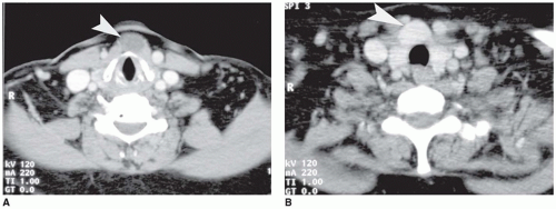
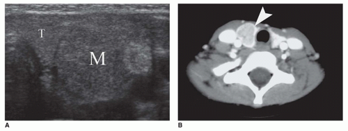
Imaging studies are an integral component of the diagnostic evaluation of a child with an enlarged thyroid or other massproducing process in the neck. US is a basic modality and provides for a confident diagnosis of TDC, which appears as a midline or paramedian cyst, possibly with debris when complicated by infection or hemorrhage (91) (Figure 21-8A, B). TDCs are usually near the hyoid bone. Branchial cleft cysts have a similar imaging appearance but are positioned in the lateral neck away from the midline (eFigure 21-48).

Ultrasound is useful in depicting thyroid nodules in patients with thyroid dysfunction or goiter. Complex cases may require MR. CT is less desirable for the evaluation of thyroid lesions because the use of iodinated contrast may preclude later radioactive thyroid ablation therapy if necessary for several weeks. Thyroid carcinomas appear well defined and heterogeneous on US, CT, or MR. Papillary thyroid carcinoma (PTC) is more likely to contain cystic-appearing, necrotic areas compared to follicular thyroid carcinoma (FTC) (Figure 21-9A). Most MTCs are solid and may contain coarse calcifications (Figure 21-9B).


FIGURE 21-8 • Thyroglossal duct cyst in an adult complicated by PTC. A: Axial contrastenhanced CT image shows a midline cyst (arrowhead). B: Axial CT image caudal to (a) shows markedly enhancing, midline mass (arrowhead).

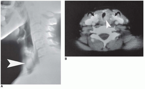
Radionuclide scintigraphy with 99mTc pertechnetate or 123I is very useful in the evaluation of thyroid dysfunction and nodules or in the localization of ectopic thyroid tissue (eFigure 21-49A to C). Nodules with decreased radiotracer uptake (“cold” nodules) are more likely to be malignant than nodules that are similar to surrounding normal thyroid or take up more radiotracer than normal thyroid (“hot,” hyperfunctioning nodules). When ectopic thyroid tissue is identified, it is important to evaluate the neck base and base of the tongue for orthotopic thyroid gland (Figure 21-10A, B, eFigure 21-49A to C).


Developmental Disorders

Dysmorphism of the thyroid gland is a structural phenomenon with several morphologic variations including absence or incomplete formation of a normal gland, failure of the normal anatomic localization of the gland, or persistence of embryologic remnants with a branching lobular pattern of immature follicles rather than dense formation of individual follicles (Table 21-5). Recessively inherited defects in enzymes responsible for thyroid hormone synthesis are other developmental disorders that are not characterized by primary structural anomalies of the thyroid gland (eFigure 21-50); however, elevated TSH levels lead to multinodular hyperplasia in the form of dyshormonogenic goiter (Figure 21-7). Clinically, these various developmental disorders present with congenital hypothyroidism, a mass at the base of the tongue or in the neck, or congenital hypothyroidism with development of a goiter. Many of these developmental anomalies also affect first-degree relatives, indicating a familial component.

Dysgenesis of the thyroid is a generic designation for various developmental anatomic anomalies that include complete failure in gland formation (agenesis), decreased amount
of thyroid tissue (hypoplasia), absence of a lobe (hemiagenesis), or ectopic location. Dysgenesis is an important etiology of congenital hypothyroidism, with prevalence in the United States of 1:3000 to 5000 live births. Most causes of congenital hypothyroidism are due to dysgenesis or inherited defects in thyroid hormone synthesis (Table 21-5). Congenital hypothyroidism has been also been observed in Williams and Down syndromes.


FIGURE 21-9 • Papillary thyroid carcinoma in 15-year-old girl. A: Transverse sonographic image showing a heterogeneous mass (M) within the homogeneous thyroid gland (T). B: MTC in an 8-year-old girl with family history of MEN2a. Axial contrast-enhanced CT shows a mass within the left thyroid lobe, which enhances less than the surrounding thyroid gland (arrowhead).

Congenital hypothyroidism in 80% to 85% of cases is associated with one of several types of dysgenesis. The prevalence of hypothyroidism in the neonatal period is 1:4000 live births with thyroid dysgenesis, 1:30,000 live births with dyshormonogenesis, 1:40,000 live births with transient hypothyroidism, and 1:100,000 live births with central hypothyroidism or hypothalamic-pituitary defect (81,89). In a study of 230 children with congenital hypothyroidism, scintigraphy revealed the following findings: ectopia in 61%, goiter in 18%, agenesis in 16%, normal in 4%, and hemiagenesis in less than 1%. In a series of 800,000 neonates with increased TSH and normally positioned thyroid glands, an enlarged gland or goiter was observed in 55% of cases, a normal gland in 29% of cases, and hypoplasia in 16% of cases. If the thyroid
gland is anatomically orthotopic in the presence of congenital hypothyroidism, a defect exists in thyroid hormone biosynthesis with the development of a dyshormonogenic nodular goiter or an inability of the gland to respond to TSH. Dysgenesis is more common in females than in males (3:1) and is sporadic in most cases (85%) (79). Affected infants with agenesis or hypoplasia have permanently elevated levels of TSH and low levels of circulating thyroid hormone. A number of mutations have been identified in the genes responsible for thyroid development, including PAX8, TTF-1 (thyroid transcription factor-1), TTF-2 (thyroid transcription factor-2), TSHR (TSH receptor), thyroglobulin (8q24), thyroperoxidase (2p25), sodium-iodide symporter (19p13), GNAS 1 (20q13), pendrin (7q31), DUOX2 (15q15), and DUOXA2 (15q15) and DHAL1 (6q24) and are pathogenetically involved in thyroid dysgenesis (79,80,81,82,83,92). These genetic defects and their association with other diseases are reviewed elsewhere (79).


FIGURE 21-10 • Ectopic thyroid gland in the trachea of an adult. A: Lateral tomogram shows an ovoid mass within the tracheal air column (arrowhead). B: Axial CT image shows markedly enhancing eccentric mass in the trachea (arrowhead) and normal thyroid lobes in the orthotopic location (curved arrows).


TABLE 21-5 ETIOLOGIC CLASSIFICATION OF CONGENITAL HYPOTHYROIDISM


I.


Primary hypothyroidism


A.


Dysgenesis (85%) (1:4,000) Idiopathic or genetic (TTF-1, TTF-2, FOXE1, PAX-8, and TSHR defects)


Agenesis


Hemiagenesis


Hypoplasia


Ectopia


Lingual thyroid (90% of thyroid ectopia) (1:10,000)


B.


Dyshormonogenesis (10-15%) (1:30,000)


Iodide transport (sodium-iodide symporter defect (NIS gene)


Iodide organification and coupling defect


Thyroid peroxidase defect (TPO gene) (Pendred defect)


Thyroid oxidase 2 defect (DUOX1/THOX1 DUOX/THOX2 genes)


Defect in thyroglobulin synthesis or transport (Tg gene)


Iodotyrosine deiodinase defect (DEHAL1 gene)


C.


Others (5%)


II.


Secondary/tertiary hypothyroidism (hypothalamic-pituitary-thyroid axis dysfunction) (1:100,000)


Genetic defects involving LHX3, LHX4, PROP 1, POU1F1, HESX1, TRHR, TSHB


III.


Peripheral thyroid hormone resistance


Genetic defects involving MCT8, THRB


IV.


Transient hypothyroidism (1:40,000)


Maternal antithyroid antibodies, goitrogenic drugs, iodine deficiency


Based on data from Peter, F, Muzsnai A. Congenital disorders of the thyroid: hypo/hyper. Endocrinol Metab Clin North Am. 2009;38:491-507; LaFranchi S. Section 2: Disorders of the thyroid gland. In: Kliegman RM, Behrman RE, Jenson HB, Stanton BF, eds., Nelson Textbook of Pediatrics, 18th ed. Philadelphia, PA: Elsevier, 2007; Bettendorf M. Thyroid disorders in children from birth to adolescence. Eur J Nucl Med Mol Imaging. 2002;29(Suppl 2):S439-S446.


Hemiagenesis is another form of dysgenesis with failure in the formation of the left lobe in most cases. This anomaly occurs in less than 0.5% of the population and is more common in females. Thyroid function is within normal limits (93).

Ectopia of the thyroid gland is more thoroughly documented on a morphologic basis than the other types of dysgenesis, as judged by the descriptions in the literature (eFigure 21-51). Ectopia has a female predominance. Lingual thyroid (base of tongue) occurs in approximately 1:10,000 individuals and is detected in most cases during a diagnostic evaluation for congenital hypothyroidism or as an incidentally discovered mass (Figure 21-11A, eFigure 21-49). Lingual thyroid accounts for approximately 90% of all thyroid ectopias. Most lingual thyroids are accompanied by an orthotopic thyroid; however, a minority of lingual thyroids constitutes the only site of thyroid tissue. Some cases classified as agenesis have a lingual remnant. Ectopic thyroid tissue including dual ectopia (location at different sites), with exclusion of occurrence in a teratoma, has been documented in the submandibular region, trachea, heart, mediastinum, and various intra-abdominal sites. The presence of thyroid follicles in lymph nodes as so-called lateral aberrant thyroid is controversial and is favored to represent metastatic thyroid carcinoma in most, if not all, cases (84). Thyroid neoplasia arising in ectopic thyroid, usually in a TDC, is recognized in children.

Ectopic thyroid may be represented by individual microfollicles or small foci of multiple microfollicles or solid nests of follicular cells without apparent colloid formation. The follicles are interspersed between bundles of skeletal muscle in the tongue or within the tissues of the other ectopic sites (Figure 21-11B, eFigure 21-52). In some instances, the epithelial structures are not readily identifiable as thyroid tissue and may require immunohistochemical staining for thyroglobulin, thyroid transcription factor 1, or PAX-8. In addition to the immature or nonfunctioning appearance of ectopic follicles, the ectopia is also hypoplastic because the total tissue volume of thyroid is less than normal for the age and gender.

Another form of thyroid dysgenesis is an enlarged lobe composed of immature lobules of fetal-appearing follicles separated by an immature mesenchyme. Nodules of immature cartilage or other heterologous tissues present within the lobule may suggest a teratoma.

Thyroglossal duct cyst (TDC) is the consequence in the failure of the thyroglossal duct to undergo complete obliteration and regression during fetal life (56). Approximately 15% of all neck masses in children are TDCs, presenting as a midline anterior neck mass overlying the hyoid bone (56,94). Rather than a midline location, 10% to 25% of TDCs are found
laterally, usually on the left side, and a minority occurs at the base of the tongue, floor of the mouth, or within the thyroid itself. The TDC differential includes branchial cleft cyst (lymphoepithelial cyst), lymphangioma, lymphadenopathy, lymphoma, epidermal inclusion cyst, and other thyroid malformations (94). Most cysts are diagnosed at or before 5 years of age as a painless cystic midline cervical mass, but may be recognized throughout life (56,94). Because of possible communication with the oral cavity through the foramen cecum, TDCs may become infected, and there may be periodic oral drainage with halitosis. Cutaneous draining sinuses may also occur in the midline of the neck through a fistulous or sinus tract. A familial association has been reported. A rare presentation of TDC is sudden death due to asphyxiation (56). Infected cysts may lead to fistula formation to the skin surface or pharynx (56).


FIGURE 21-11 • Lingual thyroid. A: Sagittal section through the tongue, which shows a smooth, ovoid, 2-cm-diameter mass in the posterior third of the tongue (arrow). Small and large cysts with adjacent red-brown thyroid tissue are present in the mass. Incidental finding at autopsy in a 69-year-old man who died from cerebral hemorrhage. (From Turk JL, Fletcher CDM, eds. Endocrine System. Royal College of Surgeons of England Slide Atlas of Pathology, 1985. Originally published by Gower Medical Publishing, Ltd. Reprinted with permission of Elsevier Inc. and CDM Fletcher, M.D.). B: This section of tongue shows the presence of thyroid follicles between the muscle fibers (arrows). This was an incidental finding at autopsy in a stillborn infant (H&E stain, original magnification 100×).

The pathologic findings of TDC vary from case to case with a dominant cyst or several smaller cysts in the soft tissues superior, inferior, or anterior to the hyoid bone (Figure 21-8A, eFigure 21-53). The dominant cyst usually measures 1 to 2 cm; however it may be in excess of 4 to 5 cm in diameter. The contents may have a mucoid or purulent appearance. TDCs can become infected. In some cases, it may be difficult to identify cysts, but rather a firm, ill-defined fibrotic area that represents prior episodes of chronic inflammation is present in the soft tissues (Figure 21-12A). Thyroid tissue is generally not appreciated in the gross examination and can be difficult to identify even microscopically. Individual follicles or larger islands of well-formed follicles are found in less than 50% of cases. Cuboidal to stratified columnar epithelium with cilia lines the cysts in 50% or more of cases (Figure 21-12B). Nonkeratinizing squamous epithelium is present in 25% of cases. The type of epithelium may vary from one cystic structure to another in any one specimen. The background stroma varies from mucoid to densely fibrotic. Lymphoid aggregates adjacent to the cyst or cysts and the ciliated respiratory-type epithelium resemble branchial cleft cyst; however, the branchial cleft cyst typically occurs in the lateral neck. Psammomatous calcification may be found in TDCs without accompanying PTC. Fine-needle aspiration biopsy (FNAB) had a positive predictive value of almost 70% in cases of TDC. In 1.5% of children with TDCs, a solid midline ectopic thyroid gland is present within the TDC. Some may have ectopic lingual thyroid tissue located in close proximity to the foramen cecum in the posterior dorsum of the tongue. Careful evaluation for a functional thyroid gland in its expected location is important prior to surgical excision of the TDC. Follicular adenomas and PTCs are reported in 1% to 4% of TDCs (Figure 21-8B) (56,94).

Branchial apparatus-associated anomalies are represented principally by the branchial cleft cyst (eFigures 21-48 and 21-54). A similar lesion, the lymphoepithelial cyst, is recognized in the thyroid (95). The cyst is accompanied by CLT in most cases. A bronchogenic cyst may also occur in the thyroid. Another type of branchial anomaly is the cyst or sinus from the oropharynx and/or hypopharynx with extension into the thyroid with the complication of recurrent acute thyroiditis.

Heterotopias in the thyroid gland include parathyroid, salivary gland, and thymic tissue (eFigures 21-55 and 21-56).


FIGURE 21-12 • Thyroglossal duct cyst. A: This midline cyst was filled with tan-white mucoid fluid on gross examination. Fibrosis of soft tissue adjacent to the cyst was present. B: This composite image shows the resected hyoid bone on the left with entrapped thyroid follicles (arrow). The area within the rectangle is magnified on the right side and shows a cuboidal epithelium (arrowheads) lining the cystic spaces. A thyroid follicle is also present in this image (arrow). Lymphoid aggregates not shown were also present (H&E stain).


Acquired Disorders

Persistent diffuse or nodular enlargement of the thyroid gland, regardless of its underlying nature, is referred to clinically as a goiter, without any specific pathologic implications. Through a variety of noninvasive and invasive techniques, including FNAB, an attempt is generally made to ascertain whether the pathologic process is inflammatory, hyperplastic, or neoplastic in nature before a decision is made about the need for surgical intervention. US is helpful in the characterization of a nodule or nodules as predominantly cystic, cystic and solid, or solid.

Thyroid nodules are detected in 1% to 1.5% of children with the entire range of pathology from developmental to neoplastic processes (congenital hypothyroidism due to dyshormonogenesis or ectopia, hemiagenesis, TDC, simple goiter, cystic lesions, nodular hyperplasia, follicular adenoma, Graves disease, and chronic lymphocytic [Hashimoto] thyroiditis) (86,96,97,98,99). Nodular hyperplasia (adenomatous hyperplasia) with a dominant nodule, followed by follicular adenoma, is the most common cause of a thyroid nodule(s) in children (100). Studies have suggested that approximately 20% to 25% of solitary thyroid nodules are malignant with the overwhelming majority representing PTCs (73,99). Management of the solitary thyroid nodule is reviewed elsewhere (96,99,100,101). Several studies have addressed the efficacy of ultrasound-guided FNAB of the thyroid in the pediatric age group with comparable results to those in adults with a diagnostic accuracy in excess of 85% in most cases.

One of the most common referrals to a pediatric endocrinologist is an enlarged thyroid gland (goiter). Most cases of a diffusely enlarged thyroid gland (nontoxic goiter) on physical examination in children are due to autoimmune-associated inflammatory conditions of the thyroid: CLT, juvenile lymphocytic thyroiditis, juvenile variant of Hashimoto thyroiditis, autoimmune thyroiditis, and diffuse toxic hyperplasia (Graves disease).

Chronic lymphocytic thyroiditis (CLT), which accounts for 40% of goiters in adolescents, affects females more commonly than males with a male to female ratio of 1:2 to 1:4, compared to a 1:10 male to female ratio in adults (81,89). The mean age at diagnosis is 11 to 12 years (range: 1 to 19 years). The thyroid gland is tender on palpation and has a granular to pebbly texture.

Nodularity may be present in 25% to 30% of cases. Most children (50% to 70%) are euthyroid, or asymptomatic with laboratory values in the hypothyroid range, whereas 20% to 40% are clinically hypothyroid. Thyrotoxicosis is present in less than 5% of cases. Antithyroid peroxidase (TPO) antibodies are present in 80% to 90% of cases and antithyroglobulin antibodies in 50% to 60% of cases. Several mechanisms including T-cell-mediated cytotoxicity, cytokine-mediated, and antibody-dependent cell-mediated cytotoxicity directed against follicular epithelial cells are implicated in the pathogenesis of CLT (eFigure 21-57).

Most cases of CLT in children are sporadic, but there is an increased association of CLT with HLA haplotypes, DR3, DR4, and DR5 (81,89). HLA-DR2 and HLA-DQ1 apparently have a protective effect against autoimmune thyroid disease (84). Polyglandular autoimmune syndrome type I, due to a defect in the autoimmune regulatory gene on chromosome 21q22.3, is defined in part by the presence of CLT; polyglandular autoimmune syndrome type II and type III are uncommon in the pediatric population (78). Systemic lupus erythematosus, chronic juvenile arthritis, Sjögren syndrome, celiac disease, vitiligo, alopecia, mixed connective tissue disease, Bannayan-Riley-Ruvalcaba syndrome, and type I diabetes mellitus may be accompanied by CLT as part of an
autoimmune diathesis . Approximately 4% of children with type I diabetes mellitus have CLT. Trisomy 21 syndrome, Klinefelter syndrome, and Turner syndrome are three chromosomal disorders associated with CLT. Approximately 25% of young individuals with Turner syndrome have antithyroid antibodies and 10% have enlarged thyroids.

The pathologic diagnosis of CLT is more often established by FNAB than by histologic examination. Surgical resection is reserved for specific clinical circumstances, such as a possible thyroid neoplasm. The thyroid is symmetrically enlarged and weighs more than 25 to 30 g. A pale, vaguely nodular, tan-gray appearance with a resemblance to lymph nodal tissue is noted on cross-section after fixation (Figure 21-13A). On occasion, one or the other lateral lobe or the pyramidal lobe is larger with the loss of symmetry. Any areas of discrete firmness, sclerosis, or nodularity may indicate the presence of PTC or scarring as in the fibrosing stage of CLT. Microscopically, lymphoid follicles with reactive germinal centers are interspersed throughout the gland with destructive replacement of parenchyma (Figure 21-13B, eFigure 21-58B, C). An intermixture of mature plasma cells is also apparent in a predominant population of B and T lymphocytes. The follicles are typically small and uniform, although some larger follicles with papillary infoldings may be seen. Some of the intact thyroid follicles may contain intrafollicular histiocytes and giant cells, as evidence of so-called palpation thyroiditis or the presence of giant cells and lymphoid aggregates where follicles once resided. The diminutive follicles are lined by cuboidal or flattened epithelial cells or by epithelial cells with optically clear nuclei and grooves as seen in PTC. The diagnosis of PTC is made in the presence of a discrete lesion(s). Classic Hurthle or oncocytic follicular cells as a diffuse finding are uncommon in CLT in children and, in this respect, do not fulfill the classic morphologic definition of Hashimoto thyroiditis (eFigure 21-59). However, CLT and Hashimoto thyroiditis are pathogenetically identical forms of autoimmune thyroiditis in all other respects. Mizukami et al. found no morphologic difference in the types of chronic thyroiditis between adults and children less than 10 years old.


FIGURE 21-13 • Chronic lymphocytic thyroiditis. A: This specimen shows the characteristic diffuse thyroid gland enlargement seen in CLT on gross examination. A vaguely nodular pattern corresponding to the presence of lymphoid follicles is seen in this cut section. B: CLT in this low-power magnification image shows prominent lymphoid aggregates interspersed between the thyroid follicles. Plasma cells and lymphocytes were present in the interstitium (H&E stain).

The fibrosing or end stage of CLT with marked loss and atrophy of follicles, fibrosis with a finely nodular pattern, and a diminution of the lymphocytic infiltrate are infrequently encountered in children. As noted earlier, the morphologic diagnosis of CLT is usually based on FNAB. The typical cytologic finding is a mixture of individual and small nonpapillary groups of benign-appearing follicular epithelial cells in a background of many dispersed small lymphocytes, some plasma cells, and histiocytes. Hurthle cells are infrequent, and even less common are papillae, whose presence should raise the possibility of PTC. Approximately 30% of cases of CLT in children had distinct nodules and 3% had a PTC.

Other types of thyroiditis, infectious and noninfectious, occur infrequently in children. Abscess of the thyroid has been reported in children, and opportunistic infections are seen in the immunocompromised setting. Recurrent acute suppurative thyroiditis with or without abscess formation should suggest the presence of a branchial pouch anomaly, such as a pyriform sinus cyst or TDC remnant (56). Most cultures demonstrate a mixed flora, containing a Streptococcus species. Common features of acute suppurative thyroiditis include a painful/tender neck mass associated with fever. Involvement of the left lobe is more common. A left hemithyroidectomy may need to be performed for recurrent infections. An infectious etiology should be excluded in granulomatous thyroiditis in a child, because subacute giant cell or de Quervain thyroiditis is extremely rare in childhood.

Hyperplasia of the thyroid gland is either diffuse or multinodular. Diffuse hyperplasia is often associated with hyperthyroidism or thyrotoxicosis. The so-called simple goiter is defined clinically as diffuse or nodular enlargement of the
thyroid gland without obvious evidence of hyperthyroidism. Children with a simple goiter are predominantly young adolescent females and do not experience any further gland enlargement. A small percentage, however, may develop CLT.

The simple or colloid goiter is a more or less symmetrically enlarged thyroid gland with a diffuse or multinodular appearance (eFigure 21-60). The follicles vary in size with one or more colloid-filled macrofollicles lined by a flattened layer of epithelial cells (eFigure 21-61). Formation of colloid cysts occurs in some cases. Conversely, nodular hyperplasia or adenomatous nodules manifest as multiple variably sized follicular nodules with or without dense fibrous bands, cystic degeneration, hemorrhage, and nonspecific chronic inflammation. Papillary hyperplasia is a source of concern in areas of degeneration, but the follicular cells usually do not have well-developed nuclear features of PTC. A peripheral (socalled parasitic or exophytic) nodule may be found in the perithyroidal soft tissues and even embedded in skeletal muscle, especially at the isthmus.

Multinodular hyperplasia is the pathologic finding associated with dyshormonogenic goiter (Figure 21-7). One example is Pendred syndrome, manifesting as goiter and hearing loss in adolescence due to a defect in the PDS gene (SLC26A4 gene) on chromosome 7 that encodes for the protein pendrin, which is involved in iodide transport across the cell membranes, and whose absence results in decreased organification of iodide with disruption in thyroid hormone synthesis. The follicular nodules of a dyshormonogenic goiter tend to be hypercellular due to the formation of microfollicles, trabeculae, and papillae (eFigure 21-62). Cytologic atypia may be present, usually in the form of “random” endocrine atypia, rather than PTC-like atypia. Endocrine-type atypia includes isolated nuclear enlargement, hyperchromasia, and mitotic activity without diffuse enlargement, clearing, or grooves. Well-differentiated thyroid carcinoma has been reported in dyshormonogenic goiters, but it is difficult to determine whether the risk of malignancy is increased in these glands.

Diffuse hyperplasia with clinical hyperthyroidism (Graves disease) is an autoimmune disorder of the thyroid, with some overlapping immunologic and pathologic findings with CLT. Hyperthyroidism also occurs infrequently on the basis of “toxic” nodular hyperplasia, functioning follicular adenoma, autosomal dominant nonimmune hyperthyroidism, and congenital hyperthyroidism. The latter two disorders have been reported with activating germline mutations in the TSH receptor gene. Sporadic congenital hyperthyroidism occurs in the presence of maternal autoimmune thyroid disease with the transplacental passage of maternal thyroidstimulating immunoglobulins. Only 1% of neonates whose mothers have active Graves disease during pregnancy have evidence of hyperthyroidism at birth. Most cases of hyperthyroidism in children are on the basis of Graves disease (81). Other etiologies of hyperthyroidism in children have been tabulated by LaFranchi (82,89).

A screening study of school-aged population children between 11 and 18 years of age revealed that almost 4% had clinical or laboratory evidence of “thyroid abnormalities” and approximately 5% of those with abnormalities had hyperthyroidism. This figure compares with other studies in which 10% to 15% of all pediatric thyroid disease is diagnosed as hyperthyroidism. Juvenile hyperthyroidism typically presents in girls (6:1, female to male ratio) who are usually 11 years of age and older (11 to 18 years) and have diffuse enlargement of the thyroid (95% of cases) or less often have a dominant “toxic” or autonomous nodule (81). Hyperthyroidism occurs in families and is associated with MAS activating mutations in the stimulatory G protein domain. Germline mutations in the TSH receptor account for cases of toxic multinodular goiter and toxic thyroid adenoma.

Graves disease (diffuse toxic goiter) is characterized by hyperthyroidism, ophthalmopathy (exophthalmos), and dermopathy (pretibial myxedema) in the pediatric population. Its peak prevalence is in adolescence (11 to 15 years of age) and is three to five times more common in girls (81). The clinical symptoms include weight loss, heat intolerance, sweating, palpitations, emotional lability, nervousness, and intellectual decline. Congenital diffuse toxic goiter occurs in a small percentage of infants (1%) born to mothers with active Graves disease. The thyroid on physical examination is goiterous, smooth, firm, and nontender. The pathogenesis of Graves disease involves T- and B-cell dysregulation leading to the production of several anti-TSH receptor, thyroidstimulating, thyroid growth-stimulating, and TSH-binding inhibitor antibodies (eFigure 21-57). Thyroid-stimulating immunoglobulin mimics TSH and binds to the follicular cell TSH receptor leading to hypersecretion of thyroid hormones. The thyroid growth-stimulating immunoglobulin also binds to the TSH receptor and stimulates follicular cell hyperplasia with the development of increased serum levels of thyroxine or triiodothyronine and decreased TSH. The presence of anti-TSH receptor antibodies confirms the diagnosis of Graves disease versus other causes of hyperthyroidism. Total or subtotal thyroidectomy is performed in cases of medical failure or intolerance. The clinical management of Graves disease in children is the subject of continued study and controversy.

Pathologically, the thyroid gland is symmetrically enlarged without apparent nodules in most cases (Figure 21-14A, eFigure 21-63A). A red-brown color without an appreciation of translucent colloid is noted on cut surface. The weight of the gland is generally more than 25 to 30 g, but this varies somewhat with the age of the patient. In the unsuppressed gland, the follicular cells have a tall columnar appearance. Crowding of these cells leads to intrafollicular papillary infoldings on histologic examination (Figure 21-14B, eFigure 21-63B). Marked follicular cell pleomorphism can be seen in pretreated glands. The colloid has a pale watery appearance and is absent in some follicles. Those follicles with colloid often show peripheral scalloping of the colloid. These latter findings are usually attenuated with preoperative suppression to diminish the function and vascularity of the gland (eFigure 21-64). Epithelial hyperplasia through the action of TSH, leading to more prominent intrafollicular papillary
infoldings, is seen in the gland treated by thiouracil. Iodine administration before surgery results in the accumulation of colloid and the formation of macrofollicles, often with associated accentuation of thyroid lobules by thin fibrous bands. Rather than cuboidal to columnar epithelium, flattened epithelial cells cover the slender papillae.


FIGURE 21-14 • Graves disease. A: This image shows diffuse symmetrical enlargement of the thyroid gland from a patient with Graves disease. The parenchyma has a deep red color due to increased vascularity within the gland. (Reprinted with permission from Lloyd RV, Douglas BR, Young WF. Endocrine Diseases. Atlas of Nontumor Pathology. Washington, DC: American Registry of Pathology, 2001.) B: This section of thyroid gland from a patient with untreated Graves disease shows follicles with hyperplastic epithelium and papillary infoldings. Pale watery colloid and an interstitial lymphocytic infiltrate (not pictured) were observed. The papillary infoldings (inset) lack the optically clear nuclei seen in PTC (H&E stain).

Lymphocytic infiltrates in the interstitium and lymphoid nodules with reactive germinal centers are prominent in some glands. Without the clinical history of Graves disease, a diagnosis of CLT may be the preferred interpretation based on histologic examination. The intrafollicular papillae may cause concern about PTC; however, the follicular cells lack the well-developed nuclear atypia of PTC. At least in the pediatric age population, PTC is rarely found in the midst of diffuse toxic hyperplasia.


Neoplasms

The World Health Organization classification of thyroid tumors contains a number of histologic types, but the overwhelming majority of differentiated carcinomas of the thyroid in children are PTC. Institutional referral patterns may affect the proportion of MTC in children with RET mutations in affected kindreds with MEN2a or MEN2b. Almost 30% of children with differentiated carcinomas at St. Louis Children’s Hospital are MTCs because of MEN2 referrals to the institution. FTC and MTC comprise less than 10% of thyroid carcinomas in the experience of most other institutions. Undifferentiated (anaplastic) carcinomas are rare in children in contrast to adults.

Only gold members can continue reading. Log In or Register to continue

Stay updated, free articles. Join our Telegram channel

Sep 23, 2016 | Posted by in PATHOLOGY & LABORATORY MEDICINE | Comments Off on The Pineal, Pituitary, Parathyroid, Thyroid, and Adrenal Glands

Full access? Get Clinical Tree

Get Clinical Tree app for offline access