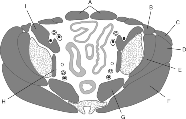
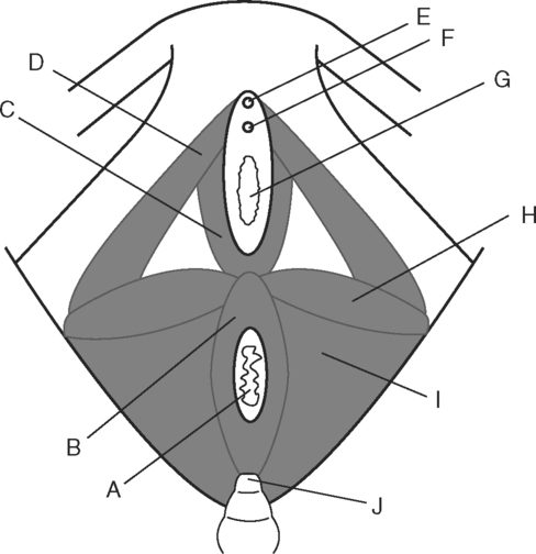
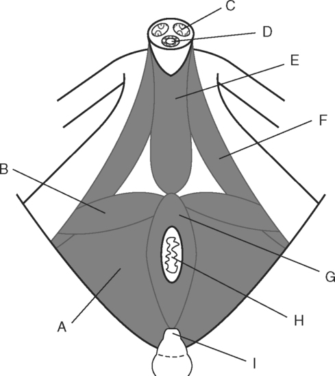
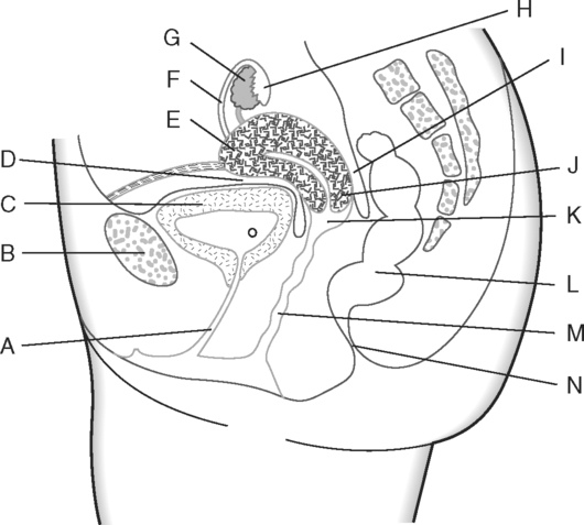
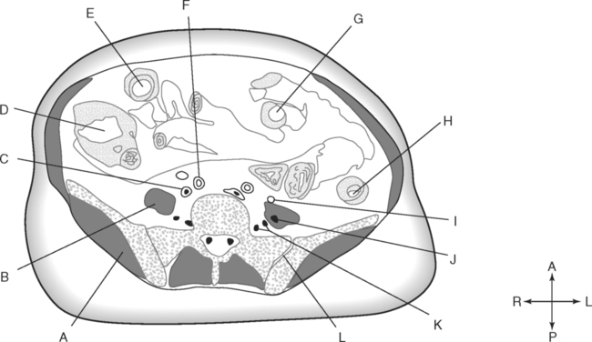
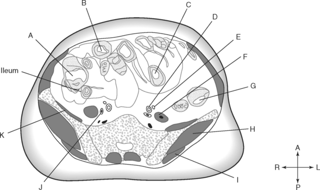
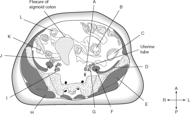
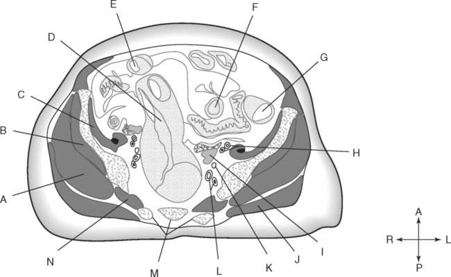
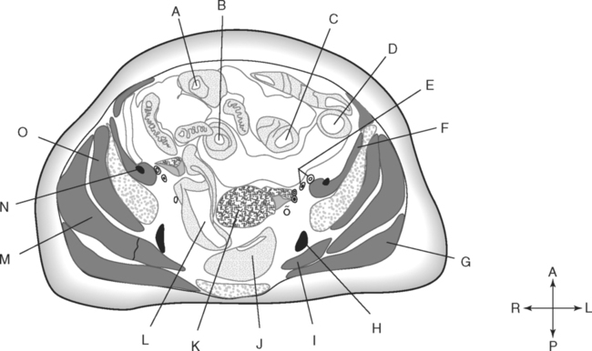
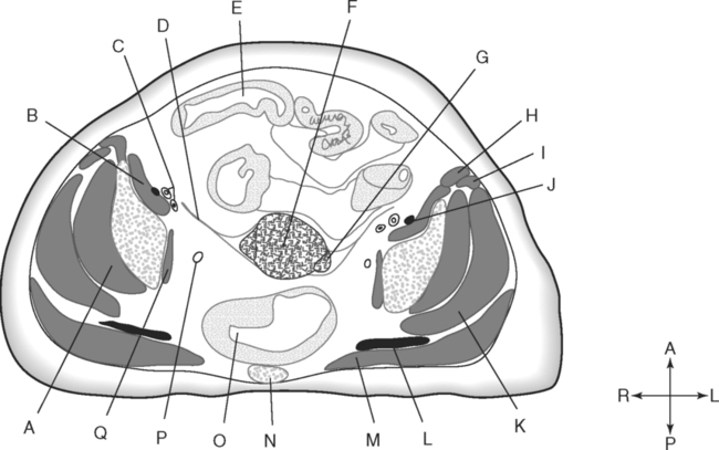
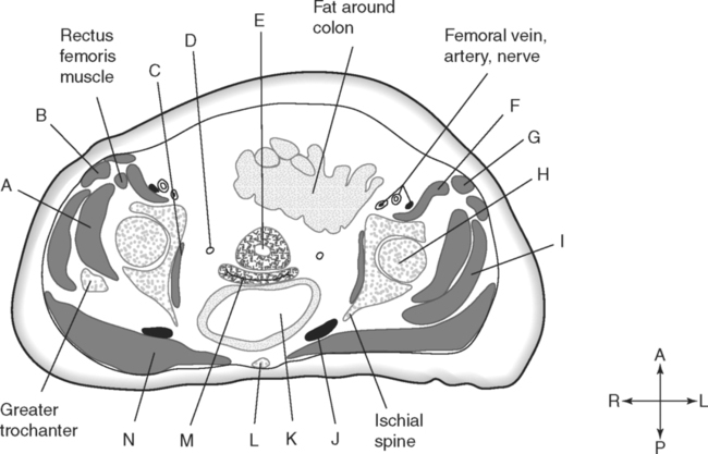
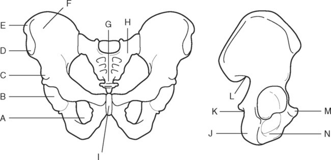
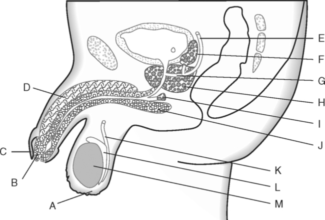
Identify the Features Indicated in Fig. 4-1: 1. What is the articulation between the two pubic bones called? 2. Where do the three bones of the os coxae meet? Identify the Muscles Indicated in Fig. 4-2: 1. What muscle lines the lateral wall of the true pelvis? 2. What muscle is the deepest of the three gluteal muscles? 3. What muscle fills the space between the ilium and sacrum? Identify the Structures and Regions Indicated in Fig. 4-3: 1. What three structures or openings are enclosed by the bulbospongiosus muscle in the female perineum? 2. Which is the larger of the two muscles in the anal region of the female perineum? 3. What structure is at the apex of the urogenital triangle of the female perineum? Identify the Structures Indicated in Fig. 4-4: 1. What muscle forms the horizontal division between the two triangles of the male perineum? 2. What is the largest muscle of the male perineum? 3. What muscle forms the lateral margins of the urogenital triangle of the perineum? Identify the Structures Indicated in Fig. 4-5: 1. What is the most posterior portion of the uterus? 2. Which is more posterior, the rectouterine space or the vesicouterine space? 3. What name is given to the vaginal space surrounding the cervix of the uterus? Identify the Structures Indicated in Fig. 4-6: 1. Name the three accessory glands of the male reproductive system. 2. What is the name given to the expanded distal end of the corpus spongiosum in the male penis? 3. What duct carries sperm from the epididymis to the ejaculatory duct? Identify the Structures Indicated in Fig. 4-7: 1. Which of the gluteal muscles is most superior? 2. What portion of small intestine joins with the ascending colon? 3. As the ureters descend from the kidneys to the urinary bladder, with what muscles are they usually associated? 4. Which is more anterior, the left common iliac artery or the left common iliac vein? 5. Within the pelvic region, which is more posterior, the ascending colon or the descending colon? Identify the Structures Indicated in Fig. 4-8: 1. What muscle originates on the iliac crest and lines the iliac fossa? 2. Why is the descending colon in a relatively constant location? 3. What nerve is closely associated with the psoas major muscle? 4. What portion of the large intestine is inferior to the ileocecal valve? Identify the Structures Indicated in Fig. 4-9: 1. Which is more anterior, the external iliac vein or the internal iliac vein? 2. Which is more medial, the ureter or the external iliac artery? 3. Which is more posterior, the external iliac vein or the external iliac artery? 4. What bone articulates with the lateral margin of the sacrum? In Fig. 4-10 Color or Outline: Identify the Structures Indicated in Fig. 4-10: 1. What portion of the large intestine is immediately anterior to the sacrum? 2. Which of the iliac vessels is closely related to the psoas portion of the iliopsoas muscle? 3. Which of the three gluteal muscles is the deepest muscle? 4. Which of the three gluteal muscles originates in the most inferior location? 5. What muscle occupies the space of the greater sciatic notch? In Fig. 4-11 Color or Outline: Identify the Structures Indicated in Fig. 4-11: 1. Which branch of the sacral plexus is deep to the piriformis muscle? 2. True or false: loops of small intestine extend into the true pelvis. 3. Which is more superior, the uterus or the urinary bladder? 4. What is the name of the peritoneal space posterior to the uterus? In Fig. 4-12 Color or Outline: Sartorius and gluteus medius—orange Tensor fasciae latae and gluteus minimus—brown Obturator internus and iliopsoas—pink Identify the Structures Indicated in Fig. 4-12: 1. What membrane encloses the proximal end of the uterine tube? 2. What large nerve is deep to the gluteus maximus muscle at this level? 3. What is the superficial muscle anterior and lateral to the gluteus medius muscle? 4. Using this section, determine the advantage of giving intramuscular injections into the superior portion of the gluteus medius muscle rather than into the gluteus maximus muscle. In Fig. 4-13 Color or Outline:
The Pelvis
Stay updated, free articles. Join our Telegram channel

Full access? Get Clinical Tree


