The nervous system

Table 11.1). The patient should be allowed to describe the symptoms in his or her own words to begin with, and then the clinician needs to ask questions to clarify information and obtain more detail. It is particularly important to ascertain the temporal course of the illness, as this may give important information about the underlying aetiology.


TABLE 11.1 Neurological history


Presenting symptoms*
Headache, facial pain

Neck or back pain

Fits, faints or funny turns

Dizziness or vertigo

Disturbances of vision, hearing or smell

Disturbances of gait

Loss of or disturbed sensation, or weakness in limb(s)

Disturbances of sphincter control (bladder, bowels)

Involuntary movements or tremor

Speech and swallowing disturbance

Altered cognition
Risk factors for cerebrovascular disease
Hypertension

Smoking

Diabetes mellitus

Hyperlipidaemia

Atrial fibrillation, bacterial endocarditis, myocardial infarction (emboli)

Haematological disease

Family history of stroke

* Note particularly the temporal course of the illness, whether symptoms suggest focal or diffuse disease, and the likely level of involvement of the nervous system.


An acute onset of symptoms (within minutes to an hour) is suggestive of a vascular or convulsive problem (e.g. the explosive severe headache of subarachnoid haemorrhage or the rapid onset of a seizure).


For these episodes of sudden onset, a precipitating event (e.g. exercise) or warning (aura) may be present. The aura that precedes a seizure may be localising (e.g. auditory hallucinations, an unusual smell or taste, loss of speech, or motor changes) or non-localising (e.g. a feeling of apprehension). The occurrence of an aura followed by sudden unconsciousness is very suggestive of the diagnosis of a major seizure or complex partial seizure.


A stroke or cerebrovascular accident usually causes symptoms which appear over minutes or are present when the patient wakes from sleep. There is a focal problem with function of the brain. Patients may be unable to move one side of the body (hemiplegia) or have difficulty with speech or swallowing, There may have been previous episodes. When there is resolution of the symptoms within 24 hours the episode is called a transient ischaemic attack—TIA). The rapid onset of focal symptoms almost always has a vascular cause—embolism, infarction or haemorrhage. If the patient can answer questions it is important to ask about the onset of the symptoms and about risk factors for stroke (Questions box 11.1).


image


Questions box 11.1


Questions to ask the (non-aphasic) patient with a possible stroke or transient ischaemic attack


1. What have you noticed has been wrong?

2. How quickly did it come on? How long ago?

3. Has it improved or gone away now?

4. Have you ever had a stroke before? How did that affect you?

5. Have you had high blood pressure or cholesterol (risk factors)?

6. Are you a diabetic (risk factor)?

7. Do you smoke (risk factor)?

8. Is there a history of strokes in the family?

9. Have you had palpitations or been told you have atrial fibrillation?

10. Have you been treated with blood-thinning drugs such as aspirin or warfarin?

image


The sudden onset of weakness on one side of the body followed by resolution and a severe headache is characteristic of hemiplegic migraine. Sudden resolution without headache suggests a transient ischaemic episode. The very gradual onset of muscle weakness suggests a muscle abnormality such as myopathy rather than a vascular event.


A subacute onset (hours to days) occurs with inflammatory disorders (e.g. meningitis, cerebral abscess or the Guillain-Barréa syndrome—acute inflammatory polyradiculoneuropathy).


A more chronic symptom course suggests that the underlying disorder may be related to either a tumour (weeks to months) or a degenerative process (months to years). Metabolic or toxic disorders may present with any of these time courses.


Based on the history (and physical examination), a judgment is made as to whether the disease process is localised or diffuse, and which levels of the nervous system are involved (the nervous system may be thought of as having four different levels: the peripheral nervous system, the spinal cord, the posterior fossa, and the cerebral hemispheres). Consideration of the time course and the levels of involvement will usually lead to a logical differential diagnosis of the patient’s symptoms. After detailed questions about the presenting problem, ask about previous neurological symptoms and about previous neurological diagnoses or investigations. The patient may know the results of CT or magnetic resonance imaging brain scans performed in the past. A thorough neurological history will include routine questions about possible neurological symptoms (Questions box 11.2). If the patient answers ‘yes’ to any of these, more-detailed questions about the nature of the problem and its time course are indicated.


image


Questions box 11.2


Questions to ask the patient with a possible neurological problem


1. Can you tell me what has been happening to you?

2. Are you right- or left-handed?

3. Have you had problems with headaches?

4. Have you been dizzy or had problems with your balance?

5. Have you noticed trouble with your speech?

6. Have you had problems with your vision?

7. Have you had weakness in an arm or leg?

8. Have you ever had a seizure or a blackout?

9. Have you ever had a head injury?

10. Have you had any back problems?

11. Have you had any scans of your brain or spinal cord?

12. What medications have you been taking?

13. Have you had high blood pressure?

14. Is there a history of neurological or muscle problems in the family?

15. Do you drink alcohol?

image


Headache and facial pain


Headache is a very common symptom (Questions box 11.3). It is important, as with any type of pain, to determine the character, severity, site, duration, frequency, radiation, aggravating and relieving factors and associated symptoms.1,2 Unilateral headache that is preceded by flashing lights or zigzag lines and is associated with light hurting the eyes (photophobia) is likely to be a migraine with an aura (‘classical migraine’); common migraine has no aura. Pain over one eye (or over the temple) lasting for minutes to hours, associated with lacrimation, rhinorrhoea and flushing of the forehead, and occurring in bouts that last several weeks a few times a year or less, is suggestive of cluster headache. This occurs predominantly in males and patients can’t stay still. Headache over the occiput and associated with neck stiffness may be from cervical spondylosis. Coital headache occurs during intercourse close to orgasm.


image


Questions box 11.3


Questions to ask the patient with headache


1. What is it like, e.g. dull, sharp, throbbing or tight?

2. Where do you feel it—at the front or back, on one side or in the face?

3. How severe is it and how long does it last?

4. Has it begun very suddenly and severely?—Subarachnoid haemorrhage

5. Do you get any warning that it is about to start, e.g. flashing lights or zigzag lines in your vision?—Migraine

6. Is it associated with sensitivity to light (photophobia)?—Migraine

7. Do you feel drowsy or nauseated?—Raised intracranial pressure

8. Is the pain on one side over the temple and have you had any blurred vision?—Temporal arteritis

9. Is the pain worst over your cheek bones?—Sinusitis

10. Are the attacks likely to occur in clusters and associated with watering of one eye?—Cluster headache

11. Is there a prolonged feeling of tightness over the head but no other symptoms?—Tension headache

12. Did you drink large amounts of alcohol last night?—Hangover

image


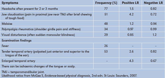
A generalised headache that is worse in the morning and is associated with drowsiness or vomiting may reflect raised intracranial pressure, while generalised headache associated with photophobia and fever as well as with a stiff neck of more gradual onset may be due to meningitis. A persistent unilateral headache over the temporal area associated with tenderness over the temporal artery and blurring of vision suggests temporal arteritis.3,4 This condition (Table 11.2) is often associated with jaw claudication, or jaw pain during eating, which can lead to considerable loss of weight. Headache with pain or fullness behind the eyes or over the cheeks or forehead occurs in acute sinusitis. The dramatic and usually instantaneous onset of severe headache that is initially localised but becomes generalised and is associated with neck stiffness may be due to a subarachnoid haemorrhage. Morning headaches worse with coughing, especially in an obese patient, may be due to idiopathic intracranial hypertension; visual loss may occur.


TABLE 11.2 Symptoms and signs of temporal arteritis

image

Finally, the most frequent type of headache is episodic or chronic tension-type headache; this is commonly bilateral, occurs over the frontal, occipital or temporal areas, and may be described as a sensation of tightness that lasts for hours and recurs often. There are usually no associated symptoms such as nausea, vomiting, weakness or paraesthesiae (tingling in the limbs), and the headache does not usually wake the patient at night from sleep.


Pain in the face can result from trigeminal neuralgia, temporomandibular arthritis, glaucoma, cluster headache, temporal arteritis, psychiatric disease, aneurysm of the internal carotid or posterior communicating artery, or the superior orbital fissure syndrome.


Faints and fits (see also page 41)


It is important to try to differentiate syncope (transient loss of consciousness) from epilepsy (Questions box 11.4). However, primary syncopal events can cause a few clonic jerks in a significant number of cases. Generalised tonic-clonic seizures (grand mal epilepsy) cause abrupt loss of consciousness, which may be preceded by an aura. Often the patient is incontinent of urine and faeces, and the tongue may be bitten. A witness may be able to describe the type of attack that occurred. It is important to try to determine whether any seizure is generalised or localised to one side of the body: a seizure affecting part of the body may indicate a focal lesion in the central nervous system, such as a tumour or abscess. If consciousness is impaired, these partial seizures are described as ‘complex’; if consciousness is unimpaired they are termed ‘simple’. Idiopathic absence seizures (‘petit mal’) occur in children. These are frequent brief episodes of loss of awareness often associated with staring. Major motor movements do not occur with this type of epilepsy.


image


Questions box 11.4


Questions to ask the patient with syncope or dizziness


1. Have you lost consciousness completely? How long for?

2. Do you black out or feel dizzy when you stand up quickly?—Postural hypotension

3. How often have episodes occurred?

4. Was the sensation more one of spinning?—Vertigo

5. Did the episode occur during heavy exercise or when you got up to pass urine at night? Exercise—suggests a left ventricular outflow tract obstruction such as aortic stenosis. Pass urine at night—micturition syncope

6. Have you injured yourself?

7. Do you get any warning?—A feeling of nausea and being in a stuffy room suggests a vasovagal episode; a strange smell or feeling of deja-vu suggests an aura and therefore a seizure

8. Have you passed urine during the episode?—Seizure

9. Have you bitten your tongue?—Seizure

10. Has anyone seen an episode and noticed jerking movements (tonic-clonic movements)?—Makes a seizure more likely but can also occur with cardiac syncope

11. Do you wake up feeling normal or drowsy? Normal—cardiac syncope. Drowsy—seizure

12. What medications are you taking—any antihypertensive medications, cardiac anti-arrhythmic drugs or anti-epileptic drugs?

image


Transient ischaemic attacks (TIAs) affecting the brainstem can occasionally cause blackouts. Use of the term ‘drop attacks’ means the patient falls but there is no loss of consciousness. In either case the patient falls to the ground without premonition and the attacks are of brief duration. Hypoglycaemia can lead to episodes of loss of consciousness. Patients with hypoglycaemia may also report sweating, weakness and confusion before losing consciousness. Bizarre attacks of loss of consciousness occur with hysteria.b During such attacks the patient may slump to the ground without sustaining any injury and there may be apparent fluctuations in the level of consciousness for a prolonged period.


Dizziness


If a patient complains of dizziness, it is important to determine what is meant by this term. In true vertigo, there is actually a sense of motion, usually of the surroundings but also of the head itself (page 41).5 When vertigo is severe it may not be possible for the patient to stand or walk, and associated symptoms of nausea, vomiting, pallor, sweating and headache may be present. Causes of vertigo include the ‘peripheral vestibular lesions’:


benign positioning (positional) vertigo—recurrent brief episodes of vertigo precipitated by a change of head position, due to crystals in the saccule and utricle

vestibular neuronitis—non-positional vertigo due to inflammation of the acoustic nerve with normal hearing, and

acute labyrinthitis—associated with hearing loss.

Other causes of vertigo include:


ototoxic drugs (e.g. aminoglycosides), associated with deafness or tinnitus;

Ménière’s disease,c which occurs in those over 50 years of age and presents with the triad of episodic vertigo and tinnitus (ringing in the ears) with progressive deafness;

acoustic neuroma (where patients may also have deafness and tinnitus);

central causes such as vertebrobasilar TIAs—these may be associated with diplopia (double vision; page 427), visual loss and ataxia; and

rarely, internal auditory artery occlusion.


Visual disturbances and deafness


Problems with vision can include double vision (diplopia), blurred vision (amblyopia), light intolerance (photophobia) and visual loss. The causes of deafness are summarised on page 348.


Disturbances of gait


Many neurological conditions can make walking difficult. These are described on page 376. Walking may also be abnormal when orthopaedic disease affects the lower limbs or spine. A bizarrely abnormal gait can sometimes be a sign of a hysterical reaction.


Disturbed sensation or weakness in the limbs


Pins and needles in the hands or feet may indicate nerve entrapment or a peripheral neuropathy (page 386) but can result from sensory pathway involvement at any level. The carpal tunnel syndrome is common; here there is median nerve entrapment, and patients experience pain and paraesthesiae in the hand and wrist. Sometimes pain may extend to the arm and even to the shoulder, but paraesthesiae are felt only in the fingers. These symptoms are usually worse at night and may be relieved by dangling the arm over the side of the bed or shaking the hand.


Nerve root, spinal cord and cerebral abnormalities can all cause disturbance of sensation and weakness.


Limb weakness can be caused by lesions at different levels in the motor system. There are a number of patterns of limb and muscle weakness:


Upper motor neurone (UMN) weakness (page 383) is due to interruption of a neural pathway at a level above the anterior horn cell. The result is an increase in tone and peripheral reflexes. Interruption of this pathway has the greatest effect on the antigravity muscles and is called pyramidal weakness. There is little or no muscle wasting.

Lower motor neurone (LMN) weakness (page 385) is due to a lesion that interrupts the reflex arc between the anterior horn cell and the muscle. There is a reduction in tone and reflexes, fasciculation (irregular contractions of small areas of muscle) may be seen and muscle wasting is prominent.

Muscle disease causes weakness in a particular muscle or group of muscles. There is wasting, decreased tone, and the reflexes are reduced or absent.

Disease at the neuromuscular junction (e.g. myasthenia gravis, page 394) causes generalised weakness, which worsens with repetition. The reflexes and tone are often normal.

Non-organic weakness (e.g. due to hysteria) causes a non-anatomical pattern of weakness in association with normal tone and power and, unless there has been prolonged disuse, normal muscle bulk.


Tremor and involuntary movements


Tremor is a rhythmical movement (Table 11.3). A slow tremor has, by definition, a rate between 3 Hz and 5 Hz. Rapid tremors are faster than 10 Hz. Resting tremors are present mostly during relaxation of the muscles, while intention tremors occur with deliberate movement and become more pronounced towards the end of the action. Tremors become worse with fatigue or anxiety. Shivering is a type of tremor brought on by cold. It is normal for there to be a fine tremor associated with holding a posture or performing a movement slowly. This is called a physiological tremor. It becomes more obvious with fright and fatigue. It is often increased by the beta-agonist drugs used to treat asthma or by caffeine. Thyrotoxicosis is a cause of exaggeration of physiological tremor. These movements are very fine and may be difficult to see unless looked for specifically. Benign essential (familial) tremor is an inherited disorder which causes tremor, but no other signs. The tremor is most easily seen when the patient’s arms are stretched out; it can become worse during voluntary movements. It usually disappears when the muscles are at complete rest. Parkinson’s diseased may present with a resting tremor (page 397). Intention (or target-seeking) tremor is due to cerebellar disease (page 398). Chorea involves involuntary jerky movements (page 399). Definitions of the terms used to describe movement disorders are shown in Table 11.4.


TABLE 11.3 Rates of tremors


Parkinson’s disease 3 to 5 Hz
Essential/familial 4 to 7 Hz
Physiological 8 to 13 Hz

TABLE 11.4 Definitions of terms used to describe movement disorders


Akithesia Motor restlessness; constant semi-purposeful movements of the arms and legs
Asterixis Sudden loss of muscle tone during sustained contraction of an outstretched limb
Athetosis Writhing, slow sinuous movements, especially of the hands and wrists
Chorea Jerky small rapid movements, often disguised by the patient with a purposeful final movement: e.g. the jerky upward arm movement is transformed into a voluntary movement to scratch the head
Dyskinesia Purposeless and continuous movements, often of the face and mouth; often a result of treatment with major tranquillisers for psychotic illness
Dystonia Sustained contractions of groups of agonist and antagonist muscles, usually in flexion or extremes of extension; it results in bizarre postures
Hemiballismus An exaggerated form of chorea involving one side of the body: there are wild flinging movements which can injure the patient (or bystanders)
Myoclonic jerk A brief muscle contraction which causes a sudden purposeless jerking of a limb
Myokymia A repeated contraction of a small muscle group; often involves the orbicularis oculi muscles
Tic A repetitive irresistible movement which is purposeful or semi-purposeful
Tremor A rhythmical alternating movement


Speech and mental status


Speech may be disturbed by many different neurological diseases and is discussed on page 377. A number of different diseases can also result in delirium or dementia, as described on page 377 and in Chapter 12.


Past health


Inquire about a past history of meningitis or encephalitis, head or spinal injuries, a history of epilepsy or convulsions and any previous operations. Any past history of sexually transmitted disease (e.g. risk factors for HIV infection or syphilis) should be obtained. Ask about risk factors that may predispose to the development of cerebrovascular disease (Table 11.1). A previous diagnosis of peripheral vascular disease or of coronary artery disease indicates an increased risk of cerebrovascular disease. Chronic or paroxysmal atrial fibrillation is associated with a greatly increased risk of embolic stroke, especially for people over the age of 70.


Medication history


Previous and current medications may be the cause of certain neurological or apparently neurological syndromes (Table 11.5).


TABLE 11.5 Drugs and neurology


1 Anti-hypertensives
Therapeutic use: reduction of risk of stroke

Side-effects: postural dizziness, syncope, depression (methyldopa)
2 Anti-platelet drugs and anti-coagulants
Therapeutic use: reduction of risk of stroke

Side-effect: cerebral haemorrhage
3 Statins
Therapeutic use: reduction in stroke risk

Side-effect: myopathy
4 Major tranquillisers
Therapeutic use: treatment of psychoses

Side-effects: ataxia, sedation, Parkinsonian tremor
5 Other neurological symptoms associated with drugs
Headache: nitrates, sildenafil

Deafness: aminoglycoside antibiotics, aspirin, frusemide

Peripheral neuropathy: amiodarone, isoniazid, metronidazole

Non-Parkinsonian tremor: bronchodilators, amphetamines

Dysphagia: bisphosphonates

Confusion and loss of memory: minor tranquillisers

Seizures: lignocaine

Ask about treatment for neurological disorders with anticonvulsants, anti-Parkinsonian drugs, steroids, immunosuppressants, biological agents, anticoagulants, antiplatelet agents and for other drug treatment that may be associated with neurological problems; use of the contraceptive pill, antihypertensive agents or drugs for other disorders needs to be documented.


Social history


As smoking predisposes to cerebrovascular disease, the smoking history is relevant. It is useful to ask about occupation and exposure to toxins (e.g. heavy metals). Alcohol can also result in a number of neurological diseases (see Table 1.3, page 7).


Family history


Any history of neurological or mental disease should be documented. A number of important neurological conditions are inherited (Table 11.6).


TABLE 11.6 Inherited neurological conditions


X-linked Colour blindness, Duchenne’s and Becker’s muscular dystrophy, Leber’s* optic atrophy
Autosomal dominant Huntington’s chorea, tuberose sclerosis, dystrophia myotonica
Autosomal recessive Wilson’s disease, Refsum’s disease, Freiderich’s ataxia, Tay-Sachs’ disease
Increased incidence in families Alzheimer’s§ disease

* Theodor Karl von Leber (1840–1917). German ophthalmologist, professor of ophthalmology at Heidelberg. He began studying chemistry but was advised by Bunsen that there were too many chemists and so changed to studying medicine.


Siguald Refsum (1907–91), Norwegian neurologist. He described this in 1945, calling it heredotaxia hemerlopica polyneuritiformis. This may be an argument for the use of eponymous names.


Warren Tay (1843–1927), English ophthalmologist, described the ophthalmological abnormalities of the condition. Bernard Sachs (1858–1944), American neurologist and psychiatrist, described the neurological features in 1187. He studied in Germany and was a pupil of von Recklinghausen. The condition was originally called amaurotic family idiocy. This may be another reason to use eponymous names.


§ Alois Alzheimer (1864–1915), Bavarian neuropathologist, described the condition in 1906. His doctoral thesis was on the wax-producing glands of the ear.


The neurological examination


Examination anatomy


More than for any other system of the body, neurological diagnosis depends on localising the anatomical site of the lesion—in the brain, spinal cord or peripheral nerve. Figure 11.1 shows the gross anatomy of the brain and the major functional areas.


image

Figure 11.1 The functional areas of the brain


As a preliminary to the neurological assessment, the clinician should obtain some biographical information from the patient, including the age, place of birth, handedness, occupation and level of education.


The examination of the nervous system and the interpretation of findings require a lot of practice. In a viva voce examination, this system more than any other system requires a polished technique. The signs need to be elicited carefully because the precise anatomical localisation of any lesions can often be determined this way. It is important, therefore, to remember some elementary neuroanatomy.


Examination can be long and difficult and it is said to take much of a day if absolutely everything that can be done (including psychometric assessment) is done. This is obviously impractical, but a screening examination that will uncover most signs takes only a relatively short time.


In brief, the following aspects of the examination must be attended to:


1. General, including examination for neck stiffness, assessment of the higher centres, speech, and abnormal movements.

2. The cranial nerves II to XII.

3. The upper limbs. Motor system: inspection, tone, power, reflexes, coordination. Sensory system: pinprick sensation, proprioception, vibration sense, light touch.

4. The lower limbs: as for the upper limbs, but including assessment of walking (gait).

5. The skull and spine for local disease.

6. The carotid arteries for bruits.


General signs


Consciousness


Note the level of consciousness. If the patient is unconscious look for responses to various stimuli (page 401).


Neck stiffness


Any patient with an acute neurological illness, or who is febrile or has altered mental status must be assessed for signs of meningism.6


With the patient lying flat in bed, the examiner slips a hand under the occiput and gently flexes the neck passively (i.e. without assistance from the patient). The chin is brought up to approach the chest wall. Meningism may be caused by pyogenic or other infection of the meninges, or by blood in the subarachnoid space secondary to subarachnoid haemorrhage. There is resistance to neck flexion due to painful spasm of the extensor muscles of the neck. Other causes of resistance to neck flexion are characterised by an equal resistance to head rotation. They include: (i) cervical spondylosis; (ii) after cervical fusion; (iii) Parkinson’s disease; and (iv) raised intracranial pressure, especially if there is impending tonsillar herniation. The Brudzinski signe is spontaneous flexion of the hips during flexion of the neck by the examiner and indicates meningism.


Kernigs signf should also be elicited if meningitis is suspected. Flex each hip in turn, then attempt to straighten the knee while keeping the hip flexed. This is greatly limited by spasm of the hamstrings (which in turn causes pain) when there is meningism due to an inflammatory exudate around the lumbar spinal roots.


Although the diagnostic value has been questioned (combined meningeal signs had a positive LR of 0.92 and a negative LR of 0.88),6 we have found these signs useful clinically (and they have excellent specificity).


Handedness


Shake the patient’s hand and ask if he or she is right- or left-handed. This is polite and allows the examiner to assess the likely dominant hemisphere. Ninety-four per cent of right-handed people and about 50% of left-handed people have a dominant left hemisphere. There is division of function between the two hemispheres, the most obvious distinction being that the dominant hemisphere controls language and mathematical functions.


Orientation


Test orientation in person, place and time by asking the patient his or her name, present location and the date (normal patients who have been in hospital for long periods often get the day wrong as one day seems very much like another in hospital). Disorientation is not a specific localising sign and may be acute and reversible (delirium) or chronic and irreversible (dementia). The mini-mental state examination (
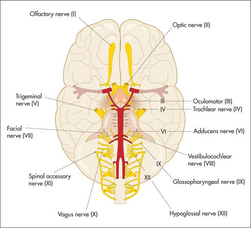
Table 12.7, page 420) is a useful way to document the progress of a confusional state or dementia over time.


The cranial nervesg


Examination anatomy


The cranial nerves (Figure 11.2) arise as direct extensions of the brain (I and II) or from the brainstem (midbrain, pons and medulla)—Figures 11.10 (page 337), 11.13 (page 340) and 11.14 (page 341).


image

Figure 11.2 Cranial nerves

Only gold members can continue reading. Log In or Register to continue

Stay updated, free articles. Join our Telegram channel

Mar 25, 2017 | Posted by in PHYSIOLOGY | Comments Off on The nervous system

Full access? Get Clinical Tree

Get Clinical Tree app for offline access