The Mediastinum


The Mediastinum


Myron R. Melamed


ANATOMY AND CLINICAL CONSIDERATIONS

The mediastinum is the median portion of the thoracic cavity, occupying the space between the lungs and containing all of the thoracic structures except the lungs. It is limited by the pleura of the right and left lungs laterally, by the sternum anteriorly, and by the vertebrae and ribs posteriorly. Despite its limited extent, the mediastinum is the site of a variety of inflammatory and neoplastic processes. Most can be recognized by percutaneous needle biopsy, as was initially demonstrated by Dahlgren and Nordenstrøm (1966) and again in subsequent publications. Cytology of sputum and bronchial secretions, with rare exceptions, has no significant diagnostic role.

Many mediastinal lesions are asymptomatic and discovered as an incidental finding on chest roentgenograms. Topography is an important factor in making a diagnosis because the different compartments of the mediastinum are likely to harbor different tumors or other lesions (Fig. 37-1 and Table 37-1). Also of great importance is the age of the patient, since many of the mediastinal lesions are age-related. The most important clinical questions to be answered by aspiration cytology are: (1) is the space-occupying lesion neoplastic, inflammatory, or a malformation; (2) if the lesion is neoplastic, is it benign or malignant; and (3) if it is malignant, is it best treated by surgical resection or by irradiation and chemotherapy? At all times the cytologic findings must be complemented by knowledge of the clinical, roentgenologic, and laboratory data.

The anterior and middle compartments are the most common sites of mediastinal tumors and benign, space-occupying lesions that include hyperplastic lymph nodes, granulomatous inflammation, and sclerosing mediastinitis.

Although space-occupying benign lesions and primary tumors of the mediastinum are less common than metastatic tumors, they usually present a much greater diagnostic challenge, and therefore are the most likely target of a transcutaneous needle aspiration biopsy. It is essential to accurately identify these lesions before appropriate therapy can be instituted. As mentioned above, close attention to the clinical findings, the location of a lesion in one of the compartments of the mediastinum (see Table 37-1), and the age of the patient are important factors in reaching the correct diagnosis.


Histology of Normal Thymus

The normal thymus varies in size according to age. It is large in infants and children, increases slowly in size until puberty (but relatively more slowly than the growth of the child), and then undergoes progressive atrophy by apoptosis of component cells. In adults it is a vestigial structure, although it retains its basic histologic features.

The thymus is a lymphoid organ supported by a network of anastomizing large epithelial cells (Fig. 37-2A-C) joined by desmosomes. Structures known as Hassall’s corpuscles,
which are similar to squamous pearls and are dispersed throughout the thymus, are formed by epithelial cells (Fig. 37-2D). During the maturation in the thymus, T-lymphocytes (so named because of their thymic origin) are located within spaces formed by the epithelial cells. The lymphocytes, enveloped by the epithelial cells, form so-called lymphoepithelial complexes. T-lymphocytes, unlike the B-lymphocytes, express both CD-4 and CD-8 antigens. In an interesting observation by Nerurkar and Krishnamurthy (2000), which has not yet been confirmed by others, penetration of lymphocytes into the epithelial cells (or emperipolesis) was demonstrated in imprint cytology of the normal thymus.


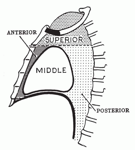
Figure 37-1 Diagrammatic representation of mediastinal compartments.


PRIMARY TUMORS


Thymic Hyperplasia

As already noted, a large thymus is physiologic and normal in prepubertal children. Benign hyperplasia of the thymus may occur in healthy children or young adults, or as a rebound phenomenon following chemotherapy of malignant tumors, including lymphomas. In such cases, a clinical differential diagnosis with malignant lymphoma and germ cell tumor may be of critical importance. Needle aspirates of the hyperplastic thymus yield a mixed population of lymphocytes that is unlikely to contain more than a few, if any, benign epithelial or spindle cells. As reported by Bangerter et al (2000), the cytologic smears resemble those of reactive lymph nodes and differ from the monotypic presentation of lymphoma (see Chap. 31), as well as from seminoma and other germ cell tumors (see below). A large thymus in a child may present as a mediastinal mass on chest x-ray, but is not likely to be a thymoma.


TABLE 37-1 MOST COMMON PRIMARY TUMORS AND SPACE-OCCUPYING LESIONS ACCORDING TO COMPARTMENTS OF THE MEDIASTINUM


Superior compartment (i.e., superior to the heart):


Thymoma


Thymic cysts


Lymphoma


Thyroid tumor


Parathyroid tumor


Aneurysms


Hyperplastic and inflammatory lymphadenopathy


Anterior compartment (i.e., anterior to the heart and great vessels):


Thymic hyperplasia


Thymoma


Thymic cysts


Lymphoma


Germ cell tumors


Thyroid tumor


Parathyroid tumor


Hyperplastic and inflammatory lymphadenopathy


Paraganglioma


Soft-tissue tumors (rare)


Posterior compartment (i.e., posterior to the heart and great vessels):


Neurofibroma


Neuroma and ganglioneuroma


Schwannoma


Neuroblastoma and ganglioneuroblastoma


Paraganglioma


Enteric cysts


Middle compartment (i.e., at the level of the heart and great vessels):


Bronchogenic cysts


Enteric cysts


Pericardial cysts


Lymphoma


Aneurysm


Hyperplastic and inflammatory lymphadenopathy


All compartments: metastatic tumors


Thymomas

Thymomas are tumors of adults. They may be associated with a variety of clinical syndromes that include myasthenia gravis, which was found in 46% of patients with thymoma at the Mayo Clinic (Lewis et al, 1987), and in 35% of such patients in Lyon, France (Chalabreysse et al, 2002). Other, less frequent associations include hypogammaglobulinemia, pancytopenia, red cell hypoplasia, and certain very
rare immune disorders. Like myasthenia gravis, these disorders may be the first manifestation of thymoma.


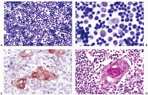
Figure 37-2 Normal thymus. A. Histology showing a network of large epithelial cells among many small lymphocytes (thymocytes). B. Imprint of a normal thymus under oil immersion. The epithelial cells have large, smoothly contoured nuclei with a delicate chromatin structure and occasional chromocenters or nucleoli. Cytoplasm is abundant and lightly stained, with indistinct cytoplasmic borders. C. Cytokeratin stain brings out the cytoplasm of the epithelial cells; lymphocytes are negative. D. Hassall’s corpuscle, a benign squamous pearl. (A,B: Diff-Quik stain.)


Histology

The dual population of cells observed in the normal thymus is reflected in thymomas, which are composed of epithelial cells and lymphocytes in varying proportions. Most thymomas are encapsulated and benign (Fig. 37-3). The epithelial component of these tumors may vary from polygonal (Fig. 37-4A-D) to spindly cells (Fig. 37-5A-D). Rarely, rosettes and other variants have been described. An elaborate classification of thymomas is not relevant to the context of this book, and the interested reader is referred to other sources for additional information (Shimosato and Mukai, 1997; Rosai, 1999).

Some thymomas that appear histologically benign may invade adjacent lung and even metastasize to other sites. There has been considerable controversy over whether the histologic structure of the epithelial component is predictive of the behavior of the tumor. Thymomas with a predominantly spindly epithelial component are generally believed to be benign, whereas those with polygonal epithelial cells are said to be more aggressive; however, so far there is no conclusive evidence of this behavior pattern. In a review of 90 thymic tumors, Chalabreysse et al (2002) concluded that favorable prognostic factors are noninvasiveness, completeness of resection, younger age, and myasthenia gravis. Thymomas with clear-cut cytologic atypia and numerous mitoses, which are classified as thymic carcinomas by World Health Organization (WHO) criteria, are more aggressive
and invasive. In our experience, the best evidence of malignant potential is invasion through the capsule and into the lung or other tissues. The cytology of frankly malignant thymomas (carcinomas) is considered separately below.


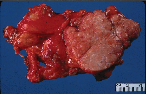
Figure 37-3 Gross appearance of a thymoma. The tumor is sharply circumscribed and soft, pale brown or tan on the cut surface. It is adherent to the adjacent lung (left) but noninvasive.


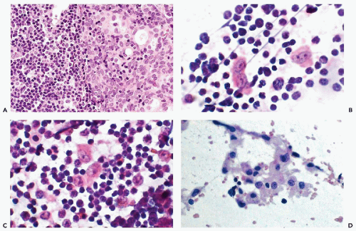
Figure 37-4 Thymoma. A. Histologic section showing sheets of epithelial cells with large round or ovoid vesicular nuclei and pale staining cytoplasm with indistinct nuclear borders. Compare with the many small lymphocytes present. B,C. At higher magnification, a tumor imprint showing several single thymic epithelial cells with abundant cytoplasm and large round or ovoid nuclei with delicate nuclear chromatin and prominent chromocenters or small nucleoli. D. FNA of another thymoma showing epithelial cells in coherent clusters, in strands of cells and single cells.


Cytology of Thymomas, Excluding Thymic Carcinoma

The aspirates of thymic tumors are typically cellular and, as expected, they demonstrate a dual population of epithelial and lymphoid cells in variable proportions (Tao et al, 1984; Sherman and Black-Schaffer, 1990; Koss et al, 1992). The epithelial cells may be present singly (see Fig. 37-4B,C), in clusters (see Fig. 37-4D), or even as tissue fragments (Fig. 37-5C,D). A highly variable number of lymphocytes is intermingled with the epithelial cells. Fragments of fibrovascular tissue may be present. The epithelial cells are polygonal, oval (see Fig. 37-4), or spindly (Fig. 37-5), with moderate to abundant pale cytoplasm and bland, vesicular nuclei that are noticeably larger than the adjacent lymphocytes (see Fig. 37-4B-D). Chhieng et al (2000) estimated the size difference at three- to fourfold. Some degree of squamous differentiation of the epithelial cells may be apparent in an aspirate. We have seldom seen well-formed Hassall’s corpuscles in thymic tumors, and their presence in an aspirate is in favor of a benign hyperplastic thymus. Chhieng et al (2000) found no Hassall’s corpuscles in a recent review of aspirates from 31 patients with thymoma. They reported that thymomas with epithelial cells with round or oval nuclei appeared to be more aggressive than tumors with epithelial cells with spindle nuclei. Chalabreysse (2002) found no such difference, and we also have no evidence that the cell or nuclear shape of the epithelial cells has any prognostic significance (Koss et al, 1992). We agree with Chhieng et al (2000) that cytologic features in the aspirate do not correlate well with any of the histologic patterns.

Although the population of lymphoid cells in most thymomas consists of small, resting lymphocytes, it should be noted that intermixed reactive lymphoblasts may be present (Pak et al, 1982; Ali and Erozan, 1998; Shin and Katz, 1998). If the latter are numerous and prominent, one must consider the possibility of a non-Hodgkin’s malignant lymphoma, which may mimic thymoma, particularly in a child or adolescent (Friedman et al, 1996). For further discussion of thymic lymphomas, see below. In the absence of lymphocytes, the epithelial cells of thymoma may be mistaken for a primary or metastatic carcinoma.

Finally, while the presence of a spindle cell population,
intermixed with lymphocytes, should suggest the possibility of thymoma, Slagel et al (1997) reviewed 22 cases of mediastinal tumors in which the needle aspirate contained a predominant spindle cell component, and found only two thymomas and two thymic cysts. The remaining cases included nerve sheath tumors, granulomatous inflammation, Hodgkin’s disease, and a heterogenous group of other tumors.


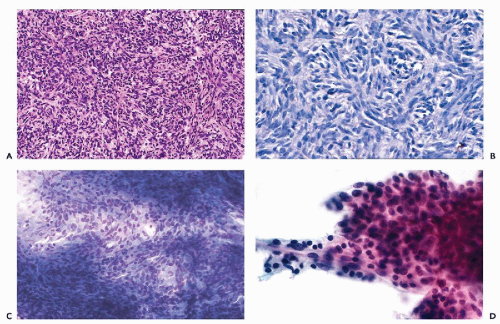
Figure 37-5 Spindle cell thymoma. A. The thymic epithelial cells form interlacing bands of elongated cells with thin, spindle-shaped nuclei. B. High magnification to demonstrate the spindle cell configuration and pattern of thymic tumor cells. C. Diff-Quik stain of the cytologic specimen. D. FNA of another case. Note that the tumor cells, which are oval or spindly and uniform, are in clusters or tissue fragments. A spindle cell tumor in the needle aspirate of an anterior mediastinal mass should be considered thymoma until proved otherwise.


Malignant Thymoma (Thymic Carcinoma)

Although the behavior of most thymomas cannot be predicted from their histologic or cytologic presentation, there is a small group of malignant tumors originating in the thymus that deserve to be classified as thymic carcinomas. They are characterized by clearly malignant epithelial cells that sometimes mimic squamous carcinoma (Fig. 37-6A-C). The cells are of irregular configuration with abundant opaque eosinophilic cytoplasm and large nuclei with coarsely clumped chromatin, visible or even prominent nucleoli, and sometimes mitoses. Necrosis is common. Rarely, these tumors are pure squamous carcinomas (Kaw and Esparza, 1993). A few mucoepidermoid carcinomas of thymus have been described (Moran and Suster, 1995). Because of their rarity, the behavior of these malignant tumors is not well documented. The tumor illustrated in Figure 37-6 had persisted for a year without major change. In general, poorly differentiated tumors are aggressive and capable of invasion and metastases.


Thymic Carcinoid

Mediastinal carcinoids are uncommon and usually amenable to successful surgical resection. They may have endocrine activity (Wick et al, 1980; Moran and Suster, 2000). The histology of these tumors and the fine-needle aspirates of thymic carcinoid do not differ from those of carcinoids of pulmonary or intestinal origin (see Chaps. 20 and 24). Wang et al (1995) and Nichols et al (1997) described cellular specimens composed of loosely clustered and single small cells with scant, finely granular cytoplasm and smoothly configured, uniform, round or oval nuclei with granular chromatin and visible small nucleoli. Unlike small-cell carcinoma, there is no necrosis and no significant nuclear molding of tumor cells. We have encountered a spindle cell carcinoid of the mediastinum. Carcinoids with oncocytic features, which we have seen in the lung (see Chap. 20), were observed in the thymus by Moran and Suster (2000).
Positive chromogranin staining of tumor cells can be used to confirm the diagnosis when in doubt.


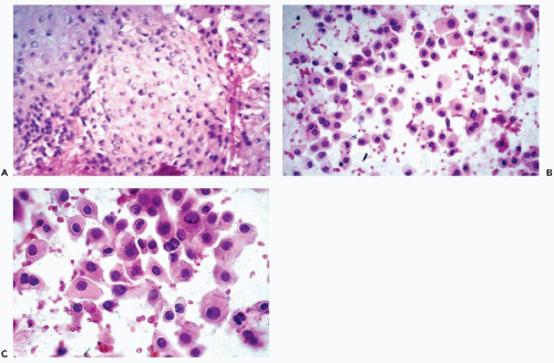
Figure 37-6 Thymic carcinoma. A. Histologic section showing an epidermoid pattern of carcinoma arising in thymus. B,C. Needle aspirate showing abundant malignant cells similar to those of a squamous carcinoma.


Malignant Lymphomas

Although the cytologic presentation of malignant lymphomas in the mediastinum is identical to that described for lymph nodes and other organs (see Chaps. 20 and 31

Only gold members can continue reading. Log In or Register to continue

Stay updated, free articles. Join our Telegram channel

Jun 8, 2016 | Posted by in PATHOLOGY & LABORATORY MEDICINE | Comments Off on The Mediastinum

Full access? Get Clinical Tree

Get Clinical Tree app for offline access