The Lymph Nodes, Spleen, and Thymus


The Lymph Nodes, Spleen, and Thymus


Kamran M. Mirza, M.D., Ph.D.

Choladda V. Curry, M.D.

Andrea N. Marcogliese, M.D.


LYMPH NODES

The lymph node is a remarkable structure that serves as (a) a meeting place for antigen, antigen-presenting cells, and naive B and T cells to initiate the adaptive immune response and (b) the site of clonal expansion and differentiation of effector B and T lymphocytes (1). These processes so essential to normal immune function also require a number of genetic events (DNA replication, class switching, somatic mutation, receptor editing, etc.) that, when deranged, lead to mutations and translocations that underlie most lymphomas (both Hodgkin and non-Hodgkin) (2).


Lymph Nodes (Normal Structure and Function)

The normal lymph node is a round or an ovoid encapsulated structure. It is usually small (2 to 3 mm) to modest (approximately 1 cm) in size, but it may attain dramatic dimensions if the lymph draining into it is particularly rich in immunogenic material. Macroscopically, normal or reactive lymph nodes are tan or creamy white in color, and the cut surface may be homogeneous or vaguely nodular (Figure 23-1). In reactive lymph nodes, the hilum may be visible on gross examination. Microscopically, three anatomic compartments can be recognized: cortex, paracortex, and medulla (3) (Figure 23-2). Residing in the cortex are the primary and/or secondary follicles, which are spherical collections of small and large B lymphocytes. The primary follicles are composed primarily of aggregates of small lymphocytes (naive B cells). The secondary follicles, formed through the process of antigen stimulation, are composed of germinal centers surrounded by mantle zones. The former is composed of a dark zone, consisting primarily of large B cells (centroblasts) and tingible body macrophages, where proliferation (clonal expansion) and somatic mutation occur, and an adjacent light zone, consisting primarily of small B cells (centrocytes), where selection of high-affinity B cells and differentiation to plasma cells (PC) and memory B cells occur (Figure 23-3). Although rich in B cells, the follicles also contain T helper cells and follicular dendritic cells. Surrounding the germinal center is a rim of uniformly sized small lymphocytes, known as the mantle, which is polarized toward the subcapsular sinus and blends imperceptibly into the cortex. Like the germinal center, the mantle is composed largely of B cells. The paracortex is a T-cell-rich zone, which contains a heterogeneous population of cells, including macrophages, interdigitating reticulum cells, scattered B cells, and abundant T cells in various stages of activation. High endothelial venules in this area serve as the site of entry for naive T cells and B cells. The medulla (not always evident in tissue sections as a discrete zone) is located in the central portion of the lymph node, composed of the medullary cords and medullary sinuses. The sinuses, endothelium-bounded spaces containing PCs, effector T cells, macrophages, and antigenpresenting cells, converge on the hilum from multiple points along the subcapsular sinus.


Clinical Significance of Lymphadenopathy in Children

Palpable lymphadenopathy is more common in children and adolescents than in adults. The most common cause of adenopathy in children is a benign lymphoid proliferation, and in many patients, some evidence of a self-limiting infectious or inflammatory process can be found to explain the enlarged nodes. The presence of certain clinical factors suggests that biopsy may disclose a condition requiring specific treatment, including fevers unresponsive to antibiotics, generalized adenopathy or massive localized adenopathy, mediastinal disease, weight loss, peripheral blood cytopenias, and elevated serum levels of lactate dehydrogenase. Slap et al. (4) defined three simple variables that identify lymph nodes that should be biopsied: (a) size greater than 2 cm in diameter, (b) abnormalities on chest x-ray, and (c) absence of symptoms of recent otolaryngologic disease in patients with cervical adenopathy. Soldes et al. (5) reported that the risk of malignancy in childhood peripheral lymphadenopathy increased with age and increasing size and number of locations of adenopathy. Other factors included supraclavicular location, an abnormal chest x-ray, and fixed nodes. A lymph node biopsy is warranted to exclude malignancy if there is
no response to antibiotic therapy or a failure to regress after 6 weeks without identification of an infectious etiology (6).


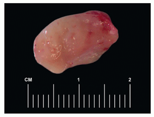
FIGURE 23-1 • Gross examination of a reactive lymph node in a child shows tan vaguely nodular cut surface.


Approach to Diagnosis in Patients with Lymphadenopathy

The availability of a broad array of new diagnostic techniques in hematopathology offers the opportunity for making faster diagnoses with more precision on smaller samples using less invasive procedures such as fine needle aspiration (FNA) cytology and core needle biopsy. The success of such efforts (if they are to be cost effective) depends in part on the quality of communication between hematologists and hematopathologists so that the most appropriate studies are ordered in a timely fashion.

In our practice, we examine touch preps or smears on biopsies or aspirates of lymph nodes immediately after staining (Table 23-1). Since the most common pediatric non-Hodgkin hematopoietic lymphoid neoplasms are recognizable as malignant on touch preps/smears (anaplastic large-cell lymphoma [ALCL], diffuse large B-cell lymphoma [DLBCL], Burkitt lymphoma, lymphoblastic lymphoma), these entities can be quickly triaged to flow cytometry and cytogenetic analysis. Similarly, most reactive proliferations, metastatic tumors, and Hodgkin lymphoma (HL) would not benefit from flow cytometry but may require cultures, cytogenetics, or other studies. This approach often allows the definitive diagnosis of many malignancies within hours and prevents substantial waste in resources.


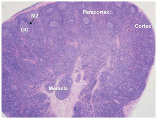
FIGURE 23-2 • The lymph node consists of three recognizable compartments: cortex (B-cell-rich zone), paracortex (T-cell-rich zone), and medulla. The secondary follicles located in the cortical areas show germinal center (GC) and well-demarcated mantle zone (MZ). (Hematoxylin and eosin stain 1.25× magnification.)


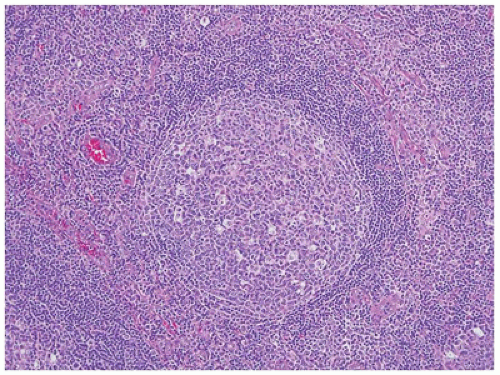
FIGURE 23-3 • The secondary follicles show a reactive germinal center, surrounded by a well-demarcated mantle zone. Polarization of the benign germinal center reflects the segregation of centrocytes to the light zone and mitotically active centroblasts to the dark zone. (Hematoxylin and eosin stain 10× magnification.)


Immunophenotypic Studies of Lymph Node Biopsy Specimens

Flow cytometry is an essential part of the diagnosis and classification of non-Hodgkin lymphoma (NHL) involving lymph nodes. The rapid availability of results (1 to 3 hours) allows triage to appropriate ancillary genetic studies while viable material is still available (7). In HL and in cases of NHL in which flow cytometry is not performed, the wide variety of antibody reagents that recognize most clinically relevant CD markers, which mark well in fixed tissue, allow immunohistochemical characterization of most hematopoietic lymphoid neoplasms (Table 23-2).


Cytogenetic Studies of Lymph Node Biopsy Specimens

For routine cytogenetic analysis, which is helpful in securing an accurate diagnosis in some cases, viable, fresh tissue must be taken by sterile technique at the time of biopsy and placed into culture so that metaphase spreads can be


generated. In the appropriate clinical setting, some karyotypic abnormalities may be pathognomonic for certain types of malignancies (Table 23-3) and can therefore be used for diagnostic/classification purposes (8,9). In other settings, especially precursor B-cell lymphoblastic lymphoma/leukemia, the results of cytogenetic studies can also be used for prognostic purposes (10). The most common karyotypic changes related to lymphoproliferative disorders in children include translocations of immunoglobulin and T-cell receptor loci, which are frequently paired with loci involved in normal development and hematopoiesis (11,12). A major advance in diagnostic cytogenetics is the widespread availability of fluorescent in situ hybridization (FISH) studies for these translocations. Two major advantages of FISH techniques over routine cytogenetics are (a) rapid turnaround (<24 hours) and (b) ability to utilize fixed tissues including touch preparations, cytospin preparations, or sections of paraffin-embedded tissues (13,14).


TABLE 23-1 APPROACH TO DIAGNOSIS AT THE TIME OF BIOPSY


image


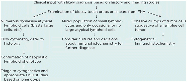
TABLE 23-2 MARKERS USEFUL IN DIAGNOSIS OF HEMATOLYMPHOID NEOPLASMS


Cluster Designation/Antigen


Utility


General markers (not lineage specific)


CD45 (LCA)


Leukocyte common antigen (+ in almost all NHL, negative/dim in most acute leukemias, negative in CHL and many ALCL)


TdT


Terminal deoxynucleotidyl transferase (+ >95% LBL, 20% AML, negative in all mature NHL)


CD34


Human progenitor cell antibody (50% of blasts in ALL/AML), endothelial cells


CD30


Activation marker, positive in CHL, ALCL, PMBCL


ALK


Positive in ALK+ ALCL and ALK+ DLBCL, negative in all other hematopoietic tumors


BCL2


Antiapoptotic protein, negative in normal germinal centers, positive in T-cells and mantle zone B cells; negative in BL and most pediatric FL, positive in subset of DLBCL


Ki-67


(MIB-1) Proliferation marker, >95% positivity in BL


B-cell lineage markers


CD10


CALLA; positive in germinal center B-cells (+ >95% B-ALL, 100% BL, 25%-80% of DLBCL)


CD19


Pan B-cell marker (present in all B-cell NHL including B-LBL)


CD20


Mature B-cell marker (present in DLBCL and BL, negative/dim in most B-LBL); + in NLPHL, variable positivity in CHL


CD22


Pan B-cell marker (present in all B-cell NHL including B-LBL)


PAX-5


B-cell transcription factor (present in all B-cell NHL including B-LBL), weak positivity in HRS cells


CD79a


Pan B-cell marker (present in all B-cell NHL including B-LBL)


CD23


Activated B cells; follicular dendritic cells, low-affinity FcR for IgE, + in most PMBCL


BCL6


Germinal center B-cells, + in BL and most DLBCL


T-/NK-cell lineage markers


CD1a


Thymic T cells, Langerhans cells, T-LBL


CD2


T cells and NK cells; E-rosette receptor, + in some AML


CD3


T cells; TcR complex component NK cells (cytoplasm only)


CD4


Helper/suppressor T cells; MHC class II receptor, monocyte/macrophages, 60% of AML


CD5


Preferential T-cell; FcR for IgM, 25% of AML


CD7


T cells and NK cells; FcR for IgM, 25% of AML


CD8


Cytotoxic T cells and some NK cells; MHC class I receptor


CD16


NK cells, granulocytes; IgG FcR III


CD43


Pan T-cell marker, macrophages, monocytes blasts in AML, T-LBL and B-LBL


CD45RO


T cells, some macrophages


CD56


NK cells and cytotoxic T cells; N-CAM isoform, neuroendocrine tumors


CD57


Cytotoxic T cells and NK cells


Myeloid/monocytic/histiocytic markers


CD1a


Immature T cells, Langerhans cells


CD15


Granulocytes, also positive in HRS cell in classic Hodgkin lymphoma


CD21


Follicular dendritic cells some B cells; C3d/EBV receptor


CD23


Follicular dendritic cells, mature B cells, macrophages low-affinity FcR for IgE


CD68


(KP-1) Macrophages, monocytes, myeloid cells, blasts in AML


CD163


Macrophages, monocytes


S100


Langerhans cells, melanoma, other cell types; + in SHML


CD207


Langerin; positive on Langerhans cells


Myeloperoxidase


Positive in myeloid cells; useful to establish myeloid lineage in acute leukemias


ALCL, anaplastic large-cell lymphoma; AML, acute myeloid leukemia; BL, Burkitt lymphoma; B-LBL, B-cell lymphoblastic lymphoma; CALLA, common acute lymphoblastic leukemia antigen; CHL, classical Hodgkin lymphoma; EBV, Epstein-Barr virus; FcR, Fc receptor; MHC, major histocompatibility complex; NHL, non-Hodgkin lymphoma; NK, natural killer; PMBCL, primary mediastinal large B cell lymphoma; TcR, T-cell receptor; T-LBL, T lymphoblastic lymphoma.


TABLE 23-3 KARYOTYPIC AND GENETIC CHANGES ASSOCIATED WITH NON-HODGKIN LYMPHOMA


Disease


Abnormality


Implicated Genes


B-cell LBL/B-cell ALL (see Chapter 24 on Bone Marrow for further discussion)


t(1;19)(q23;p13)


PBX-E2A


t(4;11)(p21;q23)


AF4-MLL


Hyperdiploidy (>50 chr)


Hyperdiploidy (47;50 chr)


Hypodiploidy


t(12;21)


ETV6-RUNX1


t(5;14)


IL-3-IgH


t(9;22)


BCR-ABL1


iAMP21


Diffuse large B-cell lymphoma


t(3q27;var)


BCL6


Burkitt lymphoma


t(2;8) (p11;q24)


IgL-lambda-myc


t(8;14) (q24; q32)


c-myc-IgH


t(8;22) (q24;q11)


c-myc-IgL-kappa


T-cell LBL/T-cell ALL


t(1;14-15)(p12;q11)


TAL1;TcR gamma


t(1;14)(p32;p14-15)


TAL1;TcR delta


t(1;7)(p32;q34)


TAL1;TcR beta


t(10;11)(p13;q14)


CALM-AF10


t(7;10)(q34;q24), t(10;14)(q24;q11)


HOX11


ALK+ anaplastic large-cell lymphoma


t(2;5)(p23;q35)


NPM-ALK, most cases of ALK+ ALCL


t(2;var)


Variety of other partners described; all result in ALK positivity by IHC


ALL, acute lymphoblastic leukemia; IgH, immunoglobulin heavy chain; IgL, immunoglobulin light chain; LBL, lymphoblastic lymphoma; TcR, T-cell receptor, IHC, immunohistochemistry.


Reactive Lymphadenopathy

The two major patterns of reactive lymphadenopathy are usually dominated either by follicular hyperplasia or by interfollicular expansion (immunoblastic, granulomatous, or histiocytic). Occasional reactive processes produce diffuse alteration of architecture and may mimic a lymphoma (Table 23-4). In practice, a single lymph node is constantly exposed to a diversity of immunogens and therefore exhibits more than one pattern of response; however, when the degree of adenopathy is sufficient to warrant a biopsy, a single pattern usually dominates. Additional factors that assist in the differential diagnosis include the age-specific nature of some disorders; certain reactive processes affecting lymph nodes are rare in children (e.g., luetic lymphadenitis, Kikuchi disease), whereas others affect them primarily (e.g., acute infectious mononucleosis, autoimmune lymphoproliferative disorder [ALPS]).


TABLE 23-4 REACTIVE LYMPHADENOPATHY IN CHILDREN


Follicular Pattern


Nonspecific Reactive Follicular Hyperplasia


HIV


Progressive transformation of follicular centers


Toxoplasmosis


Castleman disease/angiofollicular hyperplasia


Interfollicular Pattern


Paracortical immunoblastic reactions


EBV


Hypersensitivity reactions (phenytoin)


Juvenile rheumatoid arthritis 1


Systemic lupus erythematosus 1,3


Kikuchi histiocytic necrotizing lymphadenitis 1,3


Autoimmune lymphoproliferative syndrome (ALPS) 1


Kawasaki disease


Granulomatous


Mycobacterial infection (MTB and atypical mycobacterial) 1,2


Cat-scratch disease 1,2,4


Fungal infection


Histiocytic


Sinus histocytosis


Lysosomal storage disorders


Hemophagocytic syndromes (hemophagocytic lymphohistiocytosis)


Rosai-Dorfman


Dermatopathia


Langerhans cell histocytosis


Diffuse alteration of architecture


Sarcoidosis


Posttransplant lymphoproliferative disorder


Notations for other features


  1. Follicular hyperplasia


  2. Necrosis with neutrophils


  3. Necrosis with apoptosis


  4. Capsulitis


FOLLICULAR HYPERPLASIAS


Nonspecific Reactive Follicular Hyperplasia

Nonspecific reactive follicular hyperplasia is the most common pattern of reactive lymphadenopathy. The lymph node shows increased follicular density with variable sizes and shapes of follicles. The germinal centers show polarization and contain small, intermediate, and large mitotically active lymphocytes, tingible body macrophages, and apoptotic cells. Well-demarcated mantle zones surround germinal centers. The hyperplastic follicles tend to remain in the cortical regions, but in particularly robust cases, the paracortex and the medulla may be compressed by the process. Immunophenotypic studies show that the secondary follicles contain a predominance of CD20+, CD10+, BCL6+ B cells that are negative for BCL2. Differential diagnostic considerations in children are few but include Castleman disease, HIV-related adenopathy, progressive transformation of germinal centers (PTGC), and rarely pediatric type follicular lymphoma (15).


HIV-Related Adenopathy

Most series treating the subject of HIV-related persistent generalized lymphadenopathy are based on a patient population of homosexual young adult men at risk for HIV (16,17). Reports of this condition in children at risk for HIV because of maternal-fetal transmission or hemophilia present similar data (18). A spectrum of histologic findings may be seen in this context, with two clearly recognizable extremes. Florid follicular hyperplasia, the earliest change of HIV-related persistent generalized lymphadenopathy, has many features in common with nonspecific follicular hyperplasia, although the germinal centers are larger and often serpiginous and tend to fuse with focal follicular lysis (Figure 23-4). Regressively transformed germinal centers typify late persistent generalized lymphadenopathy and are characterized by small size, lymphoid depletion, and numerous dendritic cells, vessels, and amorphous eosinophilic deposits (19). The mantle is absent or very poorly formed, and the paracortex is proportionally rich in histiocytes, PCs, and high endothelial venules because
of the paucity of lymphocytes. The morphologic features associated with persistent generalized lymphadenopathy (i.e., large and irregularly shaped follicles, follicular lysis, follicular involution) are distinctive but not specific for HIV infection, as they have been seen in 5% to 10% of otherwise entirely unremarkable lymph nodes obtained as part of carcinoma staging before the beginning of the AIDS era (20).


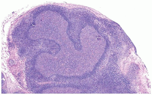
FIGURE 23-4 • Serpentine follicular center in a patient with HIV infection and persistent generalized lymph adenopathy. (Hematoxylin and eosin stain 4× magnification.)


Progressive Transformation of Germinal Centers

For unknown reasons, when clinically significant lymphadenopathy develops in some patients, the biopsy specimen shows PTGC. Most patients are asymptomatic male adolescents or young adults with isolated inguinal or cervical adenopathy (21,22). The bulk of the lymph node, which can be up to 5 cm in size, exhibits florid reactive follicular hyperplasia with focal involvement by very large (three to five times the size of surrounding reactive follicles) nodules that are dramatically expanded by an influx of small lymphocytes (Figure 23-5) with mantle cell-like morphology and phenotype (IgM+, IgD+) (23,24). Relative to normal follicles, the transformed follicles exhibit a disrupted and dispersed dendritic cell network on CD21 or CD23 staining (21). Clusters of epithelioid histiocytes are more commonly identified in children than adults (22,25). The main malignant counterpart differential diagnosis is nodular lymphocyte-predominant Hodgkin lymphoma (NLPHL), which typically demonstrates neoplastic nodules replacing the entire lymph nodes, and the presence of neoplastic lymphocyte-predominant (LP) cells (also known as “popcorn” or “L&H” cells). Two studies of PTGC that focus on the pediatric population found that HL, particularly NLPHL, occasionally may precede, follow, or be concurrent with PTGC (22,25). Approximately one-third of patients will have recurrent PTGC (25), and approximately one-fourth have associated immune disorders including immune thrombocytopenia (ITP), lupus, Castleman disease, and ALPS (22).


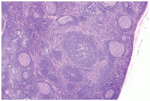
FIGURE 23-5 • Progressively transformed germinal center (with disrupted follicle infiltrated by small mantle zone lymphocytes) in a background of follicular hyperplasia. (Hematoxylin and eosin stain 4× magnification.)


Toxoplasmosis

Acute toxoplasmosis, an infectious disease, is often accompanied by lymphadenopathy. This is usually limited to the cervical lymph nodes, although occasionally patients with typical histology and serologic confirmation have isolated inguinal or axillary lymph node enlargement. Florid follicular hyperplasia dominates the histology at low power and is invariably accompanied by clusters of epithelioid histiocytes and patches of monocytoid B cells. The epithelioid histiocytic aggregates, randomly distributed throughout, abut and even infiltrate the germinal centers. The triad of florid follicular hyperplasia, monocytoid B-cell hyperplasia, and perifollicular and intrafollicular clusters of epithelioid histocytic aggregates (variably encroaching on follicular centers) has a high degree of sensitivity and specificity for diagnosis of toxoplasmosis (Figure 23-6) (26,27).


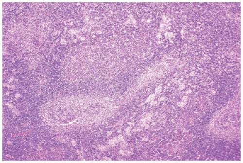
FIGURE 23-6 • Low power of toxoplasmosis demonstrating the triad of hyperplastic follicles, monocytoid B-cell proliferation, and histiocytic aggregates encroaching of follicles. (Hematoxylin and eosin stain 4× magnification.)


Castleman Disease

Castleman disease is a heterogenous group of diseases with variable clinical presentations and prognosis. The disease mainly affects adults and rarely occurs in children. Castleman disease has been previously classified based on histology into hyaline vascular (HV) type and PC type and based on clinical presentation into localized (unicentric) and multicentric (28). Human herpes virus-8 (HHV-8) has also been found to play a role in the pathogenesis of Castleman disease, and there has been a proposal to update the classification of Castleman disease to incorporate HHV-8 status (29). The entity has been recently reviewed by Talat et al. (30) In hyaline vascular Castleman disease (HV-CD), which is usually unicentric, there are increased small and involuted lymphoid follicles surrounded by broad mantle zones throughout the node (“bag of marbles”) (Figure 23-7). The expanded mantle zones are composed of tightly packed small lymphocytes often in a laminated (orbiting) or “onion-skin” pattern; this feature together with radially penetrating hyalinized sclerotic blood vessels passing into the germinal centers creating “lollipop” appearance (Figure 23-8) is characteristic of the disease (28,29). The germinal centers appear atrophic and are depleted of lymphocytes and contain both extracellular matrix and abundant follicular dendritic cells. The interfollicular areas are highly vascular with hyalinization. Large clusters or sheets of PCs are not present. The plasma cell variant of Castleman disease (PC-CD), usually multicentric, is extremely rare in children (31,32,33,34). The majority of follicles in PC-CD appear reactive, but some may show a few with hyaline vascular-like follicles. Marked interfollicular plasmacytosis is present (28,29). Castleman disease in children has different characteristics than in adults. Pediatric Castleman disease is usually unicentric and has a more favorable course. HV-CD is also more common than PC-CD. Mesenteric location of unicentric CD is relatively more common in children than in adults, who usually have intrathoracic (mediastinal) localization. Head and neck location is also uncommon in children. Similar to adults, anemia and hypergammaglobulinemia are more often present in PC variant rather than the HV variant (32,33). HHV-8-associated CD typically occurs in immunosuppressed and/or HIV-positive adults and has rarely been reported in an immunosuppressed child related to a possible inborn error of immunity to HHV-8 (35).


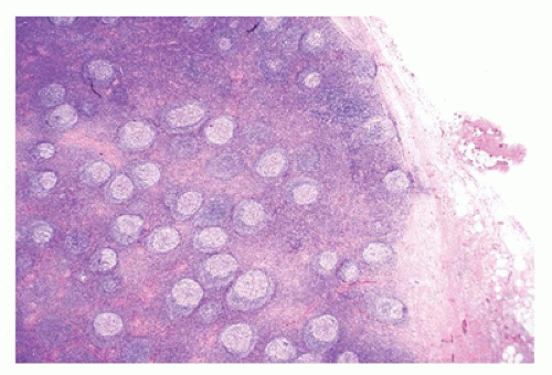
FIGURE 23-7 • Low power of hyaline vascular Castleman disease with “bag of marbles”: small uniform follicles evenly dispersed throughout the cortex and the medulla. (Hematoxylin and eosin stain 4× magnification.)


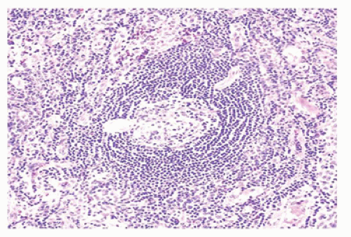
FIGURE 23-8 • The mantle zone has a laminated or “onionskin” appearance in the hyaline vascular type of Castleman disease. Note the radially penetrating vessel. (Hematoxylin and eosin stain 10× magnification.)


INTERFOLLICULAR/PARACORTICAL REACTIONS: IMMUNOBLASTIC


Epstein-Barr Virus Infection (Infectious Mononucleosis)

Acute illness secondary to Epstein-Barr virus (EBV) infection (acute infectious mononucleosis) is common in young children and is usually self-limited. In those few cases that culminate in biopsy, the clinical features are often atypical— advanced age, the presence of “B” symptoms, a negative monospot test [uncommon except in very young children (<4 years) or early in infection], localized adenopathy or persistent adenopathy, hepatosplenomegaly, or splenic rupture. Patients with X-linked lymphoproliferative disorder (Duncan syndrome) present with rapidly progressive and usually fatal disease because of an inability to mount a successful immune response against EBV-infected cells.

At low power, the architecture is obscured but generally preserved with moth-eaten follicles and prominent paracortical expansion (Figure 23-9) by immunoblasts, PCs, and plasmacytoid lymphocytes (Figure 23-10). Similar large cells pack the sinuses. Occasionally, large cells with bilobed or multilobed nuclei and prominent nucleoli are present, reminiscent of Hodgkin/Reed-Sternberg (HRS) cells. Histiocytes may be scattered singly or in small clusters, and increased numbers of capillaries and high endothelial venules also contribute to the polymorphic appearance of the paracortex.
Normal landmarks—germinal centers and subcapsular and paratrabecular sinuses—are generally present but may be compressed or distorted by the immunoblastic proliferation. In very early cases of EBV infection, monocytoid B-cell proliferation may be prominent (36). Staining with CD20 and CD3 highlights the presence of a mixture of interfollicular B and T immunoblasts, respectively, and a polytypic pattern of light-chain expression is always seen. The Reed-Sternberg-like cells are characteristically CD20+ and CD15- and show variable reactivity for the activation antigen CD30 as well as markers of EBV infection such as latent membrane protein (LMP1) or EBV-encoded RNA transcripts (EBER) (37). Difficult cases may exhibit sheet-like arrays of immunoblasts, a brisk mitotic rate, or extensive necrosis and may closely mimic large-cell lymphoma. In such cases, examination of the peripheral blood smear for atypical lymphocytes, viral serology, and immunohistochemistry to better define architectural preservation and establish the presence of EBV is helpful.


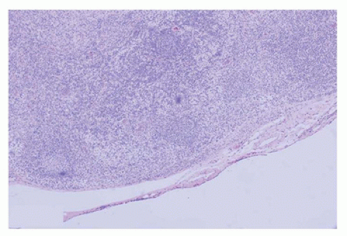
FIGURE 23-9 • The immunoblastic proliferation in acute infectious mononucleosis localizes to the paracortex and may compress or distort residual germinal centers. (Hematoxylin and eosin stain 4× magnification.)


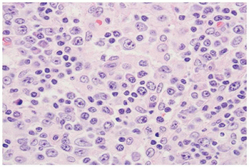
FIGURE 23-10 • Small, intermediate, and large cells fill the paracortex in acute infectious mononucleosis, often with a predominance of immunoblasts. (Hematoxylin and eosin stain 40× magnification.)


Non-EBV Viral Adenopathy

Lymphadenopathy may occur as a result of herpes simplex or cytomegalovirus infection and is seen most often in children with some form of immune deficiency. Paracortical hyperplasia with discrete foci of necrosis is the typical histologic finding in these cases. In comparison with acute EBV-related lymphadenopathy, the proportion of immunoblasts is less, and interfollicular areas are expanded by a mixture of mature lymphocytes, PCs, histiocytes, plasmacytoid monocytes, and fewer numbers of immunoblasts. Viral inclusions appearing as smudged (38) or hyperchromatic alterations of the nucleus (herpes simplex virus) or very large eosinophilic structures within the nucleus (cytomegalovirus) can be identified and may be most numerous adjacent to zones of necrosis. Immunohistochemistry is helpful in excluding neoplasia and can also document the presence of infected cells. Of note, the viral inclusions in the enlarged cells of cytomegalovirus lymphadenitis are CD15+, a potential pitfall in the exclusion of HL. Attention to clinical parameters will assist in discriminating viral lymphadenitis from other causes of necrotizing lymphadenitis, which include Kikuchi-Fujimoto disease and lupus erythematosus.


Hypersensitivity Reactions, Emphasizing Phenytoin (Dilantin) Reactions

Hypersensitivity-related lymphadenopathy is quite rare and has been associated most commonly with phenytoin (Dilantin) therapy and vaccines (small pox, measles, tetanus) but can be seen associated with a variety of drug classes. In phenytoin hypersensitivity, the architecture of the lymph node is distorted by a paracortical proliferation of immunoblasts, lymphocytes, PCs, and eosinophils. Germinal centers persist, and in some cases, a florid follicular hyperplasia may accompany the immunoblastic reaction. Rarely, a granulomatous lymphadenopathy secondary to phenytoin therapy has been reported (39). Purely diffuse architectural effacement is rare. The immunoblasts represent a mixture of CD20+ B cells and CD3+ T cells. Progression to lymphoma is well described. A similar pattern of paracortical expansion may be seen in T-cell lymphomas such as angioimmunoblastic T-cell lymphoma (40), which can be a major problem in differential diagnosis. A detailed history as well as immunophenotypic studies is usually helpful.


Juvenile-Onset Rheumatoid Arthritis Emphasizing Still Disease

Several different histologies have been described in juvenile rheumatoid arthritis (JRA), including the classic findings associated with adult RA of follicular hyperplasia, interfollicular PCs, and intrasinusoidal neutrophils. In Still disease, a pattern of paracortical immunoblastic proliferation mimicking lymphoma is occasionally described as well as interfollicular necrosis similar to that seen in Kikuchi-Fujimoto disease (41,42).


Systemic Lupus Erythematosus

Systemic lupus erythematosus (SLE) is an autoimmune disease that affects both adolescents and young adults. Lymphadenopathy is frequently observed in children with SLE and may occasionally be the presenting feature. When lymphadenopathy is part of the clinical picture, it is typically peripheral and multifocal or generalized (43). Classically, follicular hyperplasia with patchy paracortical necrosis dominates the low-power appearance of the lymph node. The germinal centers are well formed with a discrete mantle zone, and they are separated by an expanded paracortex in which pockets of necrosis are randomly distributed. The necrotic foci are composed of amorphous eosinophilic material and apoptotic debris. Neutrophils and PCs are scarce. In addition, “hematoxylin bodies” (round or oblong blue structures, 5 to 15 µm long, which stain with periodic acid-Schiff and the Feulgen method) and vascular encrustations (Azzopardi effect) may be present (44). Less commonly, necrosis dominates the morphology, and follicles may be few and more widely spaced; in these circumstances, the lymph node findings may resemble those of Kikuchi-Fujimoto disease. Other patterns described in SLE include follicular hyperplasia without necrosis resembling Castleman disease. SLE presenting with granulomatous changes has also been reported (45).


Histiocytic Necrotizing Lymphadenitis/Kikuchi-Fujimoto Disease

Kikuchi-Fujimoto disease (histiocytic necrotizing lymphadenitis) is a self-limited regional lymphadenopathy with prolonged fever that is uncommon in children. The median age is in the third decade in most large series, although the age range is wide. Rare fatal cases in children have been reported, often associated hemophagocytic syndrome. Recurrent Kikuchi-Fujimoto disease has been reported in the pediatric population (46). Histologically, these nodes exhibit follicular hyperplasia, but the histologic hallmark of this disease is zonal karyorrhexis with scant neutrophil response. At low power, the paracortex is distorted by palestaining patchy zones of necrotic debris with a cellular rim composed of apoptotic cells, histiocytes, small lymphocytes, plasmacytoid dendritic cells, and immunoblasts. The areas of necrosis may coalesce, but a serpiginous contour rarely develops. Beyond the necrotic zone is a mottled paracortex, rich in small lymphocytes, immunoblasts, apoptotic debris, plasmacytoid dendritic cells, and high endothelial venules (Figure 23-11). In children, the differential diagnostic considerations for the early proliferative lesions of histiocytic necrotizing lymphadenitis include NHL and mixed cellularity Hodgkin lymphoma (MCHL). Fully developed lesions may mimic Kawasaki disease or herpetic lymphadenitis, and lupus-related lymphadenitis may be difficult or even impossible to exclude. A third pattern is characterized by dominance of foamy histiocytes. Immunophenotypic characterization of the large cells at the periphery of karyorrhectic areas shows that they represent CD8+ T cells and plasmacytoid dendritic cells (47). One report suggests that the sparsity of CD8+ T cells in SLE may be helpful in differential diagnosis (48).


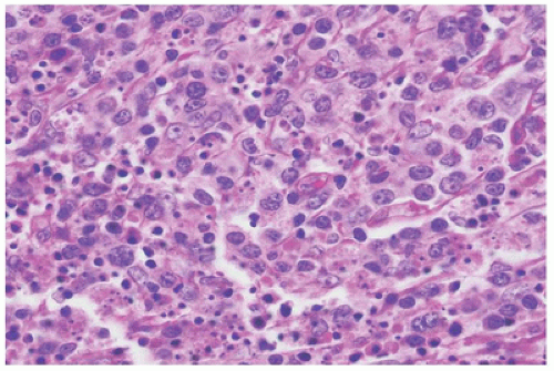
FIGURE 23-11 • The border of necrosis shows apoptotic debris admixed with histiocytes, small lymphocytes, plasmacytoid dendritic cells, and immunoblasts in Kikuchi disease. (Hematoxylin and eosin stain 40× magnification.)


Autoimmune Lymphoproliferative Syndrome

Lymphadenopathy secondary to loss of Fas or Fas ligandmediated apoptosis is a rare cause of nonneoplastic lymphadenopathy, known as autoimmune lymphoproliferative disorder (ALPS). Patients with ALPS present within the first 2 years of life with bulky generalized adenopathy and hepatosplenomegaly and chronic multilineage cytopenias due to autoimmune peripheral destruction or splenic sequestration and have an increased risk of B-cell lymphoma (49,50,51). Enlarged lymph nodes show generally intact architecture with follicles ranging from floridly hyperplastic to small involuting follicles with compressed mantle zones like those seen in HV-CD. The proliferation that occurs in the interfollicular areas consists of immunoblasts and transformed large cells with scant-to-moderate cytoplasm (52). Small lymphocytes, PCs, and histiocytes may be present. Some studies have found an overlap of histologic features with sinus histiocytosis with massive lymphadenopathy (SHML) (Rosai-Dorfman disease) (53) and sarcoidosis (54)

On flow cytometry, CD2+, CD3+, CD4, CD8 T cells with an α-β T-cell receptor are increased (“double-negative T cells” or DNT), and increased DNT in the peripheral blood (>1% of T cells) is one of the diagnostic criteria. B cells are phenotypically normal. On tissue sections, the immunoblasts in the interfollicular zones are virtually all CD3+, double-negative T cells, with only a few showing reactivity for CD4, CD8, or CD20 (55). Peripheral T-cell lymphoma may mimic ALPS, but the former typically contains more smallto-intermediate cells and rarely has a CD4-, CD8- phenotype. Gene sequencing is necessary to confirm the diagnosis of ALPS, which may be caused by mutations in Fas, Fas ligand, or the caspase 10 gene (50,56).


Kawasaki Disease

Kawasaki disease is endemic in Japan but rare in Western countries. A slight male predominance has been noted, and the peak incidence is in children 3 to 4 years old. Histologic descriptions are quite variable, and lymph node biopsy seldom leads to a firm diagnosis of Kawasaki disease without correlation with clinical parameters. The main findings are patchy paracortical necrosis with phlebitis and fibrin microthrombi. Germinal centers are inconstantly present, as is an immunoblast-rich paracortical expansion. If perinodal tissues are present for review, an acute necrotizing arteritis similar to infantile polyarteritis nodosa may be identified even in early phases; in established cases, a measure of luminal dilation is also present in larger-caliber vessels, with medial destruction (see Chapter 13).


INTERFOLLICULAR GRANULOMATOUS PROCESSES


Cat-Scratch Disease

Cat-scratch disease frequently affects children and adolescents, although in large, population-based studies, nearly half of all patients have been over the age of 20 (57). The adenopathic phase of the disease is dominated by follicular hyperplasia, capsulitis, paracortical monocytoid B-cell hyperplasia, and small, neutrophil-rich microabscesses. As the lesions develop, the microabscesses coalesce, forming serpiginous and stellate zones of eosinophilic necrosis (Figure 23-12). In the late stage, the microabscesses take on a granulomatous appearance, with a well-formed rim of palisading histiocytes and scattered multinucleated giant cells (Figure 23-13). Warthin-Starry staining may highlight pleomorphic and bow-shaped rods and cocci, both in the center of the abscesses and around blood vessels in the early phases of disease. However, Warthin-Starry staining is technically difficult and often problematic to interpret. Additionally, patients may be seronegative. Therefore, PCR techniques that detect the organism in the tissue with a high degree of specificity are preferred for confirmation of diagnosis (58). Histologically, similar lesions may also be seen in Yersinia infection, lymphogranuloma venereum, tularemia, and infection with Mycobacterium avium-intracellulare (MAI) in young children (59,60,61).


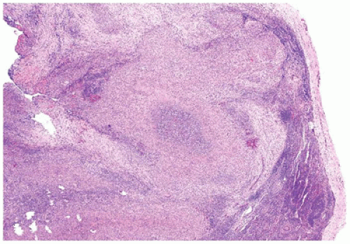
FIGURE 23-12 • The microabscesses in cat-scratch disease have a serpiginous or stellate contour. (Hematoxylin and eosin stain 4× magnification.)


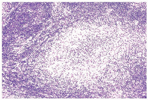
FIGURE 23-13 • In well-developed cases, the abscesses of cat-scratch disease have a broad rim of palisading histiocytes surrounding abundant neutrophils forming a so-called pyogranuloma. (Hematoxylin and eosin stain 10× magnification.)


Mycobacterial Infections

The most common cause of granulomatous lymphadenitis in small children (1- to 5-year old) is infection by nontuberculous “atypical” mycobacteria (NTM), most commonly MAI or Mycobacterium scrofulaceum (62,63). Diagnosis may be made by FNA or excisional lymph node biopsy. Cytologically, smears show epithelioid histiocytes and granulomata with reactive lymphocytes and PCs in the background as well as amorphous necrosis or necrosis associated with abundant neutrophils. Histologically, the nodal architecture is partially distorted or entirely effaced by follicular hyperplasia with well-formed granulomata composed of epithelioid histiocytes and multinucleated giant cells, which rim central areas of caseous necrosis (eFigures 23-1 and 23-2) or necrosis containing abundant neutrophils similar to lesions in cat-scratch disease (eFigures 23-3 and 23-4) (64). Although there is significant overlap between Mycobacterium tuberculosis (MTB) and NTM lymphadenopathy, some studies suggest that welldefined granulomata with caseous necrosis and numerous giant cells are more characteristic of MTB, while microabscesses are more predictive of NTM. Small lymphocytes are evenly distributed throughout lesional areas, and immunoblasts are rare or absent. Immunocompromised patients may not be able to mount responses to the infection and lack the classical granulomata, instead showing looser aggregates of histiocytes with a foamy appearance and more abundant organisms on special stains (eFigures 23-5 and 22-6) (63). Rare cases in immunocompetent patients may show mycobacterial organisms on acid-fast stain, although greater sensitivity may be obtained using fluorescence microscopy using auramine orange (65) or immunohistochemistry against
the MPT64 mycobacterial antigen (more specific for the Mycobacterium tuberculosis complex) (66,67). The remainder of cases may be diagnosed via one of several polymerase chain reaction-based techniques (68,69,70,71,72) or microbiologic culture. Most of these techniques, except for culture, have the advantage of being applicable to fresh as well as paraffin-embedded tissue and may be performed using cytology specimens, core needle biopsies, or whole lymph node biopsies. Other causes of caseating and noncaseating granulomatous lymphadenitis include nonmycobacterial infections and neoplastic disease, including peripheral T-cell lymphoma, NLPHL, and classical Hodgkin lymphoma (CHL).


Chronic Granulomatous Disease

Chronic granulomatous disease (CGD) of childhood is a congenital disorder caused by defective components of the NADPH (reduced nicotinamide adenine dinucleotide phosphate) oxidase pathway (73). This extremely rare form of granulomatous lymphadenitis presents with lymph nodes and other tissues that are extensively infiltrated by granulomata and neutrophil-rich abscess-like foci. Catalase-positive organisms, specifically Staphylococcus aureus, gram-negative bacilli, and Aspergillus, are the most common agents to be recovered in culture (73). A test for nitroblue tetrazolium reduction or other assessment of the respiratory burst by peripheral blood leukocytes using either chemiluminescence or flow cytometry should be performed in suspected cases. Molecular diagnostic testing may be performed to confirm the gene involved (74) (see Chapter 6).

In 11% to 25% of cases, a careful review of clinical, radiologic, histologic, and laboratory data fails to identify a cause of the granuloma formation (75), and a diagnosis of idiopathic granulomatous lymphadenitis is rendered. In such circumstances, with all secondary causes excluded, sarcoidosis can be considered a possibility (76) (Chapter 12).

Although occasionally seen in adolescents, sarcoidosis is rare in children and more commonly occurs in young adults. Lymph node architecture is often totally effaced with few or no residual follicles. Necrosis is rare but when present is more commonly fibrinoid than caseating. Recent studies have demonstrated an increased CD4+ FoxP3+ regulatory T-cell (Treg) population both in the peripheral blood and lymph nodes of patients with sarcoidosis (77,78). Noncaseating granulomatous lymphadenitis may also be seen in benign lymph nodes draining organs involved by tumor, and histiocytic proliferations mimicking granulomas may be present in lymph nodes involved by lymphoma (79).


INTERFOLLICULAR PROCESSES WITH HISTIOCYTIC PROLIFERATION (NONGRANULOMATOUS)

Sinus histiocytosis is a nonspecific reactive pattern that may be seen in lymph nodes draining either inflammatory or malignant processes of the skin, bowel, or lungs. In particularly striking cases, only compressed primary follicles are seen, and germinal centers are either diminutive or absent. The subcapsular and paratrabecular sinuses are expanded by a cellular infiltrate composed of large polygonal cells with bland nuclei and abundant pale eosinophilic cytoplasm, some with phagocytosed debris (80). These histiocytes can be distinguished from Langerhans cells and Rosai-Dorfman cells by their CD68+, lysozyme-positive, S100, CD1a, CD207 phenotype.


Sinus Histiocytosis with Massive Lymphadenopathy (Rosai-Dorfman Disease)

SHML, also known as Rosai-Dorfman disease, affects young patients (81). Germinal centers are atrophic, compressed, or lacking in most cases, and the paracortex is diminished secondary to the compressive effects of the expanded sinusoids (Figure 23-14). A polymorphous array of lymphocytes, PCs, histiocytes, xanthoma cells, and “SHML” cells distend the sinusoids, with the proportions varying from case to case. The SHML cells, which are the hallmark of this disorder, have oval nuclei with condensed chromatin and abundant eosinophilic to xanthomatous cytoplasm (Figure 23-15 and eFigure 23-7). Engulfed lymphocytes (“emperipolesis”) are pathognomonic, although this feature may be focal in some cases. Several groups have demonstrated the utility of FNA in the diagnosis of SHML. Smears generally demonstrate the presence of small lymphocytes, PCs, neutrophils (few), and SHML cells (82,83,84,85). The phenotype of the SHML cell is S100+ and CD68+, but the cells lack markers for Langerhans cells (CD1a and CD207) or dendritic cells (DRC, CD23, CNA42) (86). SHML has been seen in association with neoplasms, including mixed cellularity and lymphocyte-predominant HL as well as NHLs, and in patients with immune dysregulation such as post-bone marrow transplant or in the setting of
ALPS, which should be taken into account in the evaluation of unusual cases (53,87,88,89). A subset of SHML shows features of IgG4-related disease, and an overlap between certain aspects of the two diseases has been suggested (90).


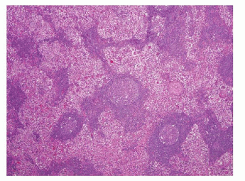
FIGURE 23-14 • As a result of massive sinusoidal expansion by histiocytes, germinal centers are compressed in Rosai-Dorfman disease (SHML). (Hematoxylin and eosin stain 4× magnification.)


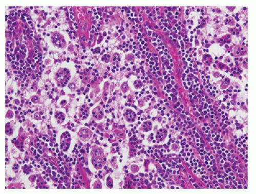
FIGURE 23-15 • The lesional cell in Rosai-Dorfman disease (SHML) has a small, cytologically bland nucleus and abundant eosinophilic cytoplasm containing one or more lymphocytes (emperipolesis). Although obvious in this case, emperipolesis may be difficult to detect on routine sections. (Hematoxylin and eosin stain 20× magnification.)


Foreign Body Sinusoidal Histiocytic Reactions

Histiocytic lymphadenopathy secondary to foreign (nonnodal) material occurs in the setting of primary metabolic disease, in lymph nodes draining tumors or ulcerated areas, adjacent to joint prostheses, or after lymphangiography (although current radiographic techniques have largely replaced lymphangiography). The adenopathy in these patients is rarely worrisome. In cases of accumulated contrast media, the sinusoids are dilated by foamy macrophages, histiocytes, and multinucleated giant cells containing large lipid vacuoles (eFigure 23-8). A similar pattern may be seen in cases of lymphadenopathy due to silicone from leaking or ruptured implants (eFigures 23-9 to 23-11). In the case of joint replacement, sinuses contain pale-staining vacuolated macrophages with refractile or birefringent material that may be Oil Red O positive, depending on whether metallic particles or polyethylene particles are present. Associated granulomata, giant cells, and fibrosis may be seen in some cases (91). Similar findings may be noted in patients with Gaucher disease or other metabolic storage diseases (Figure 23-16). In such cases, histiocytes resemble those seen in the bone marrow in storage diseases with either fibrillar “crumpled tissue paper” or bubbly foamy macrophages (see Chapter 24). Little of the material is found outside the sinusoids, and the remainder of the lymph node, although compressed or atrophic, is normal. Morbidity and mortality may be associated with the primary process causing the accumulation of foreign material, but not with the adenopathy itself.


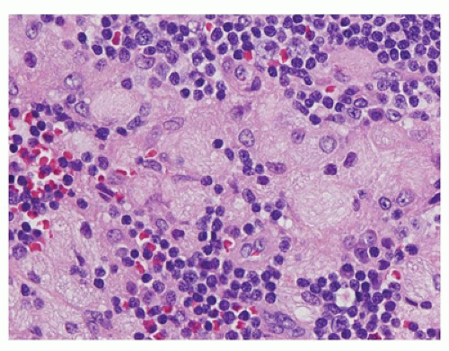
FIGURE 23-16 • Histiocytic proliferations caused by congenital storage disorders have a sinusoidal, often paracortical distribution in lymph nodes. The histiocytes seen in storage disorders often have coarsely vacuolated, (“bubbly”) or fibrillar (“crumpled tissue paper”) cytoplasm, as seen in this case of Gaucher disease. (Hematoxylin and eosin stain 40× magnification.)


Dermatopathic Lymphadenopathy

Children with dermatopathic lymphadenopathy commonly have eczema or another chronic exanthematous disorder, and they present with enlarged axillary or inguinal lymph nodes. Because of the age distribution of the predisposing dermatologic conditions (mycosis fungoides, psoriasis, eczema) (92), dermatopathic lymphadenopathy is more frequently seen in adults. At low power, involved lymph nodes show a mixed pattern of follicular hyperplasia and often marked paracortical expansion caused by an influx of histiocytes, Langerhans cells (93), and variable numbers of eosinophils (usually few) (Figure 23-17 and eFigure 23-12). The paracortical expansion
may have a pink mottled appearance because of infiltrating histiocytes and Langerhans cells (S100+, CD1a+, CD207+), some of which may contain coarsely granular brown-black melanin pigment (melanophages) that is positive on Fontana-Masson staining. Occasional hemosiderin pigment is also seen. FNA of lymph nodes with dermatopathia shows large clusters of histiocytes, histiocytes with melanin pigment, few tingible body macrophages, and histiocytes with elongated or grooved nuclei (94).


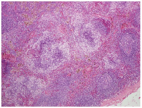
FIGURE 23-17 • The paracortical regions in dermatopathic lymphadenitis are expanded and show a pale pink swirled appearance due to the collections of abundant histocytes and Langerhans cells admixed with small lymphocytes. Some histiocytes contain brown melanin pigment. (Hematoxylin and eosin stain 4× magnification.)


Hemophagocytic Lymphohistiocytosis

Hemophagocytic lymphohistiocytosis (HLH) may be idiopathic, familial, infection-related (95,96), rheumatologic, malignancy related (97,212), related to antineoplastic therapy (98), or rarely preceded by Kikuchi disease (99,100). HLH results from uncontrolled activation of CD8+ T cells, macrophages, and histiocytes associated with decreased NK cell function and increased levels of circulating proinflammatory cytokines (97,101). HLH is diagnosed according to HLH-2004 guidelines established by the Histiocyte Society when specific clinical, laboratory, and histopathologic criteria are met (102). Clinically significant lymphadenopathy is uncommon in primary HLH, and when present, lymph node biopsy is generally performed to exclude lymphoma.

Histologically, follicles are usually small and germinal centers few in number. The paracortex is depleted and has a mottled appearance because of the presence of increased numbers of pale-staining histiocytes. The sinusoids are distended by histiocytes, many of which are phagocytic. The nuclear features are bland, and the cells have abundant eosinophilic cytoplasm containing variable numbers of red blood cells and red blood cell fragments (eFigure 23-13). Leukophagocytosis is uncommon relative to erythrophagocytosis in this condition, and in further contrast to SHML cells, the histiocytes in HLH exhibit a CD68+, S100, CD1a, CD207 phenotype. Erythrophagocytosis and hemophagocytosis can be seen as a secondary phenomenon outside the context of primary HLH, and all these conditions must be considered as part of the differential diagnostic assessment. For instance, it has been reported in patients with a robust autoimmune hemolytic anemia, active SLE (103), X-linked lymphoproliferative disease, ehrlichiosis (104), typhoid fever (105), the accelerated phase of Chediak-Higashi disease (101), SHML, acute myelogenous leukemia (AML), acute lymphoblastic leukemia (106,212), juvenile myelomonocytic leukemia (99,107), and peripheral T-cell lymphoma (108,109). In some cases, there may be a spectrum of histiocytic disorders with macrophage activation syndrome (MAS) or secondary hemophagocytic syndrome seen in patients with Langerhans cell histiocytosis (LCH) (110). Patients with T lymphoblastic leukemia have rarely shown subsequent involvement by LCH or HLH (111,112).

Only gold members can continue reading. Log In or Register to continue

Stay updated, free articles. Join our Telegram channel

Sep 23, 2016 | Posted by in PATHOLOGY & LABORATORY MEDICINE | Comments Off on The Lymph Nodes, Spleen, and Thymus

Full access? Get Clinical Tree

Get Clinical Tree app for offline access