The Laparoscopic Gastric Band Technique of Placement
Paul E. O’Brien
Introduction
Laparoscopic adjustable gastric banding was introduced in 1992 by Favretti and Cadiere as a major innovation in bariatric surgery by adding the laparoscopic approach to the key features of adjustability and ready reversibility of the Kusmak band. The Lap Band was introduced in September 2003 by Mitiku Belachew and the placement of this device is now the primary approach to bariatric surgery in most parts of the world. It is characterized by its safety and effectiveness in achieving major and durable weight loss. It reverses the comorbidities of obesity and improves quality of life. Further, it provides a gentle approach to the control of obesity because its placement is truly minimally invasive, its primary mode of action is by inducing satiety, and the degree of effect is controlled postoperatively through its adjustability. It offers the additional attraction of complete reversibility so that when, at some future time, a better method for appetite control is established, our patients have the option of changing to that method.
There are two essential components to effective use of the Lap Band. First, it must be placed correctly. This requires correct operative technique, which will be reviewed in detail in this chapter. Second and equally important to the technical detail of the operation, there must be a permanent process of care, which includes a sequence of adjustments of the tightness of the band in association with patient advice and support. It is essential that this be learned and followed if optimal results from the procedure are to be obtained.
Perioperative Management
The technique of Lap Band placement has evolved significantly over the past 12 years since its introduction. Some elements of the procedure are still subject to variation between surgeons who are achieving apparently equal outcomes, but the key positioning of the band is now essentially identical across all groups. The technique described is the product of 10 years of experience and placement of more than 1,800 Lap Bands. Throughout this time I have sought to identify a technique that is safe, quick, minimally traumatic, less expensive, and able to cope with the challenges of superobesity. It is, however, just one variation on the theme, and alternative approaches appear to be equally effective and efficient. It does provide a solid starting point for new Lap Band surgeons, who can then evolve techniques of their own as they build up their experience.
Preoperative Assessment
Clinical assessment includes full routine history and examination and specific documentation of the problems generated by the obesity, including all the comorbidities and the physical and social limitations present. Laboratory studies include all routine hematology and biochemistry plus specific screening for diabetes and insulin resistance, micronutrient deficiencies including iron studies, vitamin B12, folic acid, lipid profile, and serum homocysteine. Homocysteine is an independent risk factor for cardiovascular disease and will rise with weight loss. This rise can be prevented by taking vitamins B6, B12 and folate, which are cofactors in its metabolism. Special investigations such as upper gastrointestinal endoscopy, barium swallow and meal, ultrasound of gallbladder, esophageal manometry and 24-hour pH study, 24-hour blood pressure monitoring, and polysomnography are performed only when a specific indication exists.
Perioperative Care
All patients are given antimicrobial prophylaxis, administered at the commencement of the procedure. We use a fl-lactamase-resistant penicillin (flucloxacillin, 1 g IV stat) to cover skin contaminants and a broad spectrum to cover the enteric flora such as a third-generation cephalosporin (cefotaxime, 1 g IV stat). Prophylaxis against deep venous thrombosis consists of electrical calf stimulation throughout the procedure and subcutaneous heparin until discharge (sodium heparin, 500 units b.d. subcutaneously commencing immediately after completion of the procedure). Pain relief is principally by rofecoxib (Dynastat × 40 mg IV during anesthesia and repeated every 12 hours if needed). Oral soluble paracetamol (acetaminophen) is then used. Opiates are avoided. Ondansetron (× 4 mg IM) is available for nausea but rarely used.
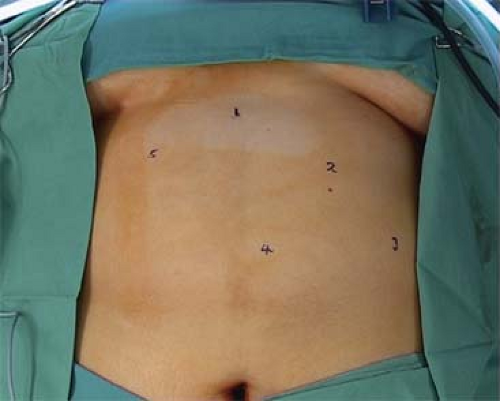 Fig. 1. Port positions. There are 3 × 5 mm ports and 1 × 15 mm ports placed. The Nathanson liver retractor (5 mm) is placed at position 2. |
Operative Position and Port Placement
The patient is placed in a reverse Trendelenburg position of a 25-degree slope, legs apart. The surgeon stands between the legs and one assistant stands on the patient’s left side. We use five ports; four of them are 5 mm in diameter and one is 15 mm in diameter (their positions are shown in Fig. 1).
We use a 5-mm end-viewing telescope for insertion of the initial port and a 5-mm, 30-degree telescope for the rest of the procedure. The video system must be a high-definition system of optimal quality (Stryker, San Jose, CA). The initial port is a 5-mm Optical Separator (Applied Medical, Rancho Santa Margarita, CA) placed at position 5, a handbreadth below the costal margin, 5 cm to the left of the midline. This port gives initial insufflation and is the path for the hook diathermy and the introduction of sutures. It becomes the site of the access port at completion. The Nathanson liver retractor is placed at port 2, in the midline just below the sternum. The two 5-mm ports are 15 cm long (Applied Medical). This extra length enables them to be passed through the abdominal wall on an angle such that they are heading toward the upper stomach. As all aspects of the procedure occur at this point, there is minimal lateral pressure needed. The first of these (position 1) is placed 1 cm below the right costal margin on the midclavicular line and is used for graspers only. The second (position 4) is 3 cm below the left costal margin at the anterior axillary line. After placing the left lateral port, the camera is moved to this port, the 5-mm Optical Separator is removed from position 5, and a 10- to 15-mm port (Separator port, Applied Medical) is placed at this position under vision. The telescope is then placed in the 15-mm port and the 5-mm Optical Separator is placed at position 3, 1 cm below the left costal margin at the midclavicular line. The telescope is moved to this site, which is the camera port for the procedure.
We use a 5-mm end-viewing telescope for insertion of the initial port and a 5-mm, 30-degree telescope for the rest of the procedure. The video system must be a high-definition system of optimal quality (Stryker, San Jose, CA). The initial port is a 5-mm Optical Separator (Applied Medical, Rancho Santa Margarita, CA) placed at position 5, a handbreadth below the costal margin, 5 cm to the left of the midline. This port gives initial insufflation and is the path for the hook diathermy and the introduction of sutures. It becomes the site of the access port at completion. The Nathanson liver retractor is placed at port 2, in the midline just below the sternum. The two 5-mm ports are 15 cm long (Applied Medical). This extra length enables them to be passed through the abdominal wall on an angle such that they are heading toward the upper stomach. As all aspects of the procedure occur at this point, there is minimal lateral pressure needed. The first of these (position 1) is placed 1 cm below the right costal margin on the midclavicular line and is used for graspers only. The second (position 4) is 3 cm below the left costal margin at the anterior axillary line. After placing the left lateral port, the camera is moved to this port, the 5-mm Optical Separator is removed from position 5, and a 10- to 15-mm port (Separator port, Applied Medical) is placed at this position under vision. The telescope is then placed in the 15-mm port and the 5-mm Optical Separator is placed at position 3, 1 cm below the left costal margin at the midclavicular line. The telescope is moved to this site, which is the camera port for the procedure.
Surgical Technique
Dissection At the Angle of His
The liver is retracted by a Nathanson liver retractor (Automated Medical Products, New Jersey, USA), which is held by an external clamp (Iron Intern, Automated Medical Products). The omentum covering the fundus of the stomach is drawn inferiorly using the graspers at positions 1 and 4. The grasper at position 4 is then passed over the grasper at position 1 to draw the fundus of the stomach downward. The grasper at position 1 then retracts the fat pad over the distal esophagus to open up the area of the angle of His. Using a hook diathermy through the 15-mm Separator port (position 5), the peritoneum over the left crus is divided over approximately a 2-cm length (Fig. 2). It is not necessary to fully expose the fibers of the left crus but essential to be at this anatomic site. That completes the dissection at this point and attention moves to the lesser curve.
Dissection on the Lesser Curve
The grasper at position 4 takes hold of the fat adjacent to the lesser curve of the stomach and draws it to the left. The pars flaccida of the lesser omentum is divided to expose the posterior wall of the lesser sac. The grasper at position 4 then draws the fat on the posterior wall of the lesser omentum to the left and the grasper at position 1 retracts the caudate lobe of the liver to expose the lower aspect of the right crus. The peritoneum of the posterior wall is opened over a 5-mm length using the hook diathermy. The point of this opening is critical. It should be 5 mm in front of the anterior margin of the right crus at its most inferior point (Fig. 3).
Stay updated, free articles. Join our Telegram channel

Full access? Get Clinical Tree


