The Kidneys, Adrenals, and Retroperitoneum


The Kidneys, Adrenals, and Retroperitoneum


Muhammad B. Zaman


THE KIDNEYS

The role of cytology of the urinary sediment in the diagnosis of renal diseases is discussed in Chapters 22 and 23. The subject of this chapter is transcutaneous fine-needle
aspiration (FNA) of the kidney. Dean (1939) first reported the treatment of a solitary cyst of kidney by aspiration. Söderström (1966) described a large experience with this method, and Von Schreeb et al (1967), Kristensen et al (1972), Thommesen and Nielsen (1975), Edgren et al (1975), and Holm et al (1975) each reported a series of cases.

It must be stressed at the outset that FNA does not replace renal core biopsies in the diagnosis of diffuse medical diseases of the kidney; however, it does play a role in the assessment of renal transplant rejection (see below).


INDICATIONS

FNA is used for the pretreatment diagnosis of space-occupying lesions of the renal parenchyma. Although renal cell carcinoma (RCC) is as common as pancreatic carcinoma, only 5% of all transcutaneous abdominal aspirations performed at the Westchester Medical Center are directed toward renal lesions, as compared to 12% for the pancreas. This is because a reliable diagnosis of renal neoplasms can be established in most patients by renal imaging studies consisting of intravenous pyelography, ultrasonography (US), computed tomography (CT), and arteriography, obviating the need for a preoperative biopsy. Each of these imaging techniques has a low, but significant, diagnostic error rate (Sherwood and Trott, 1975; Richter et al, 2000). We know of cases of nephrectomy performed for a benign neoplasm, cyst, or benign adrenal tumor mimicking renal masses.

FNA biopsy of renal abnormalities can provide a definitive diagnosis and considerably reduce surgical exploration of patients with nonneoplastic lesions, such as cysts, and may clarify the nature of solid lesions that are not clearly defined by imaging procedures (Zornoza et al, 1977a; Meier et al, 1979; Barbaric et al, 1981; Murphy et al, 1985; Nadel et al, 1986; Pilotti et al, 1988; Leiman, 1990; Cristallini et al, 1991).

The indications for FNA biopsy of space-occupying lesions of the kidney are as follows:


  • Cystic lesions (as a diagnostic and therapeutic procedure)


  • Lesions with equivocal radiologic findings


  • Confirmation of diagnosis in advanced malignant lesions prior to nonsurgical treatment


  • Confirmation of local recurrence at the site of a prior tumor or a direct extension from neighboring site (e.g., colon or adrenal)


  • Confirmation of metastatic cancer

Transcutaneous FNA biopsy has been used for the follow-up of renal allograft recipients (Häyry and von Willebrand, 1981a). The technique was described in detail by Häyry and von Willebrand (1981b, 1984), and was used successfully by Bishop et al (1989), Linsk and Franzén (1989), Egidi (1990), and Garcia-Castro et al (1993). As described in detail in Chapter 22, the aspiration specimen is used to identify subpopulations of T-lymphocytes by immunocytochemical or flow cytometric analysis.

Aspiration cytology has been used in Scandinavian countries to grade renal carcinomas, in order to select patients with high-grade (poorly differentiated) tumors for preoperative radiotherapy (Zajicek, 1979). This issue is discussed below.


TECHNIQUE OF ASPIRATION BIOPSY

The FNA technique is patterned after that used for renal core biopsies. Söderström (1966) approached this type of biopsy on purely anatomic grounds, without roentgenologic or US guidance. Others have used biplane fluoroscopic guidance, usually with intravenous pyelography (IVP). Developments in US and CT have significantly improved the guidance system in adults and children (Kristensen et al, 1974; Zeis et al, 1976; Juul et al, 1985; Nguyen, 1987; Thornburg and Weiss, 1987; Li Puma, 1988; Pilotti et al, 1988). These techniques do not depend on renal function, as does IVP, and they are helpful in separating solid from cystic lesions and cystic neoplasms from benign cysts (Pollack et al, 1982).

The principles of the aspiration procedure are discussed in Chapter 28. The special requirements for renal FNA are as follows: Regardless of the guidance modality used to target the renal lesion, the aspiration is usually undertaken with the patient in either a prone or decubitus position, which allows for the shortest possible skin-to-lesion distance. When a 22- or 23-gauge needle is used, it may be difficult to penetrate the tough renal fascia. Thus, Zajicek (1979) advocated the use of a larger-caliber needle with an obturator as a guide for the thinner needle. In our experience, this is rarely required.


Complications

In an early study, von Schreeb et al (1967) found no differences in the survival rates of patients who underwent an aspiration and those who did not. Söderström (1966) reported some instances of hematuria. A case of seeding of renal carcinomas in the needle track (with an 18-gauge needle) was described by Gibbons et al (1977). Additional reports of needle-track seeding with smaller-caliber aspiration needles have been described (Auvert et al, 1982; Wehle and Grabstald, 1986; Shenoy et al, 1991; Smith, 1991; Slywotzky and Maya, 1994).


SYNOPSIS OF ANATOMY AND HISTOLOGY

The essential features of anatomy of the kidney are briefly summarized in Chapter 22. A few additional points of special interest regarding aspiration biopsy may be added here. The right kidney is adjacent to the inferior surface of the liver, the right colic flexure, and the small intestine, and the left kidney is adjacent to the spleen, the body of the pancreas (and splenic vessels), the stomach, the left colic flexure, and the small intestine. Therefore, extrinsic cells from any of these organs, as well as mesothelial cells from peritoneal reflections and perirenal fat, are not uncommon in renal aspirates.


Each kidney is composed of an external or cortical zone (cortex), an internal medullary zone (medulla), and the renal pelvis, which opens into the ureter. Renal parenchyma consists of one to two million nephrons, interstitial connective tissue, and a very rich and complex network of blood vessels arranged in an exquisite order to carry on the function of blood filtration and excretion of waste products in the urine. Each functional unit of the kidney, or nephron, consists of a glomerulus in continuity with a renal tubule (Fig. 40-1). Each tubule is divided into a proximal convoluted segment (the progenitor of common RCC) and a distal convoluted segment (the progenitor of papillary carcinoma), the two of which are connected by a narrow segment called the loop of Henle. The tubules drain the filtered urine into collecting ducts, which are lined by two distinct types of cells. The majority of these cells are light-staining collecting duct cells (the progenitors of a rare tumor, collecting duct carcinoma (CDC)), and the minority are dark-staining intercalated cells with many mitochondria (the progenitors of oncocytoma and chromophobe RCC (ChRCC)). Collecting ducts coalesce to form the terminal ducts of Bellini, which carry the urine into the renal pelvis. For further comments on the formation of urine, see Chapter 22.


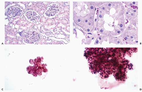
Figure 40-1 Histology and cytology of the normal kidney. A. Glomeruli and proximal convoluted tubules with pink granular cytoplasm are the predominant component of the cortex. Smaller cuboidal cells with pale cytoplasm line the distal tubule). B. Proximal tubules at high magnification. C,D. Renal glomerulus at low and high magnification. In aspiration smears, the glomeruli are seen as thick, three-dimensional, lobulated structures. The anatomic relationship among the endothelial, epithelial, and mesangial cells is lost.

The glomeruli are located mainly in the renal cortex (Fig. 40-1A). They are complex, spherical structures composed of capillary tufts with specialized endothelial cells supported by mesangial connective tissue cells. Each glomerulus is surrounded by a connective tissue envelope, known as Bowman’s capsule.

The tubules make up the largest component of the kidney parenchyma. The tubules are lined by cuboidal epithelial cells that may stain intensely pink or pale in hematoxylin and eosin (H&E) stain, and vary in size depending on their function and location in the tubular segment (Fig. 40-1B). Although several types of tubular cells may be distinguished by ultrastructural and other studies (Bander et al, 1985), they cannot be reliably subclassified in aspiration smears.

As described in detail in Chapter 22, the renal calyces and pelvis are lined by urothelium, which occasionally may be seen in aspiration smears of the lower pole of the kidney.


CYTOLOGY OF NORMAL KIDNEY

In material aspirated from renal cysts and sometimes from relatively small renal tumors, benign renal tubular cells
are often observed. However, unless a large-bore needle is used, intact glomeruli are rarely encountered (Fig. 40-1C,D).

An intact glomerulus is large enough to occupy nearly the entire 40× microscopic field. It is a sharply circumscribed, lobulated, round or oval, thick, multilayered structure composed of small cells. Because the glomeruli are squashed on the slide, their internal structure cannot be seen. Bowman’s capsule may be seen on rare occasions as a balloon-like transparent membrane.

The normal tubular cells are sometimes aspirated as intact tubules (Fig. 40-2A) or as epithelial cells, either dispersed or forming small clusters (Fig. 40-2B-D). The cells of proximal tubules, which have eosinophilic cytoplasm, are not easy to identify as such, and in fact may mimic cells derived from normal adrenal cortex and sometimes well-differentiated small malignant cells derived from papillaryor low-grade conventional RCC (see below). The normal tubular cells are generally cuboidal in shape and vary in size from small cells derived from the loop of Henle to the much more common larger cells derived from the proximal and distal tubules. Most of the larger tubular cells display a relatively abundant finely granular, transparent, pale or pink cytoplasm, depending on the stain used, and small, spherical nuclei with transparent chromatin pattern, containing tiny nucleoli.


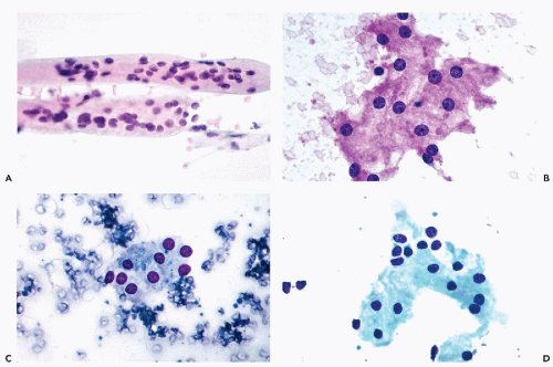
Figure 40-2 A. Whole renal tubules in aspiration smears. B. Proximal tubular cells. Note that the majority of the nuclei are separated from each other by pink granular cytoplasm. C,D. Tubular cells forming sheets in MGG-stained smears. Note the uniformity of the cells and their small nuclei.


BENIGN LESIONS


Renal Cysts

Cystic diseases of the kidney are a heterogenous group of lesions that include hereditary, congenital but nonhereditary, and acquired disorders. Renal cysts may be tiny or very large (measuring 10 cm or more in diameter), single, or multiple. In the hereditary polycystic kidney, nearly all of the adult kidney can be replaced by cysts of various sizes (which, incidentally, should not be aspirated unless there is a suspicion of a coexisting renal carcinoma).

Acquired cysts are the most common renal lesions. They are usually small and unilocular, and are formed by shrinkage of the renal parenchyma secondary to vascular insufficiency in elderly patients. They are rarely a cause for alarm. Dialysis-associated cystic disease affects patients with chronic renal failure and prolonged dialysis. End-stage kidneys are often shrunken and multicystic, and have been reported to carry an up to 50-fold increased risk of RCC (Truong et al, 1995).

Cystic nephroma, or multilocular cyst of the kidney, is a rare benign neoplasm that occurs in children or young adults (usually female). The cysts are unilateral and encapsulated. The compartments of the cyst, or locules, are lined by a single layer of epithelium with or without atypia (Eble and Bonsib, 1998).


Except for the adult form of polycystic kidney, most renal cystic lesions are asymptomatic and incidental. Sometimes, however, renal cysts may present a diagnostic dilemma and may be confused with renal carcinomas undergoing cystic degeneration (Pollack et al, 1982).

Cysts have been the most commonly aspirated renal lesions since the first report by Dean (1939) was published. If the preaspiration diagnosis of a cyst is secure, a large 18-gauge needle may be used to evacuate the cyst fluid. In less secure cases, it is preferable to use smaller-caliber needles.


Inspection of Cyst Fluid

The amount of fluid aspirated from renal cysts varies according to cyst size. In exceptional cases, 40-50 ml of fluid may be aspirated. In most instances, the fluid is clear, straw-colored, and occasionally cloudy or blood-tinged, or, rarely, chocolate brown, suggestive of a prior hemorrhage. Zajicek (1979) and Pilotti et al (1988) suggested that clear, straw-colored fluid does not require cytologic study because the fluid in cystic RCCs is usually cloudy and discolored (Rehm, 1961; Khordand, 1965; Holm, 1975; Anderson, 1977). Our policy is to study the cytologic make-up of all aspirated fluids.


Cytology

Clear cyst fluid from the diverse group of renal cysts described above may be acellular or may show a variable number of mononucleated and, rarely, multinucleated macrophages (Fig. 40-3). Rare clusters of benign epithelial cells from cyst lining or benign tubular cells from the surrounding normal renal parenchyma may be encountered. Reactive fibroblasts from the capsule of the cyst may also be seen. Occasionally, the macrophages may show significant nuclear abnormalities that at a first glance may be thought to represent cancer cells. The correct diagnosis is best established by searching for some evidence of phagocytosis in the cytoplasm of these cells. Liesegang rings, approximately spherical concentric structures, have been reported in renal cyst fluid (Sneige et al, 1988; Katz and Ehya, 1990; Raso et al, 1998) (see Chap. 25).


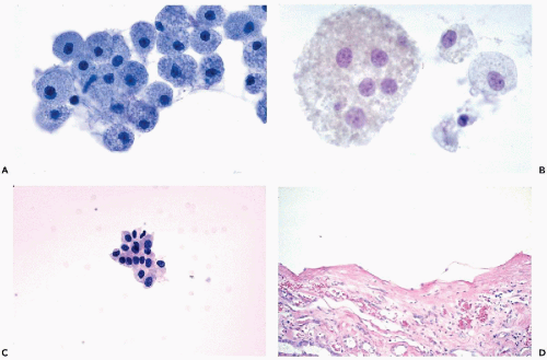
Figure 40-3 Renal cyst aspirates. A. Large mononucleated macrophages with finely vacuolated cytoplasm are the cells typically seen in clear, straw-colored cyst fluid. B. Multinucleated macrophage in cyst fluid. C. A small, tight cluster of benign epithelial cells, corresponding to the excised renal cyst shown in D.

Cloudy fluid is generally derived from an inflamed cyst and contains numerous polymorphonuclear leukocytes (see below). The cytology of cystic RCC is described under neoplasms of the kidney.


Ancillary Studies of Renal Cyst Fluids

In our experience, additional information may be obtained by determining the fat, protein, and lactic dehydrogenase (LDH) content of the renal cyst fluid. Clear fluid from acquired cysts is low in fat, protein, and LDH. Cloudy or turbid fluid is generally inflammatory and therefore high in protein and very high in LDH. A malignant cystic tumor
yields bloody fluid with high fat and protein but low LDH levels.


Renal Abscess

Patients with renal abscess are febrile and usually experience severe costovertebral angle pain, tenderness, and often pyuria. Pyelonephritis, whether of hematogenous origin or caused by an ascending infection, may result in a renal abscess. Commonly, an abscess is the result of obstruction of the ureter by calculi or tumor, leading to hydronephrosis and secondary infection in neglected patients. The abscess may be limited to the renal pelvis or may involve the entire kidney and perinephric region, in which case the collection of pus may be quite large and may mimic a cystic or a solid tumor.

The aspirate yields purulent exudate that should be submitted for microbiologic study. In endemic areas, tuberculosis must be considered as a cause of renal abscess.


NONCYSTIC BENIGN RENAL DISORDERS


Renal Infarcts

Because renal infarcts may mimic a renal tumor in imaging studies, they are occasionally aspirated. Silverman et al (1991) reported two such cases. In one case, necrotic gromeruli and tubules were observed; in the second, atypical tubular renal cells (presumably the consequence of tubular regeneration) were mistaken for an RCC. Thus, renal infarcts must be considered in the differential diagnosis of renal tumors.


Xanthogranulomatous Pyelonephritis

Xanthogranulomatous pyelonephritis is an unusual, relatively rare form of chronic pyelonephritis characterized by the accumulation of foamy macrophages intermixed with plasma cells, lymphocytes, polymorphonuclear leukocytes, and occasional giant cells. The clinical findings include flank pain, renal mass, hematuria, and recurrent urinary tract infections, usually caused by the Proteus species. Staghorn calculi may be present. The radiologic findings of a hypovascular nonfunctioning renal mass may be difficult to interpret. Grossly, there are discrete or confluent large, yellowish-orange nodules within the enlarged kidney that may be confused with RCC. On microscopic examination of tissue sections, the clusters of histiocytes with abundant, clear, foamy cytoplasm may superficially resemble clear cell renal cortical carcinoma. Akhtar and Qunib (1992) reported a case of bilateral xanthomatous pyelonephritis associated with amyloidosis and a small renal carcinoma in one of the native kidneys of a patient who had undergone a renal transplant.


Cytology

The FNA biopsy shows a polymorphous picture of macrophages and giant cells with foamy vacuolated cytoplasm in a background of necrosis and inflammatory cells. The findings may superficially suggest clear cell carcinoma, but usually are sufficiently characteristic to establish a cytologic diagnosis (Lizza et al, 1984; Sugie, 1991).


Malakoplakia

There are several cases of renal malakoplakia on record (for a summary see Esparza et al, 1989). Hurwitz et al (1992) reported a case of bilateral malakoplakia. Although to our knowledge there are no reported cases of renal malakoplakia diagnosed on FNA, this possibility should be kept in mind for future reference. The histology and cytology of this disorder are extensively discussed in Chapter 22.


Benign Renal Neoplasms


Angiomyolipoma

Renal angiomyolipoma occurs in two distinct clinical settings. It occurs either as a large, single tumor in otherwise normal patients, or as multiple bilateral smaller tumors in patients with tuberous sclerosis, a disorder characterized by mental retardation, epilepsy, and multiple sebaceous adenomas of the skin (Bennington and Beckwith, 1975; Brodsky and Granick, 1989; Silva and Childers, 1989). The tumor may also occur in the retroperitoneum (Wadih et al, 1995) and the liver (Nguyen and Catzavelos, 1990). With the widespread use of modern imaging modalities, more and more asymptomatic angiomyolipomas of the kidney have been diagnosed. The tumor has very characteristic features on CT (Fig. 40-4A). The presence of fat, accounting for clear areas, differentiates this tumor from renal carcinoma (Bosniak et al, 1988). On angiography, the tumor is vascular and may mimic RCC. However, benign and usually asymptomatic, large angiomyolipomas are often resected for fear of severe or, rarely, fatal hemorrhage.


Histology

Renal angiomyolipoma is a benign neoplasm that is composed of a mixture of smooth muscle, fat, and tortuous, thick-walled blood vessels (Fig. 40-4B,D). Some of these tumors are very cellular and may show mitotic activity in smooth muscle cells. Some of the spindly cells may show marked nuclear abnormalities in the form of large and hyperchromatic nuclei in bizarre spindly cells with abundant eosinophilic cytoplasm. In some tumors, giant cells with huge nuclei and prominent nucleoli may be observed (Eble et al, 1997). Such tumors may be confused with malignant mesenchymal tumors, leiomyosarcomas, or renal sarcomatoid carcinomas. The presence of needle- or rod-shaped crystalloids in the cytoplasm of the tumor cells was reported by Mukai et al (1992). In most instances, the characteristic histologic pattern is recognizable, particularly because of the presence of fat (Silva and Childers, 1989). The immunohistochemical profile of angiomyolipoma is rather distinctive. The cytokeratin stain is negative, whereas actin stain is strongly positive, in keeping with the smooth-muscle derivation of the tumor cells. Vimentin stain is variable, but HMB-45 (melanoma antigen) stain is positive in perivascular spindly epithelioid cells of the tumor. This panel of stains may be helpful in identifying this tumor in debatable cases.


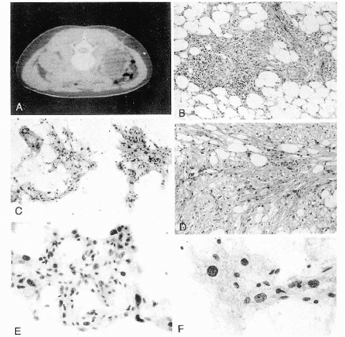
Figure 40-4 Angiomyolipoma. Angiomyolipoma of the left kidney in a 34-year-old woman with no evidence of tuberous sclerosis. A. Computed tomography (CT) revealed the tumor in the lower pole of the right kidney. The patient was in a prone position. The CT features were characteristic of angiomyolipoma, notably because of the presence of clear areas representing fat within the tumor. B,D. Tissue sections. B. Numerous thick-walled vessels, smooth muscle cells, and fat are present. D. Bundles of smooth muscle cells. Note the presence of pleomorphic, atypical nuclei. C,E,F. Aspirate. C. Irregular tissue fragments containing smooth muscle cells, fat, and vessels. E. Tissue fragment. Note the irregular arrangement of the nuclei and their variable sizes. F. Group of cells with abundant cytoplasm. Note the significant differences in nuclear sizes. Most of the nuclei are hyperchromatic, but their contour is smooth. These cytologic features may lead to an erroneous diagnosis of a carcinoma or a sarcoma. (From Koss et al. Aspiration Biopsy. Cytologic Interpretation and Histologic Bases, 2nd ed. New York: Igaku-Shoin, 1992.)


Cytology

The classical cytologic presentation of an angiomyolipoma shows small tissue fragments composed of fat and bundles of spindly cells (Koss et al, 1992). Fragments of thick-walled blood vessels may be observed (Glenthoj and Partoft, 1984; Nguyen, 1984). Gupta et al (1998b) searched for the crystalloids described by Mukai et al (1992), but could not find them with either light or electron microscopy. Even at low power, some variability in nuclear sizes within the spindly cells may be observed (Fig. 40-4C). At higher power, the nuclear variability and hyperchromasia are evident (Fig. 40-4E,F). In some cases, mitotic figures and very large, hyperchromatic nuclei may be observed, which may lead to an erroneous interpretation of the smear as an RCC (Nguyen, 1984; Granter and Renshaw, 1999). Leiomyosarcomas and other spindle cell tumors must be considered in the differential diagnosis. In most cases, however, the presence of fat should act as a deterrent to wrong interpretation of the smears (Koss et al, 1992). If adequate material is available, immunostaining (discussed above) may also be helpful in establishing the correct identity of the tumor.


Renal Adenoma


Papillary (Chromophil) Adenoma

Papillary (chromophil) adenoma is a controversial lesion, primarily defined by its size of 1 cm or less in diameter. Nearly all lesions are asymptomatic and found incidentally. It is not known whether the lesion is related to papillary renal carcinomas, even though there is some histologic similarity, and such lesions are commonly found in kidneys with clinically evident papillary RCC (Reuter and Gaudin, 1999). The tumors are well demarcated but nonencapsulated, pale gray to yellow nodules in a cortical or subcapsular location. Histologically, they are composed of densely packed tubules lined by small, regular cuboidal cells with round, uniform, bland nuclei. Tubulopapillary, purely papillary patterns and microcyst formation have been noted (Grignon and Eble, 1998).

The FNA experience with these very small lesions is very modest. Kini (1999) reported tissue fragments composed of uniform small cells with high nucleocytoplasmic (N/C) ratios.


Congenital Mesoblastic Nephroma of Infancy

Congenital mesoblastic nephroma of infancy is an uncommon benign tumor that was first described by Bolande et al (1967). The tumors may be bulky and palpable in newborns and infants, and are composed of proliferating fibroblasts and smooth muscle cells with preservation of the nephrons. Drut (1992) and Kaw (1994) described the cytologic findings in FNA in such cases. Benign spindle cells arranged in bundles were the dominant feature. Of note was the presence of epithelial cells derived from renal glomeruli and tubules that could have been mistaken for an epithelial component of a malignant stromal tumor, such as Wilms’ tumor (see below).


Metanephric (Embryonal) Adenoma

Metanephric (embryonal) adenomas are rare benign renal cortical neoplasms that are included in the new classification of renal neoplasms (Table 40-1). These tumors occur in all age groups, most often in women, and produce nonspecific symptoms of a renal mass. Hematuria may occur. Polycythemia has been reported in as many as 12% of patients (Hennigar and Beckwith, 1992; Davis et al, 1995).


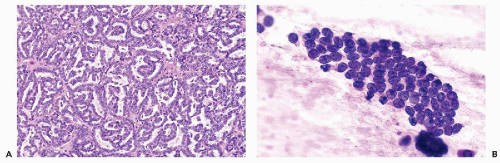
Figure 40-5 Metanephric adenoma. A. Histology. Closely packed tubular structures with papillary infolding (glomeruloid bodies). The tumor cells are monotonous and small with scanty cytoplasm. B. Touch-preparation of a nephrogenic adenofibroma (see text).


TABLE 40-1 CLASSIFICATION OF RENAL CELL NEOPLASMS


Benign neoplasms (partial listing)


Oncocytoma*


Papillary (chromophil) adenoma


Metanephric (embryonal) adenoma


Nephrogenic adenofibroma


Malignant neoplasms


Conventional (clear cell) carcinoma


Papillary (chromophil) carcinoma


Chromophobe carcinoma


Collecting duct carcinoma (CDC)


Medullary carcinoma


Renal cell carcinoma (RCC), unclassified


Tumor of undetermined malignant potential


Multilocular cystic RCC


Adapted from the Heidelberg classification of renal cell tumors (Kovacs et al, 1997).


* In this text, oncocytoma is discussed with malignant neoplasms.


Histology

Metanephric adenoma is characterized by tightly packed tubules with papillary infoldings, mimicking to some extent the configuration of glomeruli (glomeruloid bodies) (Fig. 40-5A). Similarities with Wilms’ tumor have been stressed (Davis et al, 1995; Jones et al, 1995). A cytogenetic analysis showing trisomies of chromosomes 7 and 17, and loss of sex chromosome Y suggested that these tumors are
related to papillary RCC, which shows similar findings (Brown et al, 1997). We recently encountered a case of papillary carcinoma that arose in a metanephric adenoma, confirming the relationship between the two lesions.


Cytology

Renshaw et al (1997) described the cytologic pattern of a metanephric adenoma studied by FNA in a 48-year-old woman with a tumor 4 cm in diameter. The monotonous small tumor cells with scanty cytoplasm, spherical nuclei, and occasional small nucleoli were arranged in short, tight papillary clusters and loose sheets. Stains for keratin, epithelial membrane, and carcinoembryonic antigens were negative. Zafar et al (1997) described two such cases with essentially similar findings. In one of these cases, a preoperative diagnosis of Wilms’ tumor was made. We have had experience with a touch preparation of one nephrogenic adenofibroma (see following topic), which showed only small epithelial cells, closely resembling blastema cells of Wilms’ tumor (Fig. 40-5B), in keeping with the observations of Renshaw et al (1997) and Zafar et al (1997). Thus, the similarities between the cytology of benign metanephric adenoma and that of Wilms’ tumor are striking. The monotonous population of small cells in metanephric adenoma in comparison with the triphasic morphology of Wilms’ tumor, and to some extent the older age of patients with metanephric adenomas, may serve to differentiate these two types of tumor. For a description of Wilms’ tumor, see below.


Nephrogenic Adenofibroma and Other Uncommon Benign Lesions

Nephrogenic adenofibroma is a biphasic tumor composed of an epithelial component, similar to that of metanephric adenoma (Fig. 40-5A), which is separated by bland fibro-blast-like cells (Hennigar and Beckwith, 1992; Arroyo et al, 2001). Other benign entities that may present as space-occupying lesions of the kidney include benign mesenchymal tumors (e.g., leiomyomas, fibromas, and angiomas). We have not seen any aspirated material from these lesions, and to our knowledge, no case descriptions have been published.


MALIGNANT TUMORS OF RENAL PARENCHYMA

In adults, most malignant renal tumors arise from renal tubules and are therefore designated as RCC. The old terms hypernephroma and adenocarcinoma of the kidney are no longer considered appropriate. A small number of carcinomas arise from the urothelium of the renal pelvis and are identical to carcinomas of the bladder or ureter (see below and Chap. 23). In children, Wilms’ tumor is the most common malignant renal tumor.

Nonepithelial malignant tumors (e.g., sarcomas) are exceedingly uncommon, and most arise in the renal capsule or hilum (Farrow et al, 1968).


Renal Cell Carcinoma (RCC)


Epidemiology and Genetics

RCC represents about 2% to 3% of all visceral cancers, and accounts for 85% of all renal cancers in adults, being more common in males than females. There are an estimated 31,800 new cases and 11,600 deaths per year from this disease in the United States (Jemal et al, 2002). The peak incidence is in the sixth and seventh decades of life.

A hereditary form of RCC has been described in families with an abnormality of the short arm of chromosome 3 (Cohen et al, 1979), accounting for a very small proportion of cases of this disease. Rare cases of RCC occur in people with Von Hippel-Lindau disease (i.e., the formation of abnormal vessels in the cerebellum and retinas), but most RCCs are sporadic and of unknown etiology. The customary villains—smoking and environmental factors—may play a role in the genesis of these tumors (McLaughlin et al, 1984; Yu et al, 1986). Dey et al (1996) described several cases of RCC in children.

In recent years, specific genetic abnormalities have been found in the majority of RCCs. Based on histologic, cytogenetic, and molecular studies, RCCs are now considered to be a histogenetically heterogeneous group of distinguishable tumors with significantly different prognoses (Thoenes, 1986; Kovacs, 1991; Van den Berg et al, 1993; Weiss et al, 1995). Most of these tumors are malignant, but a small minority are now recognized as benign. As a result, a new classification of renal neoplasms (shown in Tables 40-1 and 40-2) has gained acceptance (Kovacs et al, 1997; Storkel et al, 1997). This classification, in addition to documenting a close relationship between the morphologic and genetic features of renal tumors, also defines distinct clinical entities (Motzer et al, 1996). Within the malignant categories, different morphologic variants have significant differences in 5-year survival rates (Reuter and Gaudin, 1999).

It is now recommended that the old term clear cell carcinoma be replaced by conventional carcinoma, with clear cell added in parentheses as an afterthought. Although previously used terms such as granular cell and sarcomatoid type of RCC (Murphy et al, 1994) are no longer recommended, to us they define specific morphologic subgroups and will be used in this text. The rationale behind this change is based on the common origin of these morphologic variants from proximal convoluted tubules, and the shared loss of the short arm of chromosome 3 (3p-) (Table 40-2). Whether the new classification is clinically superior to the old one remains to be seen.


Staging and Grading

In 1969, Robson and associates published a staging system for RCC that has been widely accepted and correlates well with survival. The system has been repeatedly modified. In the latest modification, Fleming et al (2002) proposed that stage 1 and 2 cancers be defined as organ-confined with tumor sizes of <7 cm and >7 cm, respectively. Stage 3 tumors extend to perinephric tissue (3a), renal vein or vena cava (3b), or vena cava above the diaphragm (3c). Stage 4
tumors invade beyond Gerota’s fascia, or are metastatic to distant organs. Metastases to regional lymph nodes upstages stage 1 and 2 tumors to stage 3 tumors.


TABLE 40-2 RENAL CELL NEOPLASMS: PRESUMED CELL OF ORIGIN AND CYTOGENETIC ABNORMALITIES*


Cell of Origin


Tumor Type


Primary Cytogenetic Abnormality


Proximal convoluted tubule


Conventional RCC (subtypes included)


−3p (up to 96%), also +5q, −14q


Distal convoluted tubule


Papillary RCC (PRCC) and metanephric “adenoma”


+7, +17, also +3q, −Y


Intercalated cells (cortex)


Oncocytoma


−Y, −1, −14, t(11)


Chromophobe RCC (ChRCC)


−Y, −1, −2, −6, −10, −13, −17, −21


Collecting duct cells (medulla)


Collecting duct carcinoma (CDC)


−1, −6, −14, −15, −22, −8p, −13q


Medullary carcinoma


Unknown (sickle cell trait)


RCC, renal cell carcinoma; p, short arm of the chromosome; q, long arm of the chromosome; t, translocation; −, loss; +, gain.


* modified from Reuter and Gaudin, 1999.


The prognosis of renal cancer depends on the stage and, to a lesser degree, the grade of the tumor (Skinner et al, 1971; Bennington and Beckwith, 1975; Colvin and Dickersin, 1978). Several grading systems have been proposed over the years, but the scheme put forth by Fuhrman et al (1982) based on nuclear size, nuclear membrane irregularity, and nucleolar prominence is the most practical and widely accepted. It is summarized in Table 40-3.

The grading system can be adopted, and the tumor grade appended to reports of FNA biopsy of conventional RCC, provided it is understood that variations in grade exist in a given tumor and even within a given field. Therefore, sampling can be a problem in limited cytologic material, and undergrading is more likely to occur than overgrading. Except for conventional RCC, the clinical utility of grading has not been demonstrated.

Attempts have been made to replace nuclear grading with measurements of DNA ploidy, although the value of DNA measurements in such tumors is uncertain (review in Koss et al, 1989). Cajulis et al (1993) reported a reasonable, but not perfect, correlation between nuclear grade and DNA ploidy patterns established by flow cytometry. In their study, all of the aneuploid tumors had a high nuclear grade, but not all of the diploid tumors had a low nuclear grade.


TABLE 40-3 FUHRMAN’S GRADING OF CONVENTIONAL RENAL CELL CARCINOMA


Grade


Nuclear size


Nuclear shape


Nucleolus


1


10


Round, uniform


Inconspicuous


2


15 μm


Irregular


Visible


3


20 μm


Obviously irregular


Large


4


>20 μm


Bizarre, spindle or giant


Macronucleolus


Conventional RCC


Classification and Clinical Data

Based on the new classification, renal carcinomas (previously classified as clear cell, granular cell, papillary, tubulopapillary, or sarcomatoid types) are included in the category of conventional RCC, and account for approximately 60% of all renal tumors.

Clinically, RCC is considered the great imitator (Ochsner et al, 1973). The classic symptoms at the time of diagnosis are hematuria, flank pain, and a palpable mass, each of which occurs in less than 50% of cases (Ritchie et al, 1983). Nonspecific symptoms may include fever, night sweats, weight loss, hypertension, and, rarely, erythrocytosis, anemia, and hypercalcemia (Skinner and deKernion, 1978; Brodsky and Gavnick, 1989). A significant percentage of cases are asymptomatic incidental findings diagnosed by abdominal US or CT during workups for other diseases (Konnack and Grossman, 1985; Thompson and Peck, 1988; Porena et al, 1992).


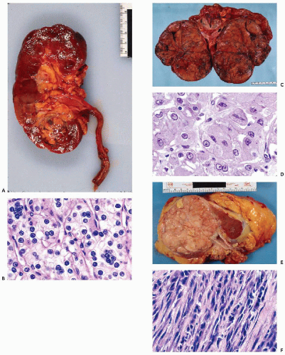
Figure 40-6 Conventional renal cell carcinoma (RCC). A. Gross picture of a 2-cm conventional RCC in the inferior pole of left kidney. The tumor was asymptomatic, an incidental finding on imaging. Note the golden-yellow cut surface. B. Histology: conventional RCC, clear cell type. Note the microtubule or acinar pattern and multiple nuclei in many cells. This is a grade 1 tumor. C. RCC with predominantly granular cells. The gross appearance is dark yellow to brick red. This large tumor (16 cm in diameter) presented as a left abdominal mass and flank pain. D. Histology of conventional RCC with predominantly granular cells. Note the presence of cytoplasmic vacuoles containing fat and glycogen. The nuclei are larger and some irregularly shaped, with visible nucleoli; therefore, this is a grade 2 tumor. E. Conventional RCC with predominantly sarcomatoid growth. The gross appearance is fleshy white. F. Histology of spindly (sarcomatoid) RCC.


The behavior of RCCs is unpredictable. In some patients, the primary tumor is occult and the patient is seen because of metastatic disease, commonly in the bone, lung, liver, or subcutaneous tissue. In other patients, metastases of RCC may occur many years after nephrectomy. Unexpected sites of metastases that can mimic a primary tumor are the thyroid, ovary, salivary gland, and, rarely, pancreas (see appropriate chapters). Finally, RCC in metastatic sites may express a morphologic pattern that is very different from the primary tumor, with the spindle-cell pattern being the most common. The recognition of metastatic renal cancer is often a diagnostic challenge.

A diagnostically valuable immunohistochemical feature of RCCs is the simultaneous expression of intermediate filaments for keratin (mainly types 8 and 18) and vimentin (Pitz et al, 1997). Although similar phenomena may occasionally be seen in other cancers, notably thyroid carcinoma, this feature is very helpful in determining the renal origin of metastatic foci in various organs (Domagala et al, 1988).


Gross Features

Surgically resected RCCs vary in size, from very small (2-3 cm in diameter) to very large (20 cm or more). The colors of the cut surface of the tumors show a reasonable correlation with histologic patterns. Thus, golden-yellow tumors (Fig. 40-6A,B) are usually composed of clear cells, brickred tumors (Fig. 40-6C,D) are granular or oncocytic, and sarcomatoid tumors are fleshy-white (Fig. 40-6E,F). The tumor may invade the calyces and the renal pelvis, in which case the voided urine cytology may be positive (see Chap. 23). RCCs have a tendency to invade the renal veins and thence the vena cava. Hemorrhage, extensive necrosis, and cavity formation within the tumor occur frequently.


Histology

Most renal carcinomas show a mixture of histologic and cytologic patterns of growth, as summarized in Table 40-4.

Although any tumor may be composed of more than one cell type, the most common renal carcinoma is the clear cell type with a classic growth pattern in solid sheets or cords. This is particularly true for relatively small tumors. Gland formation may occur. A rich network of capillaries and larger blood vessels is present. The cytoplasm of tumor cells contains lipid and glycogen, and the nucleus is disproportionately small (Fig. 40-6B).

In some of these tumors, the dominant type is a cancer cell with denser, more granular and eosinophilic cytoplasm, and more conspicuous nuclear abnormalities. These tumors were formerly classified as the granular cell type of RCC (Fig. 40-6D). They must be distinguished from chromophobe carcinoma, oncocytoma, and rare cases of CDC that may contain similar cells (see below).


TABLE 40-4 MORPHOLOGIC VARIANTS OF CONVENTIONAL RCC


Histologic Patterns


Acinar or glandular


Trabecular


Solid or alveolar


Pseudopapillary


Cell Types


Clear cell


Granular (oncocytic)


Spindly (sarcomatous)


Pleomorphic


Undifferentiated small cell


Some tumors are composed partly or wholly of spindly cancer cells (Fig. 40-6F), mimicking a variety of sarcomas (sarcomatous type of RCC). According to a large study by de Peralta-Venturina et al (2001), the sarcomatous change is most common in the conventional type of RCC, but it may also occur in other types of renal cancer, such as papillary, chromophobe, and collecting duct carcinomas. Many of the patients in that study had tumors of high stage, and their prognosis was less favorable than that of tumors without this component.

Some tumors may be composed partly or wholly of very poorly differentiated large or small cancer cells. Such poorly differentiated tumors are sometimes difficult to classify (see below).


Cytology

Aspirates of RCC often contain much blood and may show considerable necrosis with cell debris (the latter is particularly evident in tumors with cystic degeneration). Within this background, abundant tumor cells, often anchored to capillaries, are usually seen. The types of tumor cells observed in conventional RCC in FNA smears are listed in Table 40-4. It must be stressed that various types of cancer cells are often simultaneously present in the same patient. The cytologic classification of these tumors is usually based on the dominant cell type in smears, but is not always representative of the tumor type on histologic examination (Renshaw et al, 1997b). As mentioned above, a simultaneous expression of keratin and vimentin is very helpful in determining the renal origin of tumors (Domagala et al, 1988).

Aspirates of RCC usually show loosely cohesive flat groups of cancer cells with clear cytoplasm with poorly defined cell borders and many single cells. The tumor cells may also be seen anchored along capillaries, resulting in a pseudopapillary appearance (Fig. 40-7A). The clear cancer cells are large (much larger than benign tubular cells) and have abundant clear or faintly blue delicate cytoplasm
that is often filled with numerous small vacuoles containing lipid and glycogen but not mucin (Fig. 40-7B). The vacuoles are better seen in air-dried smears processed with hematologic stains. Phagocytosed hemosiderin may be seen. The nuclei are relatively small, but still much larger than those of benign tubular cells. They are only slightly pleomorphic, haphazardly placed, usually hyperchromatic, and contain readily visible nucleoli.


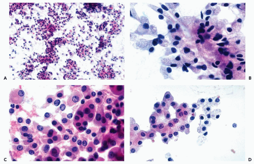
Figure 40-7 FNA appearance of conventional RCC with a mixture of clear and granular cells. A. Loosely cohesive flat groups of large cancer cells. Many single cells are present. B. High magnification reveals abundant, finely vacuolated cytoplasm and relatively small, hyperchromatic nuclei in large cancer cells of various configurations. C. Adjacent field shows predominantly granular cancer cells with eosinophilic cytoplasm. D. Another example of a renal smear showing side-by-side cancer cells with clear and granular cytoplasm.

In low-grade conventional RCCs, composed predominantly of clear cells, the FNA smears fixed in 95% ethanol may show numerous well-preserved nuclei stripped of cytoplasm (“naked” nuclei) (Fig. 40-8A,B). This phenomenon is caused by dissolution of the cytoplasmic lipids in alcohol, resulting in cytoplasmic disintegration. “Naked” nuclei may also be observed in metastatic renal cancers. Some of these nuclei contain prominent ruby-red nucleoli, consistent with renal origin.

In high-grade conventional RCCs, the malignant nature of the cells is usually quite evident (Fig. 40-8C,D). The cancer cells have large nuclei with prominent, sometimes ruby-red nucleoli, and relatively scanty cytoplasm that is either clear or eosinophilic and granular. It is usually easy to recognize such tumors, and the only point of differential diagnosis may be metastatic carcinomas, which have a different clinical and radiologic presentation.

As illustrated in Figure 40-7, most conventional RCCs show a mixture of clear cells and granular (oncocytic) cells in an ample FNA sample. The granular cancer cells have an eosinophilic, granular cytoplasm, are usually smaller and more uniform than the clear cells, and have larger and more atypical nuclei, and hence a higher N/C ratio. Cytoplasmic granules, reflecting numerous mitochondria, are well visualized in air-dried smears processed with hematologic stains. In H&E- or Papanicolaoustained smears the granules are intensely eosinophilic (Fig. 40-7C,D). In some cases, the granular cells are predominant, but so long as some clear cells are also present in the smears, the diagnosis of a conventional RCC is favored. The eosinophilic cells may resemble hepatocytes, which may be obtained in a misdirected FNA smear and mistaken for granular cells of RCC. Normal hepatocytes do not show the nuclear abnormalities seen in granular cancer cells. Lipofuscin and bile, when present in the cytoplasm, provide additional clues to the hepatic derivation of these cells. Weir and Pitman (1997) noted that the characteristic vascular pattern observed in
well-differentiated hepatomas was absent in RCC (see Chap. 38).


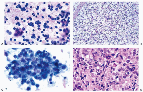
Figure 40-8 FNA of low- and high-grade renal carcinomas. A. FNA of a small RCC (shown in Fig. 40-6A) shows numerous well preserved “naked” nuclei, with some cells still retaining their eosinophilic granular cytoplasm. B. Histology of the same case shows a low-grade (grade 1) conventional RCC, clear cell type. C,D. Cells of a high-grade RCC with clear, scanty cytoplasm forming a cluster. B. Corresponding histology is from metastasis to the liver (core biopsy).

Somewhat similar cells may be observed in two relatively uncommon renal tumors: the oncocytoma and chromophobe renal carcinoma. Table 40-5 is a summary of salient clinical, cytologic, and ancillary characteristics of renal tumors with granular cytoplasm. The very rare collecting duct carcinoma is not included in this table. All of these tumors are discussed below. In addition, Reuter (1999) has reported that rare cells with granular cytoplasm may occur in angiomyolipoma and metastatic melanoma.

Among the uncommon cell types of conventional RCCs are undifferentiated cancer cells. This term implies the presence of clearly malignant cells of variable shapes and sizes with prominent nuclear abnormalities, but no specific features of RCCs. The cells occur singly or in very loosely structured aggregates, and are sometimes very large and multinucleated. In our experience, the pleomorphic cells with very large nuclei are relatively uncommon in renal FNA and always indicate the presence of a high-grade tumor (Fig. 40-9A). The interpretation of such cells depends on the company they keep. In the presence of at least some classic cancer cells with clear or granular cytoplasm, the most likely diagnosis is RCC. If the pleomorphic cells occur alone, the possibility of a metastatic carcinoma from an extrarenal site must be entertained.

In still other types of tumors, some of the undifferentiated cancer cells are quite small and occur singly and in small clusters (Fig. 40-9B). These cells, which have relatively large hyperchromatic nuclei and scanty, clear cytoplasm, may occur in conventional RCC and may be accompanied by clear or granular cancer cells. However, similar cells may also be found in Wilms’ tumor, metastatic small cell carcinoma, and a large cell lymphoma. The interpretation depends entirely on the smear pattern, which is quite different in each of these entities (see below).

In some conventional RCCs and in carcinomas composed of spindly cells, the cancer cells tend to be elongated or spindly and may be similar to fibroblasts. They have delicate, finely vacuolated, abundant, faintly granular cytoplasm and pleomorphic ovoid to elongated nuclei, usually with prominent nucleoli (Fig. 40-9C,D). Such cells correspond to areas of the primary tumor that are composed of elongated cells (see Fig. 40-6F), and may also occur in metastatic foci, mimicking a sarcoma (Koss et al, 1992).


TABLE 40-5 RENAL CELL TUMORS WITH GRANULAR CYTOPLASM


Oncocytoma


Chromophobe RCC (ChRCC)


RCC With Predominant Granular Cells


Relative incidence


3-7%


6-11%


Not known but common in higher grade lesions (all variants 60%)


Natural history


Essentially benign


Mortality 6%


Mortality 38%


Average tumor size and location


6 cm (central stellate scar in 1/3), often subcapsular


Large (9 cm), cortical


5.5 (recent series)-8 cm (older series); more common in upper pole


Smear pattern


Predominantly isolated cells or small aggregates


More aggregates
Some single cells


Loose clusters and single cells, bare nuclei


Cell size and cytoplasmic characteristics


Very large, uniform, low N/C ratio
Homogeneous, dense pink granular cytoplasm


Large, pleomorphic with high N/C ratio
Dense pink granular cytoplasm with perinuclear pale zones


Medium, uniform to pleomorphic, high N/C ratio.
Sometimes vacuolated pink granular cytoplasm


Nuclear features


Uniform, small and round (8-10 μm),
Finely granular chromatin
Inconspicuous nucleoli
No mitoses


Uniform to pleomorphic (10-15 μm)
Notched nuclear contour
Coarse chromatin
Prominent nucleoli


Large (15-20 μm) relatively uniform
Fine chromatin, large nucleoli


Ancillary tests


Stain for lipid and glycogen negative
EMA positive
LMW keratin positive
Vimentin negative


Hale’s colloidal iron stain positive
EMA positive
LMW keratin negative
Vimentin negative


Stain for lipid and glycogen positive in some cells
EMA positive
LMW keratin negative
Vimentin positive


EMA, epithelial membrane antigen; LMW, low molecular weight; N/C ratio, necleocytoplasmic ratio.


Variants of Conventional Renal Cell Carcinoma


Multilocular Cystic Renal Cell Carcinoma

Approximately 3% of conventional RCCs are multicystic. Adults on long-term dialysis constitute a high-risk group (Truong et al, 1995). The gross appearance of these tumors is that of a multiloculated cystic mass, separated from the renal parenchyma by a thick fibrous capsule. The cysts are variable in size and separated from each other by thin fibrous septa. The cyst contents are serous or bloody, and are either fluid or clotted. Golden-yellow rims of tumor may be visible in some cyst walls, but there are no visible tumor masses (Fig. 40-10A).

In histologic sections the cysts are lined by one or more layers of neoplastic epithelial cells, occasionally with papillary tufting. The golden-yellow portions of the cyst wall usually show a typical RCC composed of clear cancer cells (Fig. 40-10B). Nonneoplastic stroma shows numerous hemosiderin-laden macrophages.


Cytology

The FNAs yield a variable amount of fluid. The smears are sparsely cellular and may show macrophages and leukocytes. The malignant cells are often reduced to bare nuclei with prominent nucleoli or uniform small cells with clear cytoplasm and bland nuclei (Fig. 40-10C). In sparsely cellular specimens with uniform tumor cells, a diagnosis may be difficult to establish (Kini, 1999). The prognosis is generally excellent (Koga et al, 2000).


Renal Cortical Adenomas Versus Small Renal Cell Carcinomas

Renal epithelial tumors measuring less than 3 cm in diameter are, as a rule, an incidental finding. The tumors are generally benign and for many years have been designated as adenomas. However, as early as 1938, Bell (1938) observed that renal tumors measuring less than 3 cm in diameter are sometimes capable of forming metastases. The present consensus among urologists and pathologists is that these tumors represent small RCCs. The small tumors are either composed of clear cells or they show packed papillary structures lined by cuboidal cells. There are no light-microscopic, immunohistochemical, or electron-microscopic features to distinguish them from conventional RCC of the clear cell type.


Cytology

Because of their small size, these tumors are rarely aspirated. The cytologic presentation is identical to that of a low-grade conventional (clear cell variant) of RCC, as described above.


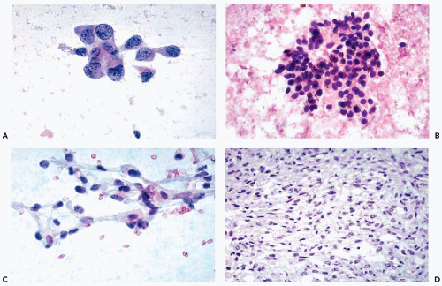
Figure 40-9 A. Renal carcinoma with pleomorphic cells. Pleomorphic cancer cells with abundant cytoplasm and large irregular nuclei. Such cells are usually observed in very high-grade tumors, but may also represent metastatic cancer. B. Conventional RCC with small cells. Undifferentiated small cells with scanty clear cytoplasm. The presence of phagocytosed hemosiderin is in favor of an RCC. C,D. Spindle cell carcinoma of the kidney. C. Elongated cancer cells with clear cytoplasm. Although the nuclei are hyperchromatic and of variable sizes, they are not conspicuous. D. Corresponding histology. Such findings are not uncommon in metastatic sites, where they may be mistaken for a sarcoma.


Rare Variants

Unger et al (1993) described a variant of RCC with eosinophilic cytoplasmic globules or inclusions, which stained magenta in Diff-Quik-stained FNA, in otherwise classic clear cancer cells. The inclusions were thought to represent a form of glycogen. Hirokawa et al (1998) described a conventional RCC with melanin-like pigment in the cytoplasm of cancer cells.


Papillary Renal Cell Carcinoma (PRCC or Chromophil Carcinoma)

Papillary RCC (PRCC) represents 7% to 14% of primary epithelial renal tumors. The mean age at the time of diagnosis (61.8 years) is similar to that for conventional RCC. The ratio of males to females is 1.8 to 1. The tumors are frequently multifocal and occasionally bilateral (Amin et al, 1997). The tumors, which are of variable sizes at the time of diagnosis, are often small and located in the poles of the kidneys, which permits surgical treatment by partial nephrectomy (Lager et al, 1995; Renshaw and Corless, 1995). This has been our experience also. These tumors are usually hypovascular on angiography, but often show intramural hemorrhage (Fig. 40-11A). Genetic data document that PRCC is a distinct entity separate from the conventional RCC (see Table 40-2).

It has been known for many years that papillary carcinoma of the kidney carries a much better prognosis than conventional renal carcinoma (Koss et al, 1992). In a large study, Amin et al (1997) reported an overall 5-year survival rate for PRCC of about 90%, which is much higher than that of conventional RCC (estimated at about 50%). The 5-year survival of patients with stage 1 papillary carcinoma is close to 100% (Robson et al, 1969).


Histology

The hallmark of this tumor is the formation of papillae with wide cores that are filled with large foamy macrophages (Fig. 40-11B). Delahunt and Eble (1997) observed two types of epithelial cells surfacing the papillae. In about two thirds of the cases, the surface of the papillae was formed by small epithelial cells with pale cytoplasm and oval nuclei. In the remaining cases, the papillae were surfaced by large eosinophilic or basophilic (chromophil) epithelial cancer cells with large nuclei, and hence a high N/C ratio. The nuclear texture is generally finely granular, and enlarged
nucleoli of moderate sizes are often present. The neoplastic cells often contain large amounts of ingested hemosiderin (Fig. 40-10). Psammoma bodies are not uncommon, particularly in tumors with large cells. Solid variants of PRCC have been described based on cytogenetic findings (Renshaw et al, 1997).


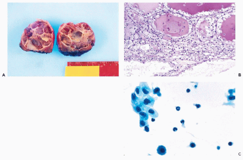
Figure 40-10 Multilocular cystic RCC. A. Gross appearance of a multilocular cystic RCC. B. Histologic sections of light-yellow areas show a grade 1 conventional RCC with cystic changes. FNA of multilocular RCC may be unrewarding (see text). C. Sparse tumor cells with clear and vacuolated cytoplasm in a background of macrophages and leukocytes.


Cytology

The cytologic presentation of papillary carcinoma in FNA is quite characteristic and corresponds to the two subtypes described by Delahunt and Eble (1997). In both subtypes, the smears contain a large number of foamy macrophages with finely vacuolated cytoplasm and small nuclei derived from the disrupted cores of the papillary fronds. The macrophages form a unique background wherein cancer cells, occurring singly or in small papillary clusters, can be observed (Fig. 40-11C). Two types of cancer cells may be observed. In most cases, the cells are small and cuboidal, and are provided with a delicate cytoplasm and relatively small nuclei with prominent nucleoli (Fig. 40-11C). In other cases, the cancer cells are larger and approximately spherical, with eosinophilic cytoplasm; crisp cytoplasmic borders; large, somewhat hyperchromatic, roughly spherical nuclei; and clearly visible nucleoli of various sizes often arranged in papillary fronds (Fig. 40-11D). Cancer cells containing brown hemosiderin crystals are common (Fig. 40-11D) and, along with macrophages, constitute a valuable clue to the diagnosis of PRCC. Psammoma bodies occur from time to time but are not a constant finding (Dekmezian et al, 1991).

The primary differential diagnosis is with conventional RCC that may sometimes exhibit a pseudopapillary growth pattern. The homogenous rather than vacuolated cytoplasm of the tumor cells, and the presence of large macrophages, hemosiderin, and occasional psammoma bodies are unique to papillary tumors and allow for an easy distinction to be made in most cases.


Chromophobe Renal Cell Carcinoma

Thoenes et al (1985, 1988) recognized this relatively uncommon variant of renal epithelial tumors and illustrated its morphologic, histochemical, and ultrastructural features (see Table 40-5). The tumor has a unique genetic make-up (see Table 40-2). The age and sex distributions are virtually identical to those seen in conventional RCCs. Most patients with this tumor (60%) are asymptomatic, approximately 30% have a palpable mass, and a minority have hematuria. Stage for stage, ChRCCs carry a significantly better prognosis than conventional RCCs.

Grossly, these are large tumors (9 cm in diameter, on average), usually located in the renal cortex. The cut surface is lobulated and gray-brown. Microscopically, the tumor is usually composed of large tumor cells with relatively small nuclei (10-15 μm in diameter), forming solid nests separated by delicate fibroconnective tissue septa (Fig. 40-12A). The cell membrane is well defined, and the cytoplasm
in most cases is very pale and transparent (typical chromophobe tumors) or, in some cases, eosinophilic and granular (Fig. 40-12D).


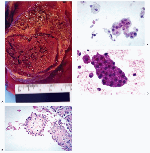
Figure 40-11 Papillary RCC (PRCC). A. Large PRCC with intramural hemorrhage. The upper portion of the tumor with a yellowtan appearance showed abundant foamy macrophages. B. Histology reveals neoplastic papillary fronds with large foamy macrophages in the center. C. Aspiration smear of a papillary renal carcinoma showing a large papillary cluster of epithelial cells with large, bland nuclei and tiny nucleoli. There are numerous large macrophages in the background. D. Numerous hemosiderin crystals are seen in this papillary frond of neoplastic cells from another case.

Ultrastructurally, the tumor cells are rich in cytoplasmic vesicles that contain acidic mucins, which accounts for the positive stain of the cytoplasm with Hale’s colloidal iron reaction. In tumor cells with granular cytoplasm, the density of mitochondria is increased (Thoenes et al, 1988). ChRCCs with eosinophilic (acidophilic), granular cytoplasm may be confused with oncocytoma (see below) and, to a lesser extent, with granular cell variants of conventional RCC. In contrast to the common RCCs, however, ChRCCs do not react with vimentin (Thoenes et al, 1988).


Cytology

These large tumor cells of variable configuration have abundant cytoplasm that is clear or granular in the center of the cell, and condensed at the periphery (Fig. 40-12B). Recognizing this cytoplasmic feature is the key to the diagnosis. The perinuclear pale zone is somewhat reminiscent of koilocytes, which are observed in squamous cells in cervical smears from patients infected with human papillomavirus (see Chap. 11). The eosinophilic or granular variant resembles oncocytes or hepatocytes. The nuclei are often peripheral in location, have irregular outlines, and vary somewhat in size but seldom exceed 15 μm in diameter (Fig. 40-12B,C). Binucleation is common. Although the nucleoli are visible in many cells, they are not conspicuous.
Characteristically, the cells stain strongly with Hale’s colloidal iron, express epithelial membrane antigen (EMA), and are vimentin-negative (see Table 40-5).


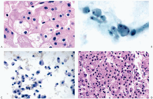
Figure 40-12 Chromophobe RCC (ChRCC). A. Histology of the classic ChRCC. Note that the pink, granular cytoplasm is condensed at the periphery, with many cells showing perinuclear clearing. B. Cytology of ChRCC. The cells show abundant fluffy cytoplasm with central clearing and peripheral nuclei. C. Eosinophilic variant of ChRCC metastatic to the liver. Note the cells with peripheral nuclei. D. Cell block of the same case. (C: Pap stain, × 400.)


Oncocytomas

Although renal oncocytomas are essentially benign, they are sufficiently similar to renal carcinomas to be described here rather than with benign lesions. This tumor was first described by Zippel (1942) and was largely ignored until the publication of 13 cases by Klein and Valensi (1976). Additional cases were described by Lieber et al (1981). It is estimated that oncocytomas comprise 3% to 7% of all primary renal neoplasms (Reuter and Gaudin, 1999). The majority of the patients are asymptomatic, and the tumor is discovered during workups for other, unrelated conditions. Most series show a wide age distribution, with peak incidence in the seventh decade of life. Men are affected nearly twice as often as women (Amin et al, 1997; Perez-Ordonez et al, 1997).

On angiography, oncocytomas show an avascular or hypovascular core, presumably secondary to the central stellate scars that are observed in about one third of the cases. The average tumor size is 6 cm. The cut surface reveals an encapsulated, uniform, mahogany-brown tumor. Necrosis and hemorrhage are absent (Fig. 40-13A).

The most common genetic abnormality is loss of chromosome 1 and Y, which is also seen in ChRCC (see Table 40-2). Both tumors are thought to arise from intercalated cells of the renal cortex, and may show striking gross and microscopic similarities (see Table 40-5).

As with oncocytic tumors of other organs, such as the salivary glands, thyroid, or parathyroid, renal oncocytomas are composed of large cells with granular, eosinophilic cytoplasm devoid of vacuoles and supported by delicate fibroconnective tissue septa. The nuclei are small, round, and uniform (Fig. 40-13B). Of all the renal tumors with eosinophilic cytoplasm (see Table 40-5), oncocytomas have the smallest nuclei, measuring 8-10 μm in diameter. Mitotic figures and papillary features are absent. Microcysts filled with blood may be present. Ultrastructurally, the cytoplasm of tumor cells is packed with mitochondria to the virtual exclusion of any organelles other than a few lysosomes. Thus, electron microscopy remains the gold standard for confirming this diagnosis. Stains for lipid and glycogen are negative.

If strict diagnostic criteria are followed and the diagnosis of oncocytoma is limited to tumors composed exclusively
of well-differentiated oncocytes with small nuclei, the prognosis is excellent and the tumor is considered to be benign. However, there have been reports that at least some tumors may invade the parenchyma of the kidney and may recur (Rodriguez et al, 1980; Lieber et al, 1981). For tumors diagnosed as oncocytomas that produce metastases, the possibility of misclassification must be considered. As an example, we recently reviewed a case that was initially classified as oncocytoma and was resected in 1985. Two years later, a mass in the liver was discovered and aspirated. The FNA smears strongly suggested that the cells represented metastatic renal tumor. The “oncocytoma” from 1985 was reclassified as ChRCC (Fig. 40-12A,B). Also, oncocytomas may be associated with conventional RCC (Klein and Valensi, 1976; Fromowitz and Bard, 1990).


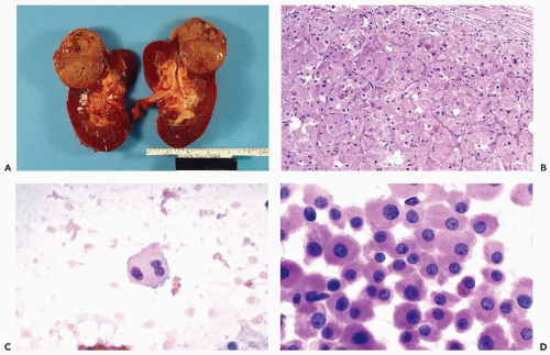
Figure 40-13 Oncocytoma. A. Gross appearance of an oncocytoma. The tumor is often subcapsular, uniformly tan to mahogany brown, and may show blood-filled microcysts but no necrosis or hemorrhage. B. Histology of the oncocytoma: very large uniform cells with dense, pink, granular cytoplasm and disproportionately small, uniform nuclei. The cells are supported by delicate fibroconnective tissue. C,D. Oncocytomas in aspiration biopsies. Note the faintly granular, eosinophilic granular cytoplasm and small nuclei.


Cytology

As first described by Rodriguez et al (1980), the FNA smears contain a uniform population of large eosinophilic granular cells (oncocytes) that are either isolated or form small, loose aggregates. The cell borders are well defined and the nuclei are quite small and round, with finely granular chromatin and inconspicuous, tiny nucleoli (Fig. 40-13C,D). Although oncocytic-type cells may also occur in the chromophobe or granular cell types of conventional RCC, these two lesions are characterized by significant nuclear abnormalities, including large nucleoli that are not observed in oncocytoma (Wiatrowska and Zakowski, 1999) (see Table 40-5).


Collecting Duct Carcinoma (CDC)

Collecting duct carcinomas (also known as carcinomas of the ducts of Bellini) are very rare, aggressive renal tumors. They account for less than 1% of malignant renal tumors, and are thought to arise within the collecting ducts of the renal medulla. Cytogenetic findings support the concept that the CDC is a distinct entity (Schoenberg et al, 1995) (see Table 40-2). CDCs are usually located in the medulla of the kidney, adjacent to the renal pelvis, and tend to develop in younger patients with symptoms of hematuria, pain, and weight loss, and the presence of a palpable mass. Fifty percent of these patients have metastatic disease to the lymph nodes, bone, and viscera at the time of diagnosis.

In tissue sections, the tumors are composed of neoplastic ducts and tubules lined by clearly malignant cells, some
of which have granular cytoplasm (Fig. 40-14A), and of nests of clear cells embedded in fibroblastic stroma that is sometimes very dense. Mucin may be present in some of the glandular structures. Adjacent renal tubules may show dysplastic changes (Kennedy et al, 1990), and psammoma bodies may be present.


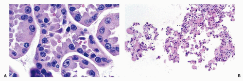
Figure 40-14 Histology of collecting (Bellini) duct carcinoma. A. Neoplastic ducts/tubules are lined by cancer cells with eosinophilic cytoplasm and large, hyperchromatic nuclei with prominent nucleoli. Mucin vacuoles may be seen. B. Cell block of FNA of medullary carcinoma. The microscopic presentation is not specific.


Cytology

Because they are located in the vicinity of the renal pelvis, CDCs may be recognized in urinary sediment (Mauri et al, 1994; Caraway et al, 1995), retrograde brushings of the renal pelvis (Zaman et al, 1996), and transcutaneous FNAs (Layfield, 1994; Caraway et al, 1995; Ono et al, 2000). The neoplastic cells are medium-sized and uniform, with scanty eosinophilic granular or vacuolated cytoplasm, very large hyperchromatic nuclei, and very large multiple nucleoli (Layfield, 1994

Only gold members can continue reading. Log In or Register to continue

Stay updated, free articles. Join our Telegram channel

Jun 8, 2016 | Posted by in PATHOLOGY & LABORATORY MEDICINE | Comments Off on The Kidneys, Adrenals, and Retroperitoneum

Full access? Get Clinical Tree

Get Clinical Tree app for offline access