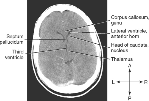
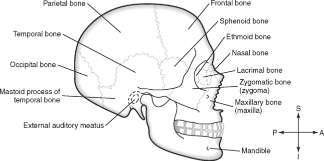
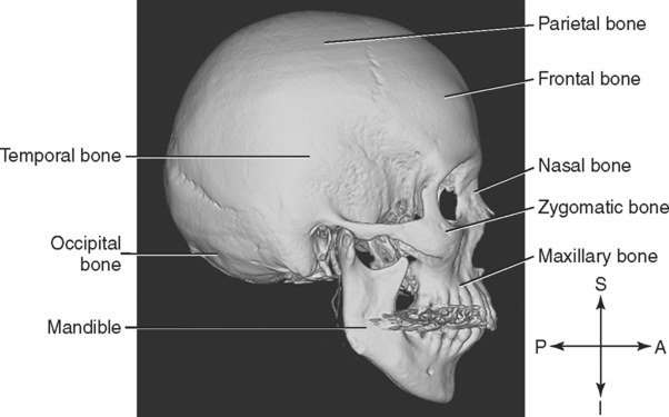
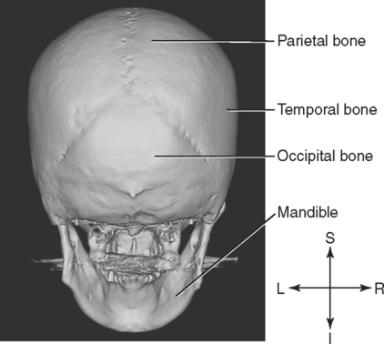
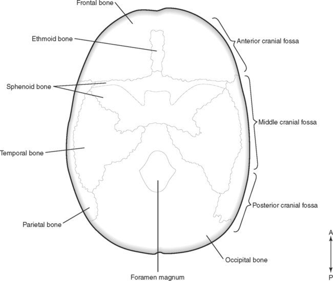
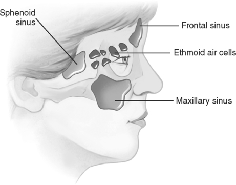
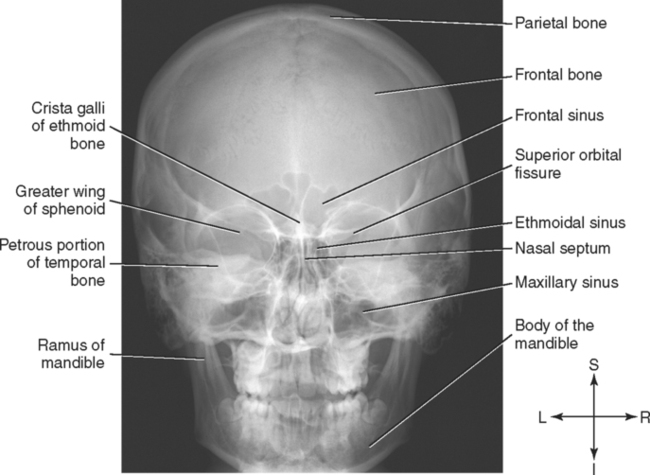
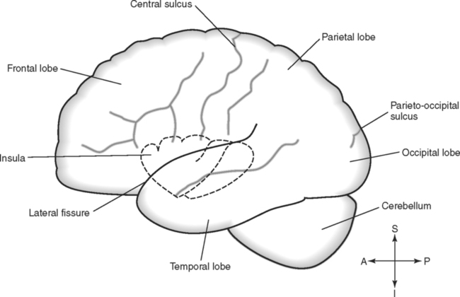
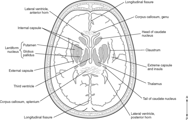
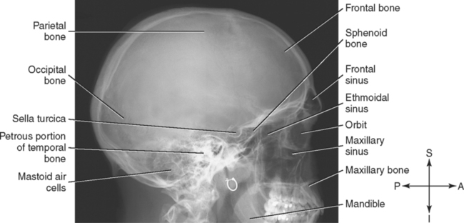
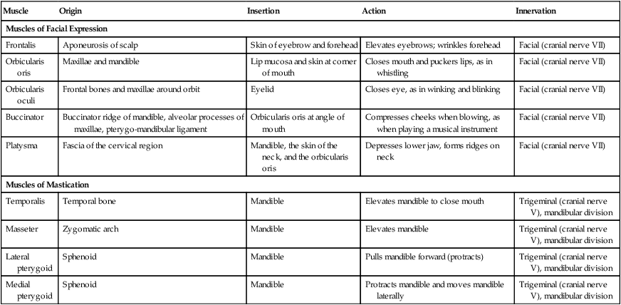
Upon completion of this chapter, the student should be able to do the following: • Name the bones of the cranium and face. • Identify the four paranasal sinuses. • Name five muscles of facial expression, describe the location of each, and state the insertion and innervation of this group. • Name four muscles of mastication, describe the location of each muscle, and state the insertion and innervation of this muscle group. • Compare the location and relationships of the three salivary glands. • Identify the five lobes of the cerebrum. • Describe the relationships of the basal ganglia. • Describe the location and structure of the diencephalon. • Locate the components of the brainstem. • Compare the cerebrum and cerebellum with respect to size, appearance, location, and structure. • Trace the flow of cerebrospinal fluid through the ventricles in the brain. • Describe the three layers of meninges. • Identify six subarachnoid cisterns by describing their location and significance. • Describe the arterial blood supply to the brain. • Identify the major venous sinuses that return blood from the brain to the internal jugular vein. • Name the 12 cranial nerves and the foramen that serves as a passageway for each and the functions of each nerve. • Describe the structure of the eye, including the bulbus oculi, musculature, vascular supply, and protective features. • Discuss the relationships of the internal jugular vein, external jugular vein, internal carotid artery, and external carotid artery to each other and to other surrounding structures, such as the parotid gland and the sternocleidomastoid muscle. The bony framework of the head is called the skull. This is the most complex osseous structure of the body, and it consists of 22 bones connected by immovable joints, called sutures. For descriptive purposes, these bones are divided into the cranium and the facial skeleton, although no distinct line of demarcation separates the two parts. Some of the bones surround a large cranial cavity that contains the brain. The superior surface of this region is covered by the scalp. Some skull bones adjacent to the nasal cavity contain air-filled spaces, called paranasal sinuses. Many of the bones in the skull have holes or openings, called foramina, that serve as passageways for nerves and blood vessels. In addition to the skull, seven other bones are associated with the head; these are the auditory ossicles and the hyoid bone. The osseous components of the head are summarized in Table 5-1 TABLE 5-1 Summary of Bones Associated With Head The eight bones of the cranium surround the cranial cavity, which houses the brain. The single frontal bone forms the forehead and superior part of the orbit of the eye. It contains the frontal sinuses, which communicate with the nasal cavity. Two parietal bones form most of the top of the cranium. Two temporal bones form a portion of the sides and base, or floor, of the cranium. Each temporal bone has a thin, flat, squamous portion that forms the inferior-lateral part of the cranium. The posterior portion of the temporal bone is the mastoid process. The external auditory canal (meatus), the tympanic membrane, the middle ear, and the inner ear are located in the petrous portion, which extends medially to form part of the base of the cranium. The zygomatic process of the temporal bone extends anteriorly to join the zygomatic bone to form the zygomatic arch. The single ethmoid bone is located between the eyes and forms most of the medial wall of each orbit. The superior surface of the ethmoid bone forms a part of the base of the cranial cavity and the roof of the nasal cavities. A thin, perpendicular plate of the ethmoid bone extends inferiorly to form part of the nasal septum. The superior and middle conchae (turbinates) in the nasal cavity are part of the ethmoid bone. The single sphenoid bone lies at the base of the skull anterior to the temporal bones. Often described as bat shaped, the sphenoid bone has “wings” that form the anterior-lateral portion of the cranium and the lateral walls of the orbits. The sella turcica is found in the center of the bone and is the location of the pituitary gland. The single occipital bone forms the posterior portion and part of the base of the cranium. It has a large hole, the foramen magnum, for passage of the spinal cord and the vertebral vessels. The bones of the cranium are illustrated in Figs. 5-1 to 5-4. The cranial cavity, a large space formed by the eight cranial bones, contains the brain. The floor of the cranial cavity is subdivided into anterior, middle, and posterior cranial fossae (FIG. 5-5). The anterior cranial fossa is formed by portions of the ethmoid, sphenoid, and frontal bones. This fossa houses the frontal lobes of the brain. The floor of the middle cranial fossa is composed of the body and greater wings of the sphenoid and the squamosal and petrous portions of the temporal bones. This region contains the temporal lobes of the brain. The posterior cranial fossa comprises the remainder of the cranial cavity and is formed by parts of the sphenoid, temporal, and occipital bones. This region contains the cerebellum, the pons, and the medulla oblongata. The inferior-most portion of the posterior cranial fossa shows the large foramen magnum, through which the spinal cord passes. The facial portion of the skull consists of 14 bones. Some of these bones are illustrated in Figs. 5-1, 5-2, and 5-4. The two maxillae (singular, maxilla) unite in the midline to form the upper jaw. A horizontal piece of each maxilla, the palatine process, forms the anterior portion of the roof of the mouth, which is the hard palate. If the palatine processes of the two maxillae fail to join during prenatal development, a cleft palate results. Each maxilla contains a maxillary sinus, which is a large air space that communicates with the nasal cavity. Two palatine bones form the posterior portion of the hard palate. Two zygomatic bones, one on each side, form the prominence of the cheek and the lateral margin of the orbit. A process of the zygomatic bone extends posteriorly and unites with the temporal bone to form the zygomatic arch. Two small, rectangular nasal bones join in the midline to form the bridge of the nose. Fractures of these bones are common facial injuries. The paranasal sinuses are air-filled spaces in some of the bones adjacent to the nasal cavity. The four sets of paranasal sinuses are named according to the bone in which they are located; they are the frontal sinus, ethmoid sinus, sphenoid sinus, and maxillary sinus. Usually developing after birth as outgrowths of the nasal cavity, the sinuses retain their original openings so that their secretions drain into the nasal cavity. The mucous membrane that lines the sinuses is continuous with the mucosa of the nasal cavity, but it is thinner and less vascular than the nasal mucosa. Fig. 5-6 illustrates the paranasal sinuses. Small maxillary sinuses, which are located in the bodies of the maxillae, are present in the newborn and grow slowly until the child reaches puberty. The accelerated development of the maxillary sinuses during adolescence contributes to the apparent change in facial features that typically occurs during this period. When fully developed, the maxillary sinuses are the largest of the paranasal sinuses. The maxillary sinus drains into the nasal cavity by way of a relatively long hiatus semilunaris, which opens into the middle meatus. A couple of factors hinder the drainage of this sinus: the hiatus traverses a superior direction when the body is erect, thus necessitating drainage “against gravity,” and the opening from the sinus into the hiatus is in a superior location. The drainage problem is further complicated because the maxillary sinus is the most inferiorly located of the paranasal sinuses, and communicating channels allow the other sinuses to drain into the maxillary sinus. For these reasons, the maxillary sinus is the sinus most often involved in infections. The frontal, ethmoid, and sphenoid sinuses are innervated by branches of the ophthalmic division of the trigeminal nerve, which is the fifth cranial nerve. The maxillary sinus is innervated by branches of the maxillary division of the fifth cranial nerve. (The cranial nerves are discussed later in the chapter.) Sinuses and other features of the skeleton of the head are illustrated by the radiographs in Figs. 5-7 and 5-8. Foramina are openings in bones that serve as passageways for nerves and blood vessels. The vessels that transport blood and the nerves that carry impulses must pass through the sutured, helmetlike bones of the skull on their way to and from the brain; therefore these bones have numerous foramina. Table 5-2 provides a summary of the foramina of the skull. TABLE 5-2 The ear has three chambers: the inner ear, the middle ear, and the external ear. The ear ossicles, the malleus, incus, and stapes, are located in the middle ear chamber in the petrous portion of the temporal bone. The ossicles transmit and amplify sound waves through the middle ear. The single hyoid bone is located in the neck just superior to the larynx. The hyoid is unique because it does not attach directly to any other bone; instead, it is suspended by ligaments. Muscles associated with the hyoid bone are described in Chapter 6. Numerous muscles are located in the head, many of them small and difficult to separate from adjacent muscles. They are even more difficult to isolate by imaging techniques. These muscles have functional significance because they deal with facial expression and the chewing of food. Only the larger and more significant muscles are presented in this text (Table 5-3). TABLE 5-3 Muscles Associated With Facial Expression and Mastication The four muscles of mastication provide chewing movements by acting on the temporomandibular joint to move the mandible (see Table 5-3). All insert on the mandible and are innervated by the mandibular division of the fifth cranial nerve. They are quite readily seen on transverse sections. The fan-shaped temporalis muscle covers the squamosal portion of the temporal bone and is a powerful muscle used to close the mouth by elevating the mandible. The masseter muscle, or chewing muscle, is located on the lateral aspect of the ramus of the mandible. Both the lateral and medial pterygoid muscles originate on the lateral pterygoid plate of the sphenoid bone and insert on the medial surface of the mandible. When these muscles are observed in transverse sections, the temporalis is seen in the more superior sections. In lower sections, starting at the lateral surface and in sequence, the masseter, the ramus of the mandible, the lateral pterygoid, and the medial pterygoid are seen. Superficially, the cerebrum is divided into lobes. A central sulcus separates the frontal lobe from the parietal lobe. Posteriorly, the parieto–occipital sulcus separates the parietal lobe from the occipital lobe. Laterally, the temporal lobe is situated below the lateral fissure (sulcus). A fifth lobe, called the insula, or island of Reil, is located deep within the lateral fissure. The lobes and fissures of the brain are illustrated in Fig. 5-9. Scattered throughout the white matter are distinct regions of gray matter called basal ganglia. Two of the larger basal ganglia are the caudate nucleus and the lentiform nucleus, or lenticular nucleus. In sectional anatomy, the caudate nucleus is usually visualized in association with the lateral ventricle. The lentiform nucleus is rather centrally located in each cerebral hemisphere. It is subdivided into the lateral, or external, putamen, and the medial, or internal, globus pallidus. The claustrum, another of the basal ganglia, is a thin layer of gray matter just lateral to the lentiform nucleus and deep to the cortex of the insula. A band of white matter that is medial to the lentiform nucleus is called the internal capsule. The white matter that is present between the lentiform nucleus and the claustrum is the external capsule. The claustrum is separated from the insula by the extreme capsule. Because of their appearance, the caudate nucleus, the internal capsule, and the lentiform nucleus are sometimes referred to as the corpus striatum. Fig. 5-10 illustrates the arrangement of the basal ganglia in a transverse section. The axial computed tomography (CT) image in Fig. 5-11 shows some of the features of the cerebrum and basal ganglia.
The Head
Anatomical Review of the Head
Osseous Components
Bone
Number
Total
Bones of the Cranium
8
Frontal
1
Parietal
2
Occipital
1
Temporal
2
Sphenoid
1
Ethmoid
1
Bones of the Face
14
Maxillae
2
Nasal
2
Zygomatic
2
Lacrimal
2
Mandible
1
Vomer
1
Inferior nasal conchae
2
Palatine
2
Middle Ear Bones
6
Malleus
2
Incus
2
Stapes
2
Hyoid Bone
1
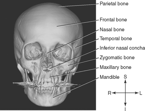
Cranium.
Face.
Paranasal Sinuses.
Foramina of the Skull.
Foramen
Location
Structures Transmitted
Carotid canal
Temporal bone
Internal carotid artery; sympathetic nerves
Hypoglossal canal
Occipital bone
Hypoglossal nerve
Infraorbital
Maxilla
Maxillary branch of trigeminal nerve; infraorbital nerve and artery
Internal auditory meatus
Temporal bone
Vestibulocochlear nerve
Jugular
Temporal bone
Internal jugular vein; vagus, glossopharyngeal, and spinal accessory nerves
Magnum
Occipital bone
Medulla oblongata/spinal cord; accessory nerves; vertebral arteries
Nasolacrimal canal
Lacrimal bone
Nasolacrimal (tear) duct
Olfactory
Ethmoid bone
Olfactory nerves
Optic
Sphenoid bone
Optic nerve; central artery and vein of retina
Ovale
Sphenoid bone
Mandibular branch of trigeminal nerve
Rotundum
Sphenoid bone
Maxillary branch of trigeminal nerve
Stylomastoid
Temporal bone
Facial nerve
Additional Bones Associated With the Skull.
Muscular Components
Muscle
Origin
Insertion
Action
Innervation
Muscles of Facial Expression
Frontalis
Aponeurosis of scalp
Skin of eyebrow and forehead
Elevates eyebrows; wrinkles forehead
Facial (cranial nerve VII)
Orbicularis oris
Maxillae and mandible
Lip mucosa and skin at corner of mouth
Closes mouth and puckers lips, as in whistling
Facial (cranial nerve VII)
Orbicularis oculi
Frontal bones and maxillae around orbit
Eyelid
Closes eye, as in winking and blinking
Facial (cranial nerve VII)
Buccinator
Buccinator ridge of mandible, alveolar processes of maxillae, pterygo-mandibular ligament
Orbicularis oris at angle of mouth
Compresses cheeks when blowing, as when playing a musical instrument
Facial (cranial nerve VII)
Platysma
Fascia of the cervical region
Mandible, the skin of the neck, and the orbicularis oris
Depresses lower jaw, forms ridges on neck
Facial (cranial nerve VII)
Muscles of Mastication
Temporalis
Temporal bone
Mandible
Elevates mandible to close mouth
Trigeminal (cranial nerve V), mandibular division
Masseter
Zygomatic arch
Mandible
Elevates mandible
Trigeminal (cranial nerve V), mandibular division
Lateral pterygoid
Sphenoid
Mandible
Pulls mandible forward (protracts)
Trigeminal (cranial nerve V), mandibular division
Medial pterygoid
Sphenoid
Mandible
Protracts mandible and moves mandible laterally
Trigeminal (cranial nerve V), mandibular division
Muscles of Mastication.
Brain
Regions of the Brain.
Cerebrum.