1,
It should be pointed out here that there is only limited evidence-based information concerning the validity of clinical signs. Many parts of the physical examination are performed as a matter of tradition. As students develop their examination skills, experience and new evidence-based data will help them refine their use of examination techniques. We have included information about the established usefulness of signs where it is available, but have also included signs that students will be expected to know about despite their unproven value.
For clinical viva voce (with live voice) examinations and objective structured clinical examinations (OSCEs), the examiners expect all candidates to have a polished and thorough examination method.
This formal approach to the physical examination leads to the examination of the parts of the body by body system. For example, examination of the cardiovascular system, which includes the heart and all the major accessible blood vessels, begins with positioning the patient correctly. This is followed by a quick general inspection and then, rather surprisingly for the uninitiated, seemingly prolonged study of the patient’s fingernails. From there, a set series of manoeuvres brings the doctor to the heart. This type of approach applies to all major systems, and is designed to discover peripheral signs of disease in the system under scrutiny. The attention of the examining doctor is directed particularly towards those systems identified in the history as possibly being diseased, but of course proper physical examination requires that all the systems be examined.
The danger of a systematic approach is that time is not taken to stand back and look at the patient’s general appearance, which may give many clues to the diagnosis. Doctors must be observant, like a detective (Conan Doyle based his character Sherlock Holmes on an outstanding Scottish surgeon).
Diagnosis has been defined as ‘the crucial process that labels patients and classifies their illnesses, that identifies (and sometimes seals) their likely fates or prognoses and that propels us towards specific treatments in the confidence (often unfounded) that they will do more good than harm’.
In normal clinical practice, the detail of the physical examination performed will be ‘targeted’ and will depend on clues from the history and whether the consultation is a follow-up or new consultation. Students however must know how to perform a complete examination of the body systems even though they will not often perform this in practice (except perhaps during examinations).
First impressions
First impressions of a patient’s condition must be deliberately sought; they cannot be passively acquired. Make a conscious point of assessing the patient’s general condition right at the start. The specific changes that occur in particular illnesses (e.g. myxoedema) will be discussed in detail in the appropriate chapters. However, certain abnormalities should be obvious to the trained or training doctor.
First, decide how sick the patient seems to be: that is, does he or she look generally ill or well? The cheerful person sitting up in bed reading Proust (
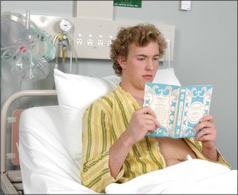
When a patient walks into the consulting room or undresses for the examination, there is an opportunity to look for problems with mobility and breathlessness.
Apart from gaining a general impression of a patient’s state of health, certain general physical signs must be sought.
Vital signs
Certain important measurements must be made during the assessment of the patient. These relate primarily to cardiac and respiratory function, and include pulse, blood pressure, temperature and respiratory rate. For example, an increasing respiratory rate has been shown to be an accurate predictor of respiratory failure.
The vital signs must be assessed at once if a patient appears unwell. Patients in hospital have these measurements taken regularly and charted. They provide important basic physiological information.
Facies
A specific diagnosis can sometimes be made by inspecting the face, its appearance giving a clue to the likely diagnosis. Other physical signs must usually be sought to confirm the diagnosis. Some facial characteristics are so typical of certain diseases that they immediately suggest the diagnosis, and are called the diagnostic facies (
TABLE 3.1 Some important diagnostic facies
| Amiodarone (anti-arrhythmic drug)—deep blue discoloration around malar area and nose |
| Acromegalic ( |
| Cushingoid ( |
| Down syndrome ( |
| Hippocratic (advanced peritonitis)—eyes are sunken, temples collapsed, nose is pinched with crusts on the lips and the forehead is clammy |
| Marfanoid ( |
| Mitral ( |
| Myopathic ( |
| Myotonic ( |
| Myxoedematous (prolonged hypothyroidism) ( |
| Pagetic ( |
| Parkinsonian ( |
| Ricketic ( |
| Thyrotoxic ( |
| Turner’s syndrome ( |
| Uraemic ( |
| Virile facies ( |
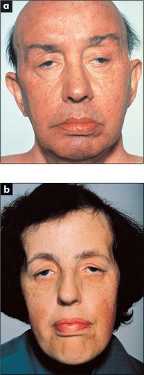
Figure 3.2 Some important diagnostic facies: (a) myopathic; (b) myotonic
From Mir MA, Atlas of Clinical Diagnosis, 2nd edn. Edinburgh: Saunders, 2003, with permission.
Jaundice
When the serum bilirubin level rises to about twice the upper limit of normal, bilirubin is deposited in the tissues of the body. It then causes yellow discoloration of the skin (jaundice) and, more dramatically, the apparent discoloration of the sclerae. The usual term scleral icterus is misleading, since the bilirubin is actually deposited in the vascular conjunctiva rather than the avascular sclerae. The sclerae (conjunctivae) are rarely affected by other pigment changes. In fact, jaundice is the only condition causing yellow sclerae. Other causes of yellow discoloration of the skin, but where the sclerae remain normal, are carotenaemia (usually due to excess consumption of carotene, often from intemperate eating of carrots or mangoes), acriflavine, fluorescein and picric acid ingestion.
Jaundice may be the result of excess production of bilirubin, usually from excessive destruction of red blood cells (termed haemolytic anaemia), when it can produce a pale lemon-yellow scleral discoloration. Alternatively, jaundice may be due to obstruction to bile flow from the liver, which, if severe, produces a dark yellow or orange tint. Scratch marks may be prominent due to associated itching (pruritus). The other main cause of jaundice is hepatocellular failure. Gilbert’s disease is also a common cause of jaundice. It causes a mild elevation of unconjugated bilirubin and is due to an inherited enzyme deficiency that limits bilirubin conjugation; it has a benign prognosis.
Jaundice is discussed in detail in
Cyanosis
This refers to a blue discoloration of the skin and mucous membranes; it is due to the presence of deoxygenated haemoglobin in superficial blood vessels. The haemoglobin molecule changes colour from blue to red when oxygen is added to it in the lungs. If more than about 50 g/L of deoxygenated haemoglobin is present in the capillary blood, the skin will have a bluish tinge.
Central cyanosis means that there is an abnormal amount of deoxygenated haemoglobin in the arteries and that a blue discoloration is present in parts of the body with a good circulation, such as the tongue. This must be distinguished from peripheral cyanosis, which occurs when the blood supply to a certain part of the body is reduced and the tissues extract more oxygen than normal from the circulating blood: for example, the lips in cold weather are often blue, but the tongue is spared. The presence of central cyanosis should lead one to a careful examination of the cardiovascular (
| Central cyanosis 1. Decreased arterial oxygen saturation • Lung disease: chronic obstructive pulmonary disease with cor pulmonale, massive pulmonary embolism |
| Peripheral cyanosis |
Pallor
A deficiency of haemoglobin (anaemia) can produce pallor of the skin and should be noticeable, especially in the mucous membranes of the sclerae if the anaemia is severe (less than 70 g/L of haemoglobin). Pull the lower eyelid down and compare the colour of the anterior part of the palpebral conjunctiva (attached to the inner surface of the eyelid) with the posterior part where it reflects off the sclera. There is usually a marked difference between the red anterior and creamy posterior parts (see
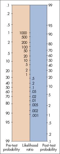
Figure 3.3 Fagan’s nomogram for interpreting a diagnostic test result
Adapted from Sackett DL, Richardson WS, Rosenberg W, Haynes RB. Evidence-based medicine: how to practice and teach EBM. Churchill-Livingstone: London, 1997.
| Sign | Positive LR | Negative LR |
| Pallor at multiple sites | 4.5 | 0.7 |
| Facial pallor | 3.8 | 0.6 |
| Palm crease pallor | 7.9 | NS |
| Conjunctival pallor | 16.7 | − |
NS = not significant.
* Positive likelihood ratio: when the finding is present, describes the probability change. The higher the LR is above 1, the more likely there is disease.
† Negative likelihood ratio: when the finding is absent, describes the probability change. The closer the LR is to 0, the more likely there is not disease.
From McGee S, Evidence-based physical diagnosis, 2nd edn. St Louis: Saunders, 2007.
Facial pallor may also be found in shock, which is usually defined as a reduction of cardiac output such that the oxygen demands of the tissues are not being met (
| 3 Massive pulmonary embolus |
| 4 Sepsis, e.g. gram-negative bacteria (endotoxin) |
| 5 Anaphylaxis |
| 6 Endocrine failure, e.g. adrenal failure, hypothyroidism |
| 7 Neuropathic—from drugs (e.g. antihypertensives, anaesthesia), spinal cord injury of autonomic neuropathy |
Hair
Bearded or bald women and hairless men not uncommonly present to doctors. These conditions may be a result of more than the rich normal variations of life, and occasionally are due to endocrine disease (
Stay updated, free articles. Join our Telegram channel

Full access? Get Clinical Tree


