The Female Reproductive System


The Female Reproductive System


Michael K. Fritsch, M.D., Ph.D.

Mariana M. Cajaiba, M.D.


Abnormalities confined to the genital tract are quite unusual in prepubertal girls, and such disorders seldom come to the attention of pediatricians or pediatric pathologists. Major developmental abnormalities affecting the reproductive system are often eclipsed by concomitant urinary tract abnormalities, which are more immediately clinically significant. Abnormal gonadal development is an important group of diseases that may also result in abnormal development of secondary sexual characteristics. The most frequent acquired diseases of the female genital tract are infections and neoplasms. Infections confined to the female reproductive tract are seldom life threatening in childhood, yet they may result in reproductive sequelae during adulthood. Neoplasms are dominated by those arising from germ cells.


ANATOMY AND EMBRYOLOGY

The female reproductive tract consists of the gonads, a ductal system (fallopian tubes, uterus, cervix, vagina), and the external genitalia (clitoris, labia majora, labia minora, vestibule, mons pubis). The process of sexual differentiation can be divided into various phases. Chromosomal (genetic) sex is determined by the XY or XX genotype, with the potential for abnormal deletion or addition of sex chromosomal material such as in Turner (45,X) and Klinefelter syndromes (47,XXY). Gonadal sex refers to gonadal differentiation into testis or ovary and is predominantly dependent on the expression of the sex-determining region on Y gene (SRY). SRY begins a cascade of molecular signals, resulting in male gonadal differentiation; the inhibition of these male-specific signals and the presence of several female-specific molecular signals result in female gonad differentiation. Phenotypic sex refers to the differentiation of the ductal system and of the external genitalia, a process that is regulated by production of the antimüllerian hormone (AMH) by the Sertoli cells of the testis and steroid hormone production by the testis (testosterone/dihydrotestosterone) and the ovary (estrogen/progesterone). Lastly, the assigned or adopted sex is usually determined by the chromosomal, gonadal, and phenotypic sex at birth but may be altered postnatally in a variety of disorders of sex development (DSD, see below). These processes occur predominantly in utero, with the final phenotypic changes being initiated by the onset of puberty. Previously, it was thought that sex determination involved the initiation of molecular and cellular events leading to male gonad development and that if this pathway failed, there was a default to a female gonadal phenotype. We now know that there are specific male and female molecular pathways that are needed to initiate, complete, and maintain the respective sex phenotypes as well as pathways that prevent the other sex from developing. Specific alterations in these pathways during development can result in partial male and female gonads (e.g., ovotestis) and/or ambiguous genitalia.


Primordial Germ Cells

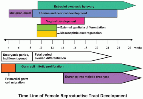
The primordial germ cells (PGCs) first appear in the wall of the yolk sac at about 3 to 4 weeks after fertilization and migrate through the hindgut and mesonephric ridge to the genital ridge beginning about week 4 to 5, a process mediated by a number of molecular factors (1,2,3,4). Germ cells that do not reach the genital ridge are thought to undergo apoptosis, but they have also been proposed as the cell of origin of extragonadal germ cell tumors. PGCs are bipotential, and complete differentiation into oogonia or prospermatogonia depends upon the local environment. The initial events in testicular and ovarian differentiation are independent of the presence or genotype of PGCs in the gonad. However, completion of appropriate ovarian development depends upon the presence of meiotic germ cells. In the absence of meiotic germ cells, the ovarian structure degenerates leaving streak ovaries (5,6). Germ cell sexual dimorphism begins at about 11 to 12 weeks of gestation. Male PGCs (prospermatogonia) enter mitotic arrest, whereas ovarian PGCs (oogonia) enter the first meiotic prophase and then arrest. We now know that retinoic acid produced locally by the ovary induces PGC entry into meiosis beginning in the medulla and slowly radiating out to the cortex. Male germ cells produce the retinoic acid-degrading enzyme CYP26B1 via paracrine FGF9 signaling from male support cells to prevent entry into meiosis.


Early Gonadal Development

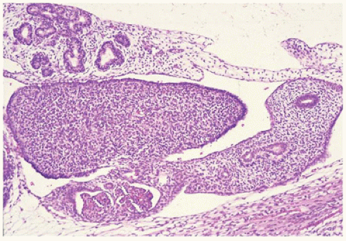
The gonadal ridge develops at the ventromedial aspect of the mesonephros by a proliferation of mesodermal cells and thickening of the overlying coelomic epithelium at 4 to 5 weeks’ gestation. Between 4.5 and 6 weeks, the indifferent gonad is indistinguishable as male or female (Figure 18-1). A number of critical genes have been identified in regulating normal genital ridge development including SF1, WT1, LHX9, CITED2, IGF signaling, and potentially EMX2 and PBX1. In humans and mice, the Wilms tumor gene isoform WT1-KTS as well as the steroidogenic factor 1 (previously referred to as SF1 and now officially designated NR5A1) genes are important in maintaining the indifferent gonad. Mutations in SF1 can lead to adrenal-gonadal failure, mostly affecting male gonad development, as maintenance of SF1 expression is also critical in testis development. The WT-KTS isoform is required for cell survival and proliferation within the bipotential gonad in both males and females (5). WT1+KTS isoform is critical later for male development, and the ratio of +KTS/-KTS seems critical for appropriate gonadal differentiation. Mutations in WT1 are associated with three syndromes characterized by gonadal dysgenesis (Wilms tumor/aniridia/gonadal dysgenesis/retardation—WAGR [OMIM 194072], Denys-Drash [OMIM 194080], and Frasier [OMIM 136680]) (see www.omim.org or www.ncbi.nlm.nih.gov/omim). The phenotype of affected individuals includes genital and kidney defects as well as an increased risk for the development of Wilms tumors. Insulin signaling occurs via IGF1 and IGF2 and is critical for cell proliferation during early gonadal development. In the absence of proper insulin signaling, the gonads can be very small or absent. At about 7 weeks’ gestation, in XY embryos, a Y-linked genetic switch, SRY, is expressed by pre-Sertoli cells (1,2,3,4,5). The expression of SRY is necessary and sufficient to trigger male testis development; however, both the timing and the level of SRY expression are critical for normal testis development. In an XY embryo, the lack of SRY results in ovarian development. The SRY protein contains a highly conserved DNA-binding domain that allows specific genes to be turned on or off. The primary target is SOX9, which is thought to be the key Sertoli cell differentiation factor. Both SRY and SF1 bind upstream of SOX9 to initiate transcription. Once SOX9 transcription is initiated, there is a feedforward loop whereby SOX9 itself replaces SRY and binds upstream to maintain its own transcription. SOX9 also induces FGF9, which stimulates more SOX9 production. SOX9 is essential for production of the first male-specific cell type, the Sertoli cell. Mutation in SOX9 leads to the human dwarfism syndrome camptomelic dysplasia (OMIM 114290), which is often associated with XY sex reversal (7). Duplication of SOX9 causes XX femaletomale sex reversal. SF1 expression in the developing testis regulates expression of several male-specific genes, including AMH (see Chapter 28). It has been recently discovered that SOX9 activation results in increased prostaglandin D2 (PGD2) production, which is essential for maintenance of the testis phenotype by facilitating Sox9 transportation into the nucleus. In animal models, there are well-documented examples of both male and female maintenance factors (e.g., PGD2), which if disrupted even postnatally can result in gonadal transdifferentiation to the alternative gonad. Leydig cells are derived from mesenchymal precursors via desert hedgehog (DHH) signaling (1,2,3,4).


FIGURE 18-1 • The undifferentiated gonad lies adjacent to the mesonephros and the wolffian and müllerian ducts. The mesonephros has a profound effect on normal gonadal differentiation.


Ovarian Differentiation

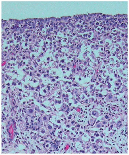
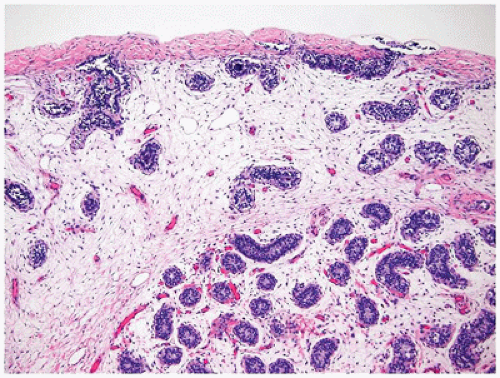
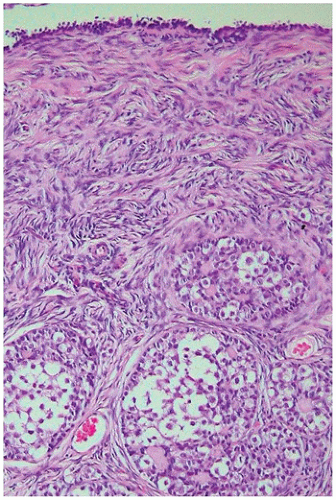
Ovarian development is thought to be characterized by active ovarian determination pathways as well as by inhibition of testis-specific pathways. In addition, it is now clear that once established, maintenance of the ovarian phenotype occurs via FOXL2 and estrogen receptor alpha and beta signaling. The early formation of the ovary can be thought of as involving three major processes: (a) entrance of oogonia into prophase of meiosis I to form primary oocytes, (b) granulosa cells surrounding these primary oocytes to form primordial follicles, and (c) differentiation of the steroid-producing theca cells. By 6 weeks of development, primordial sex cords form. The ovary can be identified at 7 to 8 weeks’ gestation by the absence of true testicular cords and possessing PGC nests. After their arrival in the gonad, germ cells in the female continue to undergo active mitotic cell division, a process that diminishingly continues into the third trimester at least in the cortex. It is estimated that approximately 3 to 4 million germ cells are present in each ovary by 20 weeks’ gestation, and then the number decreases to about 0.5 to 1 million at term. The mechanism leading to the loss of oocytes remains poorly understood. At approximately 12 weeks’ gestation, the first germ cells begin to enter into meiosis via retinoic acid signaling, a process first seen close to the medullary region (Figure 18-2). On entering meiosis, the primary oocyte will arrest at the diplotene stage of the first meiotic prophase and become enclosed by follicular cells to form primordial follicles. Follicular or granulosa cells are in direct contact with the germ cells and are thought to
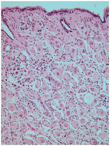
play a role in regulating continued meiotic arrest in the germ cells. Oogenesis is a gradual process that is generally complete by the third trimester, and there is no further increase in the number of primary oocytes thereafter (Figure 18-3). However, in the mouse germ or stem cell, replication may continue into adulthood. In addition to the germ cells, the ovary is populated by stromal cells that continue to remodel during the early first trimester, resulting in the formation of the ovarian cortex and the medulla. During the second trimester, a subepithelial collagenous connective tissue layer develops within the ovarian cortex beneath the basement membrane. The interstitial (thecal) cells, of unknown origin, can be detected during the first half of the second trimester, although estrogen production may begin as early as 8 to 10 weeks’ gestation. The importance of the production of estrogen by the developing fetus remains somewhat controversial with some evidence that significant ovarian estrogen production does not occur until following birth (6). The histology of the developing ovary has been previously described in detail (8).


FIGURE 18-2 • Developing ovary at approximately 22 weeks’ gestation showing formation of primordial follicles containing oocytes arrested in meiosis I in the deep ovarian cortex and premeiotic oogonia in the superficial cortex. Germ cell proliferation continues within the premeiotic oogonia until term.

While many of the molecular details of testis development have been clearly established, the details regulating molecular ovarian development are now coming to light. The NR0B1 gene, previously designated DAX-1, is a dosagesensitive gene locus on the X chromosome that encodes an orphan nuclear receptor protein. This gene was initially thought to be a specific ovary-determining gene but has since been shown to be more essential for normal testicular development and not required for normal ovarian development. NR0B1 is expressed in the normally developing ovary but turned off in the developing testis (5,6). Mutations of NR0B1 in XY males lead to hypogonadotropic hypogonadism with primary testicular defects and are associated with adrenal insufficiency (X-linked adrenal hypoplasia congenita). Loss of function of NR0B1 in XX females does not alter normal ovarian development. Overexpression of NR0B1, however, leads to ovarian development in males even if SRY is expressed. NR0B1 therefore appears to be more important in normal testis development. Unlike the SRY gene and testis development, there does not appear to be a single ovarian determining gene. The canonical WNT signaling pathway is specifically activated in XX gonads and diminished in XY gonads. WNT4 is expressed in both early gonads but is then up-regulated in ovarian somatic cells, and down-regulated in the testis. WNT4 expression seems critical for some portions of ovarian development, but not all. Constitutive activation of beta-catenin in XY gonads results in sex reversal with a XY female gonad. Without WNT4 expression in XX individuals, male-specific changes occur that include the presence of steroid-producing cells within the ovary, persistence of the wolffian ducts, loss of the müllerian ducts, and the development of a male-specific coelomic blood vessel to the ovary. In the absence of WNT4, germ cells can still enter meiosis, but there is massive apoptosis of the germ cells prior to birth. Another recently identified gene that
is involved in activating the WNT/beta-catenin pathway is a pro-ovarian gene, RSPO1 (R-spondin-1). Homozygous mutation in RSPO1 results in female-to-male sex reversal in XX individuals. It would seem that activation of the canonical WNT signaling pathways is required to fully suppress the testis-determining pathway. WNT4 signaling appears to be necessary for (a) normal development of the müllerian duct, (b) suppression of the Sertoli cell lineage in the developing ovary, and (c) oocyte maintenance (1,2,3,4,6). Another gene proposed to be involved in ovary-specific development is FOXL2. FOXL2 seems to be critical for maintenance of the ovarian phenotype by repressing SOX9 expression and therefore preventing testicular differentiation. Mutations in the forkhead transcription factor 2 (FOXL2) gene in humans are associated with eyelid defects and premature ovarian failure (blepharophimosis, ptosis, and epicanthus inversus syndrome—BPES [OMIM 110100]). FOXL2 is expressed by pregranulosa cells (9). While estrogen secretion in utero does not appear to be important for sex determination, estrogen signaling does appear to be critical in the maintenance of the maturing ovary later during puberty. Blocking estrogen signaling in puberty results in transdifferentiation of the ovary and expression of Sertoli cell markers. Estrogen signaling appears to be critical in maintaining the mature ovarian structure by suppressing testicular development during early adult development. Estrogen signaling seems to antagonize activation of the transcriptional enhancer site for SOX9, thereby helping prevent SOX9 expression. It would seem that the gonadal phenotype is maintained early and throughout life by repression of the molecular pathways of the opposite sex. This helps to explain the wide variety of gonadal and genital anomalies including ovotestis.


FIGURE 18-3 • Developing ovary at term showing numerous primordial follicles, and early development of the subepithelial stromal layer that will become more prominent with age. The number of germ cells will decrease progressively with age.

The ovary differs from the testis in that the presence of germ cells is essential for normal ovarian development. In the developing testis, the male germ cells do not play a significant role in the structural development of the organ. Female germ cells appear to follow an intrinsic clock to enter the first meiosis and arrest prior to completion. Once female germ cells have entered meiosis, they have committed to the oocyte fate. During fetal development, the oocyte becomes surrounded by a single layer of granulosa cells to form the primordial follicle. Early primordial follicle formation and maintenance involve several complex molecular pathways acting sequentially and together (reviewed in (4,10,11)). Maturation of these follicles can proceed under hormonal signaling, especially during the third trimester of pregnancy.

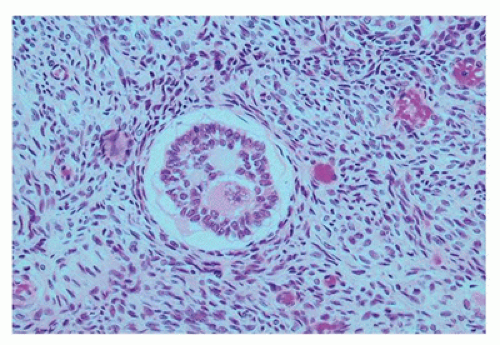
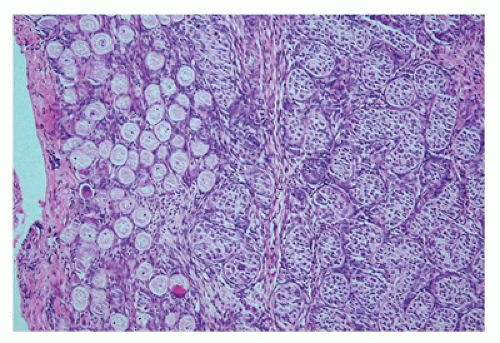
At birth, the ovary is tan, flat, and elongated and measures about 1.3 × 0.5 × 0.3 cm. and weighs less than 0.3 g. Before birth, some primordial follicles can develop further. The ovum enlarges, and the surrounding follicular cells become more cuboidal to columnar and thereby form a primary follicle. This may be followed by stratification of the granulosa cells and increased granulosa cell proliferation, resulting in a preantral follicle (Figure 18-4). The graafian follicle demonstrates a cavity within the granulosa cell layer. The granulosa cells in these follicles have scant cytoplasm and often surround cavities filled with deeply eosinophilic material, known as Call-Exner bodies that may result in microscopic structures resembling gonadoblastoma or annular tubulelike profiles. These likely represent abnormal folliculogenesis. Thecal cells, which differentiate from the stromal cells at the periphery of developing follicles, may be seen. Throughout childhood, the ovaries enlarge to reach the size and the shape of an adult ovary (4 × 2 × 1 cm, 5 to 8 g). During the prepubertal period, the number of oocytes and primordial follicles continues to decrease, and the amount of ovarian stroma increases. Like the testicular Leydig cells, ovarian hilus cells disappear during childhood and reappear during puberty.


FIGURE 18-4 • Primary follicle, preantral stage, showing a thick layer of granulosa cells surrounding the oocyte. Several Call-Exner bodies containing acellular hyaline material are present.


The Ductal System

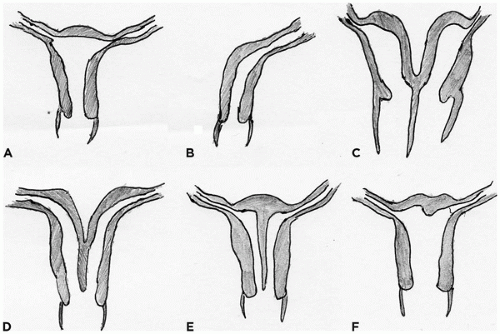
The development of the urinary and reproductive systems is highly interdependent, which explains the high incidence of coexisting genital and urinary tract anomalies in several syndromes. Early in development (approximately week 4), paired mesonephric (wolffian) ducts arise from the intermediate mesoderm and nephrogenic cord (Figure 18-5). In the female embryo, the mesonephric duct is needed for induction of paramesonephric (müllerian) duct development from invaginations of the coelomic epithelium. The mesonephric and the paramesonephric ducts are enclosed in a peritoneal fold that gives rise to the broad ligament of the uterus. In the male embryo, AMH produced by fetal Sertoli cells leads to regression of the ipsilateral paramesonephric duct between weeks 8 and 10. AMH is proposed to have multiple functions in women (12). After the sensitivity of the paramesonephric ducts to AMH has disappeared, the ovarian granulosa cells begin to produce AMH with increasing levels that peak at about 10 years of age. AMH is thought to maintain meiotic arrest in the oocyte of the developing follicle in prepubertal females (10,11).

In the female embryo, the paramesonephric ducts fuse caudally before reaching the urogenital sinus, a process that is completed by week 10 (Figure 18-5). The unfused paramesonephric ducts become fallopian tubes and the fused portions the uterus and the upper vagina. The distal tip of the müllerian duct abuts the posterior wall of the urogenital sinus
within a patch of mesoderm. This point is the future site of the hymenal membrane. The patch of mesodermal urogenital sinus epithelium begins to proliferate, forming a column of squamous cells called the vaginal plate that eventually gives rise to the vaginal epithelium. The vaginal plate and the müllerian duct become patent by canalization early in the second trimester (by week 18). Mesonephric ducts in the female embryo begin to regress if not stimulated by testosterone by about week 10; however, mesonephric remnants in the broad ligament and lateral wall of the uterus and vagina can persist as Gartner ducts. By 13 weeks’ gestation, the body (corpus) of the uterus and the cervix begin to be distinguished. In the fetus and the newborn, the cervix is twice as long as the corpus, whereas in the adult, the corpus is about two times longer than the cervix. At birth, the cervix and uterus together measure about 4 cm in length. The effects of maternal hormones (estrogens and progestins) result in a proliferative-to-weakly secretory endometrium at birth and cervical squamous cell maturation. These changes rapidly disappear after birth. During childhood, the endometrium is usually thin, with inactive glands in a spindled inactive stroma. The uterus reaches a plateau of growth in the 2nd year of life, until the premenarchal uterine growth increase. The final adult (nulliparous) uterus measures 7 to 8 cm in longest dimension and weighs between 40 and 80 g. The final maturation of the female reproductive tract is at the beginning of uterine bleeding (menarche), which occurs between 11 and 15 years of age. The early menstrual cycles are often anovulatory and can result in disordered proliferative endometrium. By mid-adolescence, regular menstrual cycles should be occurring, along with the monthly histologic changes that are well described for the adult female reproductive tract.


FIGURE 18-5 • Diagram indicating the gestational period of development of the ovary, ducts, and external genitalia.

A number of genes have been reported as important in regulating normal internal female genital development. WNT signaling is essential for normal müllerian duct development. Spatial and temporal expression patterns of members of the HOXA gene locus are essential for normal development of the fallopian tubes (HOXA9), uterus (HOXA10), cervix (HOXA11), and upper vagina (HOXA13). In mice, many transcription factors have been implicated in normal müllerian duct formation including Lim1, Pax2, Emx2, several Wnt members (Wnt4, Wnt5a, Wnt7a, Wnt7b, Wnt11), and members of the retinoic acid receptor family (Rar-alpha, beta, and gamma) (13). Estrogen receptors alpha and beta are highly expressed in the female internal genital tract and are important for attaining the final functional phenotype especially for the uterus. Knocking out both ERa and ERb results in a hypoplastic uterus (reviewed in (13,14,15,16)).


Female External Genitalia

The external female genitalia begin to form during the 4th week of embryonic development. The genital tubercle forms ventral to the cloacal plate as two stromal elevations of the ectoderm. Lateral to the cloacal plate on each side, two parallel folds develop, which will give rise to the labia majora and the minora. As the labioscrotal folds extend cranially around the genital tubercle, they fuse and become the mons pubis (17,18). Sonic hedgehog signaling is an important regulator of genital tubercle formation, as well as other downstream genes including WNT5a and FGF8. The role of estrogen in the development of the entire female reproductive tract is unknown. Androgen production in the male is a critical regulator of penis and scrotum formation. If a female fetus is exposed to elevated androgens before 10 to 12 weeks of gestation, the external genitalia may become ambiguous or resemble a phenotypic male and the vagina will often open into the membranous portion of the urethra. If androgens become elevated after week 20, the only effect will be an enlarged clitoris. Similarly, androgen insensitivity due to mutations in the androgen receptor in XY males results in female external genitalia (16,17).


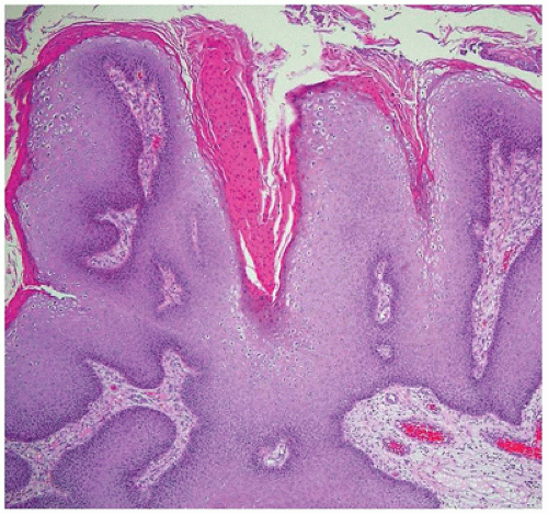
The entire vulva, with the exception of the vestibule, is lined by keratinized, stratified squamous epithelium. The vestibule and the vagina are lined by nonkeratinizing squamous epithelium, which becomes glycogenated in women of reproductive years, and should not be confused with koilocytotic change. The vaginal vestibule contains the orifices of the paraurethral (Skene) glands, the major (Bartholin) and minor vestibular glands, and the urethral meatus. The paired paraurethral glands are located on either side of the urethral meatus, are composed of pseudostratified mucus-secreting columnar epithelium, and are drained by ducts lined by transitional epithelium. The vestibular glands contain acini composed of simple columnar, mucus-secreting epithelium. The major vestibular (Bartholin) glands are drained by ducts lined proximally by mucus-secreting epithelium epithelium and more distally by transitional epithelium with terminal lining by squamous epithelium on their exit just external to the hymenal ring. The minor vestibular glands are located close to the surface and ring the vestibule.


STRUCTURAL ABNORMALITIES OF THE FEMALE REPRODUCTIVE ORGANS

The phenotypic abnormalities that can occur in the development of the female reproductive tract are numerous. These may represent isolated poorly understood variations in normal development, or they may be associated with major malformation syndromes; either may be related to chromosomal abnormalities or defects in known and unknown genes. Additionally, teratogens have been associated with abnormalities of the reproductive tract. DSDs are discussed separately.


FIGURE 18-6 • American Fertility Society classification of müllerian duct abnormalities. Class I (not shown) müllerian aplasia consists of hypoplastic or absent uterus and other müllerian structures. (A) Normal uterus; (B) class II, unicornuate uterus; (C) class III, uterus didelphys (complete failure of midline fusion of müllerian ducts with duplication of uterus and cervix +/- vagina); (D) class IV, bicornuate uterus (defect in fusion at level of uterus only); (E) class V, septate uterus (incomplete (partial) resorption of uterine septum); (F) class VI, arcuate uterus (mildest form with small residual septum in superior portion of uterus). Artwork by RB Fritsch.


The Ductal System

Müllerian duct anomalies are frequently associated with DSD. In patients with normal ovaries, müllerian duct malformations occur in about 0.5% of women and are frequently accompanied by anomalies of the urinary tract. These müllerian duct anomalies include lateral and vertical fusion defects, hypoplasia, or absence of fallopian tubes, the uterus, or the upper vagina. Uterine abnormalities can be structurally and morphologically divided into three categories. These include (a) complete failure of formation of the müllerian duct unilaterally (resulting in unicornuate uterus) or bilaterally (absent uterus), (b) arrested müllerian duct development (hypoplastic uterus), or (c) abnormal lateral fusion of the müllerian ducts to varying extents, resulting in paired uteri and cervices, accompanied by a vaginal septum (uterus didelphys), paired uteri and one cervix (bicornuate uterus), or a uterine septum (septate uterus) (19). The American Fertility Society classification scheme is shown in Figure 18-6 (20,21). A newer classification scheme to describe müllerian anomalies has been recently proposed; however, its clinical utility remains under study (22). Patients with müllerian anomalies may be asymptomatic or may present with infertility, repeated abortions, breech delivery, preterm delivery, dyspareunia, or dysmenorrhea. Because the wolffian ducts are essential for inducing the müllerian ducts, defects in wolffian duct development may likewise lead to uterine abnormalities. Isolated abnormalities of the fallopian tubes (duplication or absence) are rare. Abnormalities of the cervix are
also very rare and include atresia or hypoplasia, which is due to failure of canalization of the müllerian ducts. The molecular events regulating lateral fusion remain largely unknown.

Absence of the vagina occurs in 1 in 4000 to 5000 women and is often associated with müllerian anomalies. In müllerian agenesis, also known as Mayer-Rokitansky-Kuster-Hauser (MRKH) sequence, there is an absent vagina and often an absent or poorly formed uterus and fallopian tubes (19,23). These patients are genotypic (46,XX) and phenotypic (normal external genitalia) females with normal endocrine status, and they often present with amenorrhea. Some cases are associated with upper urinary tract abnormalities and/or spine and skeletal abnormalities. MURCS (Müllerian, renal, cervical, somite) association appears to be an extreme presentation of these clustered anomalies (OMIM 601076). It has been suggested that this sequence may be due to abnormal development of the wolffian duct, with resulting abnormal müllerian duct development. MRKH sequence is thought to have a heterogeneous molecular basis with candidate genes proposed to include those involved in normal uterine development such as WT1, PAX2, HOX genes, WNT2, WNT4, and PBX1. The MRKH sequence usually occurs in a sporadic manner, and therefore, temporally altered aberrant gene expression may be related to exogenous factors rather than to mutations. Other anomalies of the vagina result from developmental defects involving the cloaca or urogenital sinus. As previously discussed, the lower third of the vagina and the hymen are thought to be derived from the ectoderm. If the urogenital sinus develops normally, the vagina can be present as a blind pouch even in the absence of normal müllerian duct development. A variety of miscommunications between the urethra, rectum, and vagina have been described (24). Vaginal obstruction can result from absence of communication between the introitus and the vaginal canal, related to defects in vertical fusion. Etiologies include imperforate hymen, atresia of the lower vagina, or a transverse vaginal septum (failure of vertical canalization of the vaginal plate at the site of fusion between the urogenital sinus and the müllerian ducts). Imperforate hymen, formed where the urogenital sinus and canalized fused sinuvaginal bulbs meet, is the most common cause, and if there is complete obstruction, this leads to a marked dilatation of the vagina and the uterus (hydrometrocolpos—retention of secreted mucous). If the person is asymptomatic until the onset of menses, the obstruction can result in hematocolpos.


The External Genitalia

Abnormalities in the development of the perineum, labia, and clitoris are not uncommon. The normal variations and structural abnormalities of the vulva have been extensively reviewed (25). Complete absence of the external genitalia is rare and occurs as part of malformation syndromes such as sirenomelia, limb-body wall defects, Robinow syndrome, multiple pterygia syndrome, Fryns syndrome, or CHARGE association. Vulvar duplication is rare and is associated with multiple congenital anomalies. Abnormalities of the clitoris and labia may cause problems in assigning the correct sex at birth. Clitoral hypertrophy may resemble male hypospadias. Exposure of the female fetus to excess androgens, such as in congenital adrenal hyperplasia, a maternal or fetal virilizing tumor, or maternal medication, can result in clitoral hypertrophy and may result in partial fusion of the posterior portion of the labia. Complete absence of the clitoris is rare.


DISORDERS OF SEX DEVELOPMENT

DSDs, previously referred to as intersex disorders, are defined as any congenital condition presenting with aberrant development of gonads and/or genitalia resulting in discordant chromosomal (genetic) and anatomic sex, associated or not with chromosomal anomalies. An early diagnosis is warranted not only for potential early gender assignment but also for identification of patients at risk for the development of gonadal neoplasms. DSDs encompass several conditions with significant clinicopathologic overlap, and the diagnosis of a specific entity requires a multidisciplinary approach with correlation of several clinical and laboratory features, including phenotypic features (external and internal genitalia), endocrine profile, karyotype, molecular studies, and histopathology. In addition, both the lack of uniformity in the existing literature and the confusing nomenclature represent a challenge to the diagnosis of these conditions. A revised nomenclature and classification system (Table 18-1) had been proposed in an attempt to provide a more descriptive approach to DSD (26); however, this classification system is still limited in terms of overcoming all the subtleties and overlapping features associated with these conditions.

Despite these limitations, the etiologies of DSD can be broadly characterized into chromosomal, molecular, or endocrine. Chromosomal abnormalities result from numerical aberrations in sex chromosomes and include aneuploidies (Chapter 3), such as 45,X (Turner syndrome), 47,XXY (Klinefelter syndrome), or mosaic aneuploidies, such as 45,X/46,XY (mixed gonadal dysgenesis) and 46,XX/47,XY. The molecular aberrations resulting in DSD include numerous defects in genes involved in testicular development such as SRY, WT1, SOX9, DHH, AMH, or the AMH receptor, among others (2,3,27,28,29). Endocrine causes are divided into undervirilization syndromes (including androgen insensitivity syndrome and defects in androgen biosynthesis) and hypervirilization syndromes (examples include congenital adrenal hyperplasia and excess exogenous androgens) (30).

Most patients with DSD present with a wide spectrum of clinical manifestations, and significant clinical overlap is
seen among distinct entities. The clinical presentation in DSD reflects the extent of gonadal maldevelopment. The external genitalia phenotype typically is a direct result of the degree of virilization achieved by androgen exposure during fetal development. Ambiguous genitalia, usually resulting from partial virilization, is the most common presentation of DSD and encompasses a broad range of phenotypic abnormalities including hypospadias, clitoral hypertrophy, micropenis, bifid scrotum, and fused labia majora. Internal genitalia development is mostly determined by exposure to antimüllerian hormone (AMH) and androgens. AMH is responsible for regression of the müllerian (paramesonephric) structures that will give rise to the uterus and fallopian tubes. Androgens are implicated in the development of the wolffian (mesonephric) structures that will give rise to the epididymis, vas deferens, and seminal vesicles. Other clinical presentations of DSD include precocious puberty, delayed puberty, primary amenorrhea, and complete sex reversal. As an example, bilateral streak gonads in a 46,XY patient often result in a female phenotype due to absence or minimal production of androgens and AMH by the undifferentiated gonadal tissue. A female phenotype in a 46,XY individual is also usually seen in cases of complete androgen insensitivity syndrome.


TABLE 18-1 CLASSIFICATION OF DSD


  1. Normal sex chromosomes


    1. Female pseudohermaphroditism (excess androgens in females)—(46,XX DSD)


      1. Adrenogenital syndrome (21-hydroxylase deficiency, 11-β-hydroxylase deficiency)


      2. Maternal ingestion of androgenic hormones


      3. Maternal virilization


    2. Male pseudohermaphroditism (deficient androgens in males)—(46,XY DSD)


      1. Testicular regression syndrome


      2. Gonadotropin-Leydig cell defects


      3. Steroid enzyme deficiencies (testosterone/dihydrotestosterone)


      4. Androgen insensitivity syndromes


      5. Persistent müllerian duct syndrome


  2. Abnormal sex chromosomes


    1. Sexual ambiguity frequently present


      1. Mixed gonadal dysgenesis—(45,X/46,XY MGD)


      2. True hermaphroditism—(ovotesticular DSD)


    2. Sexual ambiguity infrequently present


      1. Pure gonadal dysgenesis—(46,XY complete gonadal dysgenesis)


      2. Klinefelter syndrome—(47,XXY)


      3. Turner syndrome—(45,X)


      4. XX male syndrome—(46,XX testicular DSD)


Shown in bold is the new nomenclature.


From Lee PA, Houk CP, Ahmed SF, et al. Consensus statement on management of intersex disorders.


International Consensus Conference on Intersex. Pediatrics 2006;118:e488-e500.


FIGURE 18-7 • Streak gonad composed of wavy ovarian stroma with no identifiable follicular structures.


Gonadal Pathology in DSD

Morphologically, the gonads in DSD can be divided into well or maldeveloped (dysgenetic gonads). Well-developed gonads can be anatomically normal but functionally abnormal, resulting in a DSD phenotype due to inadequate hormone production and are usually associated with endocrine etiologies of DSD such as congenital adrenal hyperplasia (adrenogenital syndrome). In contrast, dysgenetic (or maldeveloped) gonads are often associated with chromosomal, molecular, or unknown etiologies of DSD. Morphologically, dysgenetic gonads usually fall into one of the following patterns: streak gonads, dysgenetic testes, and ovotestes. Streak gonads consist of variable degrees of ovarian stroma and dense fibrous tissue and an absent tunica albuginea (31); primordial follicles/germ cells are usually scant or absent, and primitive cord-like structures are often present (Figure 18-7). In Turner syndrome patients (45,X), streak gonads are thought to represent regressive changes within ovarian tissue, whereas in patients with a 46,XY cell line, the streak gonads are thought to represent abortive testicular development. Dysgenetic testes occur in patients with a 46,XY cell line (examples include mixed gonadal dysgenesis and a few cases of gonadal dysgenesis due to germline mutations in genes involved in testicular development) and are thought to represent a milder phenotype of abortive testicular development, containing variable amounts of well-developed seminiferous cords (31). However, these seminiferous cords contain subtle abnormalities such as irregular contours and aberrant distribution of germ cells; in addition, dysgenetic testes show a poorly developed tunica albuginea with irregular thickness and deficient collagenization, occasionally penetrated by seminiferous cords, and foci of ovarian stroma (Figure 18-8). Ovotestis is the rarest type of dysgenetic gonad, characterized by recognizable testicular (containing well-formed seminiferous cords with spermatogonia) and ovarian (containing numerous follicles at various degrees of
maturation) parenchyma occurring within the same gonad (Figure 18-9) (32,33). This gonadal phenotype characterizes ovotesticular DSD (true hermaphroditism), for which different etiologies have been implied, including numerical anomalies in sex chromosomes (such as 46,XX/46,XY mosaicism) and molecular defects in genes involved in gonadal development (34).


FIGURE 18-8 • Dysgenetic testis demonstrating abnormal branching of seminiferous tubules, some of which invade the tunic albuginea. The tunica can be of variable (often thin) thickness.


Tumors Associated with Gonadal Dysgenesis

Patients with dysgenetic gonads containing Y chromosome material in their genome, specifically the GBY region encoding the TSPY gene, are at increased risk for the development of neoplasms (35). The presence of SRY or other sex-determining genes is irrelevant in this context. More than 50% of neoplasms arising in these gonads are gonadoblastomas (Figure 18-10). The frequency of occurrence of gonadoblastoma in these patients correlates with the frequency of an intact Y chromosome containing the GBY region. Gonadoblastoma is seen in approximately 30% of patients with mixed gonadal dysgenesis, less than 3% of individuals with true hermaphroditism, and over 50% of patients with 46,XY pure (complete) gonadal dysgenesis (36). The pathology of gonadoblastoma is described later. The significance of the development of gonadoblastoma is that it is associated with a very high frequency of concurrent or future development of a malignant germ cell tumor, most commonly dysgerminoma. Other less common gonadal tumors in patients with DSD include other types of germ cell tumors, juvenile granulosa cell tumor (JGCT), Sertoli-Leydig cell tumor, and epithelial tumors. DSD patients with a 46,XX karyotype and patients with Turner syndrome without 45,X/46,XY mosaicism only rarely develop gonadal tumors; however, hilus cell hyperplasia and hilus cell tumors have been reported.


FIGURE 18-9 • Ovotestis removed from a 6-year-old child with ambiguous genitalia and an undescended left testicle. Peripheral lymphocyte karyotype 46,XY. The gonad demonstrates two distinct regions: the area on the left composed of ovarian stroma and numerous primordial follicles, and the area on the right lower corner showing normally developed seminiferous tubules with reduced numbers of germ cells.


FIGURE 18-10 • Small, streak gonad from a 46,XY phenotypic female that is composed of wavy ovarian-type stroma and no primordial follicles. Deep within the cortex, well-defined nests composed of a mixture of germ cells and granulosa-like cells, consistent with gonadoblastoma.


NONNEOPLASTIC LESIONS OF THE LOWER GENITAL TRACT


Infections

Infections of the lower genitourinary tract account for the majority of genital lesions in premenarchal girls. These infections are most commonly due to a variety of bacterial organisms that do not penetrate the mucosa and are not related to specific pathologic findings. Infections common in sexually active females, such as Gardnerella vaginalis and molluscum contagiosum, are quite rare in young children and should raise the suspicion of sexual abuse (Chapter 7). Specific infections of the vulvovagina include human papillomavirus (HPV), herpes simplex virus, syphilis, and molluscum contagiosum.


Human Papillomaviruses

Condyloma acuminata are sexually transmitted lesions caused by papillomaviridae, most commonly the HPV types 6 or 11, although HPV 2 may also be seen. It is now recommended that condyloma acuminata be referred to as low-grade squamous intraepithelial lesions (LGSILs) (37). The modes of transmission of HPV include perinatal, autoinoculation, heteroinoculation, and sexual abuse. These lesions may involve the vulva, vagina, cervix, urethra, and/or perianal skin. Vulvar and vaginal lesions are commonly papillary and are almost always multiple; cervical lesions are often flat, white lesions surrounded by hyperemic mucosa. Uncommonly, the involved epithelium may extend into the endocervical glands and, therefore, have an endophytic appearance. Most lesions are asymptomatic unless secondarily infected. Histologically, parakeratosis, acanthosis, hyperkeratosis, and dyskeratosis are evident (Figure 18-11). The typical koilocytic cells with perinuclear cytoplasmic halos surrounding irregularly contoured (“raisinoid”) nuclei may be seen in the more superficial or intermediate layers. The development of high-grade intraepithelial neoplasia— HGSIL—(associated with high-risk HPV genotypes 16 and 18) is an uncommon finding in females under 21 years of age. Most cases in this age group correspond to LGSILs, whereas high-grade lesions and progression to carcinoma are rare occurrences (38). LGSIL may at times be difficult to distinguish from HGSIL or vulvar intraepithelial neoplasia. The presence of a flat, macular growth pattern, abnormal mitoses, atypical nuclei, marked variation in nuclear size and shape, and hyperchromasia are all characteristics of vulvar intraepithelial neoplasia. In addition, immunohistochemistry can be helpful as Ki67 will show increased staining up into the epidermis in LGSIL and all the way to the surface epidermis in HGSIL. Also, p16 is extremely helpful as LGSILs show only weak staining, whereas HGSIL demonstrate strong staining in the epidermis. Identification of the specific HPV genotype may be indicated in some circumstances, but is not considered routine in children.


FIGURE 18-11 • Condyloma acuminatum showing parakeratosis, acanthosis, and numerous koilocytic cells with nuclear irregularity and prominent perinuclear vacuolization. No dysplasia is present.


Herpes Simplex

Patients with genital infection with herpes simplex virus types I or II can present with dysuria and vulvar pain, often accompanied by generalized malaise and fever. The clinical picture is dominated by the appearance of vesicles and shallow ulcers that are often secondarily infected. Only two-thirds of culture-positive women show diagnostic genital lesions. Histologically, the ulcers typically demonstrate extension deep into the epidermis, with the characteristic intranuclear inclusions present at the periphery of the lesion. Late in the evolution of the ulcer, the infected cells undergo karyorrhexis and lysis, and therefore, infected cells may not be identifiable in biopsy material. Cytologic evaluation of scrapings from a fresh ulcer or freshly opened vesicle will usually show the characteristic viral cytopathic effects. Recurrent episodes of herpetic vulvitis are common; however, these episodes decrease in frequency over time whether or not acyclovir is given. Anogenital herpes in children raises the concern of sexual abuse, but is not definitive evidence. Varicella infection of the lower genital tract is rare in children and more commonly detected in postmenopausal women.


Syphilis

The primary lesion of syphilis is the chancre, a painless, shallow ulcer with raised edges that usually presents within 10 to 90 days of initial contact. These lesions often occur on inconspicuous surfaces, such as the cervix, and in about 50% of patients, the primary lesion is never seen (39). Histologically, the chancre is characterized by ulceration of the epidermis with acute and chronic inflammation within the dermis. There is a marked perivascular inflammatory response with a large number of plasma cells. The lack of specificity of these findings raises the importance of considering syphilis in the differential diagnosis of inflammatory lesions. Lymphadenopathy may develop 3 to 4 days after the
chancre appears. If the primary stage is left untreated, the secondary stage of the disease will become evident within 6 weeks to 6 months when the patient will show elevated plaques measuring up to 3 cm, especially on the vulva. These plaques are known as condylomata lata and demonstrate marked acanthosis, epithelial hyperplasia, and hyperkeratosis. The inflammatory response within the dermis is similar to that seen in the chancre. Both the chancre and the condyloma lata are rich in spirochetes, which may be detected by the Dieterle or Warthin-Starry silver stains. However, these stains may be negative even with active infection. Serologic studies should be performed if syphilis is considered clinically or pathologically; even these studies may be negative for weeks after the presentation of the primary chancre. Other methods used for detecting spirochetes include dark field examination of serum expressed from the base of the ulcer or by a fluorescent-conjugated antibody technique. These methods are more sensitive and specific than the silver stain on paraffin-embedded tissue (39).

Only gold members can continue reading. Log In or Register to continue

Stay updated, free articles. Join our Telegram channel

Sep 23, 2016 | Posted by in PATHOLOGY & LABORATORY MEDICINE | Comments Off on The Female Reproductive System

Full access? Get Clinical Tree

Get Clinical Tree app for offline access