The Eyelids, Orbit, and Eye


The Eyelids, Orbit, and Eye


Cytologic diagnosis of various disorders of the eyelids and the external eye has been in use for many years (Kimura and Thygeson, 1955). Naib (1972, 1981) was a major contributor to this knowledge. Cytologic sampling of the orbit, its adnexa, and the eye became possible with the developments of imaging modalities, such as the computed tomography (CT) and precision ultrasound. With the use of small caliber needles under imaging guidance, the aspiration of these organs became safe and accurate (Jakobiec and Chattock, 1978; Czerniak et al, 1983, 1985; Koss et al, 1992; Glasgow and Foos, 1993). In this chapter, the application of cytologic techniques to the external eye, the orbit and its contents will be discussed.


ANATOMIC AND HISTOLOGIC RECALL

The eye is located within the bony structure of the skull, the orbit. The eye is connected to the brain by the optic nerve and to the orbit by a number of striated muscles that control its movements. Besides the eye and the muscles, the orbit is filled with loose connective tissue that contains nerves, vessels, and small deposits of lymphocytes.

The anterior surface of the eye, a transparent to light lens-like structure, the cornea, is lined on its surface by a transparent, stratified, nonkeratinized squamous epithelium (Fig. 41-1A). Laterally, the cornea becomes the sclera, a fibrous structure that encloses the eye. The eye is protected anteriorly by the eyelids which, on the surface facing the eye, are lined by a stratified epithelium containing numerous mucus-producing goblet cells. This epithelium is in continuity with the squamous epithelium lining the cornea; the transition occurs at the limbus, where the peripheral cornea merges with the anterior sclera. The outer surface of the eyelids is formed by skin. The eyelids contain numerous mucus-producing glands, the largest and most important being the meibomian glands. Smaller glands are known as glands of Zeis and Moll. Extending from the eyelids into the orbit are the lacrimal glands, similar in structure to serous salivary glands, which, by a series of canals, secrete tears that lubricate the eye and the eyelids.

The internal structure of the eye is extremely complex and beyond the scope of this chapter, thus only the key structures will be mentioned. The interior of the eye is divided into two chambers: the anterior aqueous chamber, located between the cornea and the transparent crystalline lens, and the posterior chamber, filled with transparent viscous vitreous (vitreous body), demarcated anteriorly by the lens and posteriorly by the retina. The anterior chamber contains a contractile pigmented structure, the iris, forming the pupil of the eye and regulating the input of light. All parts of the eye serve the primary purpose of processing light signals by multilayered sensory complex neuronal tissue, the retina. The retina can become the site of a malignant tumor of childhood, the retinoblastoma. On the outer, orbital side, the retina is supported by a layer of melanin-containing pigment epithelium that extends anteriorly into the iris. In turn, the pigmented epithelium is separated from the sclera by an intermediate layer, the uvea, composed
of connective tissue containing blood vessels, nerves, and melanocytes. The uvea is divided into three distinct anatomic segments: the choroid, which surrounds most of the eye and transits anteriorly into the ciliary body, and the iris. The most common malignant tumors of the eye, malignant melanomas, develop in the uvea, particularly in the choroid but also, less commonly, in the ciliary body and the iris. The optic nerve may be the site of formation of orbital gliomas and meningiomas. For a detailed description of the histology of the eye, the reader is referred to a simple, yet detailed and accurate account by Stevens and Lowe (1992).


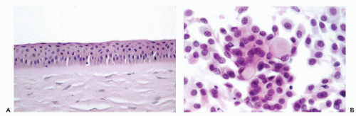
Figure 41-1 A. Normal corneal epithelium. Note orderly epithelium of squamous type without surface keratinization. B. Normal conjunctival scrape smear. Cuboidal and columnar epithelial cells and goblet cells.


THE EYELIDS AND EXTERNAL SURFACE OF THE EYE


Sampling Techniques

The external surface of the eye and the eyelids are accessible to simple cytologic procedures, within the reach of any ophthalmologist and any laboratory of cytology. Scraping the surface of the lesion under local anesthesia with an appropriate small instrument and preparation of alcoholfixed or air-dried smears is sufficient for the diagnosis of inflammatory disorders and some malignant lesions, such as carcinoma of the cornea and conjunctiva, or carcinomas of the eyelid (Naib et al, 1967; Dykstra and Dykstra, 1969; Spinak and Friedman, 1977; Naib, 1970, 1981; Koss et al, 1992). Tsubota et al (1990) and Kobayashi et al (1991) advocated the use of a modified small endocervical brush with short, soft bristles (S-Brush, Medscan, Malmö, Sweden) for securing material from the conjunctiva. Instead of smears, these authors advocated the use of a cytocentrifuge and, more recently, ThinPrep (Cytyc Corp, Boxborough, MA) for processing of the samples (Kobayashi et al, 1997). Another brush with a spherical tip (Acellon-M) was used for the same purpose by Fujihara et al (1997).

Nolan et al (1994, 1997, 2001), described an ingenious technique named “impression cytology” for the study of conjunctival samples. Following local anesthesia, a cellulose acetate strip is placed on the surface of the cornea using gentle pressure, carefully peeled off, fixed in alcohol, and stained with Papanicolaou stain. The technique was used by other investigators with impressive results (Dart, 1997; Divani et al, 1997).


Normal Cells in Conjunctival and Corneal Scrape Smears

The normal cell population in scrape smears consists of squamous cells of corneal origin and cuboidal to columnar epithelial cells of conjunctival origin. Goblet cells may be present (Fig. 41-1B). In about 20% of the patients, squamous cell nuclei may display a central chromatin bar, similar to the appearance of Anitschkow cells, first observed by Marner (1980). On scanning electron microscopy, the bars form a ridge in the nucleus, whereas in transmission electron microscopy, a tortuous folding of the nuclear membrane was observed (Kobayashi et al, 1992, 1998). Similar squamous cells have been observed in the oral cavity (see Chap. 21 and Fig. 21-1). Kumar and Manabati (1998) also observed nuclear protrusions in the form of nipples in conjunctival cells, a common finding in endocervical cells, discussed at length in Chapter 8. These nuclear variants may occur in health and disease and have no diagnostic significance.


Inflammatory Lesions


Viral Infections

Viral infections are very common and cause painful inflammation of the conjunctiva and the cornea. Their identification may be of substantial assistance in the clinical management of the patient. Naib et al (1967, 1972, 1981) have given excellent descriptions of cytologic findings in eye disorders caused by viruses. These are summarized in Table 41-1. The morphologic manifestations of various viral infections were described and illustrated in Chapters 10 and 19. Olding-Stenkvist and Brege (1975) applied immunofluorescent techniques for the diagnosis of herpetic conjunctivitis.
There are now several other diagnostic techniques available, such as polymerase chain reaction (PCR) for viral identification.


TABLE 41-1 CYTOLOGIC CHANGES IN VIRAL INFECTIONS OF THE EYE AND ADNEXA


Disease


Ocular Site


Location of Inclusions


Inclusion: Descriptive Features


Remarks


Trachoma (chlamydia)


Cornea and conjunctiva


Cytoplasmic in mature cells


Multiple small (0.5 μm), basophilic with halos


Clusters of inclusions from necrotic cells


Inclusion conjunctivitis (frequent in newborn infants)


Cytoplasmic in mature cells


Same as above


Clusters of inclusions


Adenovirus


Conjunctiva


Intranuclear in small cells


Multiple, small eosinophilic, becoming coalescent, basophilic with halos


Few cells involved


Vaccinia


Conjunctiva


Cytoplasmic


Single, large, eosinophilic


Herpes, simplex and zoster


Conjunctiva and cornea


Intranuclear, often in multinucleated cells with nuclear molding


Enlarged “ground-glass” nuclei forming eosinophilic inclusions


In herpes zoster multinucleated cells and eosinophilic inclusions are rare


Measles


Conjunctiva


Multinucleated giant cells


Multiple eosinophilic cytoplasmic inclusions with sharp halos


(Modified from Naib ZM, et al. Exfoliative cytology as an aid in the diagnosis of ophthalmic lesions. Acta Cytol 11 :295-303, 1967; and Naib ZM. Cytology of ocular lesions. Acta Cytol 16:178-185, 1972.)


Chlamydial Infections

Conjunctival infection, caused by the bacterium, Chlamydia trachomatis, results in trachoma, the most widespread cause of blindness in the developing world. The infection, usually transmitted by human contact, leads to opacification of the upper part of the cornea and can be diagnosed by cytologic scraping. Besides trachoma, chlamydia may lead to various other less dangerous infections, such as chronic inclusion conjunctivitis, characterized by formation of lymphoid follicles in the conjunctiva and lower part of the cornea. As discussed above, Kobayashi et al (1991) advocated the use of the S-brush for the diagnosis of chlamydial infection.

The cytology of the chlamydial infection of the eye is identical to the infections in the female genital tract, described and illustrated in Chapter 10. Naib (1972) stressed the presence of numerous small (0.5 μm) basophilic cytoplasmic inclusions with halos as the characteristic cytologic finding (see Table 41-1). A number of staining techniques documenting this infection in infants was discussed and illustrated by Duggan et al (1986).


Other Inflammatory Processes

Allergic conjunctivitis, a common disorder, is characterized in smears by a large number of eosinophils mixed with other inflammatory cells. Rivasi et al (1992) also observed an increase in goblet cells and the presence of various foreign materials, presumably of plant or mineral origin.

Bacterial infections result in acute conjunctivitis characterized by a dominance of neutrophils in smears. Gonorrheal conjunctivitis in newborn infants is a preventable disease with prophylactic treatment with antibiotics. A related organism, Moraxella (Branhamella) catarrhalis, an encapsulated diplococcus, may cause a purulent inflammation in adults. Eyelid infections with staphylococci (sty) rarely require cytologic confirmation. Actinomycosis of the cheek may spread to the orbit.

An occlusion of the meibomian ducts by an inflammatory process, usually secondary to inflammation of the hair follicles (blepharitis), may cause a granulomatous inflammation and palpable enlargement of the meibomian glands, known as chalazion. A chalazion may be mistaken for a tumor of the eyelids and, more importantly, tumors may be mistaken for a chalazion. Needle aspiration biopsy is the ideal diagnostic technique in these situations.

Mycotic infections are uncommon but may lead to a severe corneal disease and loss of vision, particularly with Phycomycetes (Zygomycetes), such as mucormycosis (Johnson, 2000). These infections may cause sudden blindness (Downie et al, 1993). Other fungi, such as Candida and Aspergillus species, may be observed (Naib, 1981; Johnson, 2000). The morphology of these fungi is discussed at length in Chapter 19.


Parasites


Acanthamoeba

Corneal infection (keratitis) caused by Acanthamoeba species occurs with increasing frequency, mainly in wearers of soft contact lenses (Stehr-Green et al, 1989; Moore and McCulley, 1989).
Corneal scrapings may be used for diagnosis (summary in Karayianis et al, 1988; Rivasi et al, 1995). Against a background of acute inflammation, trophozoites and the double-walled cysts of the parasite may be identified by several staining and fluorescent techniques, but also in Papanicolaou stain (Fig. 41-2). An early diagnosis and aggressive therapy are essential to prevent a loss of vision (Moore and McCulley, 1989; Rivasi et al, 1995).


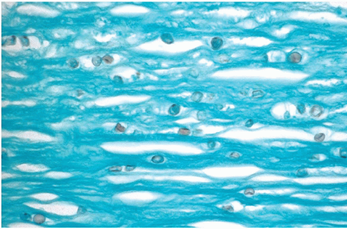
Figure 41-2 Acanthamoeba in corneal epithelium in a young wearer of contact lenses. Small spherical cysts of the parasite are stained with Gomori-methenamine silver. (Courtesy of Dr. Pearl Rosenbaum, Montefiore Medical Center, Bronx, NY.)


Microsporidiosis

Microsporidial keratitis, caused by the tiny intracellular protozoan parasite of the genus Nosema or Encephalitozoon, previously a rare event observed after corneal trauma in immunologically normal patients, is becoming increasingly prevalent in HIV-positive, immunosuppressed individuals (Wittner et al, 1993; Weber et al, 1994; Rasterelli et al, 1994; Coyle and Weiss, 1996). The epidemiology and life cycle of the parasite were summarized by Chen et al (2002). The parasite may be demonstrated in conjunctival smears and biopsies with various staining techniques or phase contrast microscopy (Fig. 41-3). Identification of specific genus is possible with electron microscopy, by a specific antibody or genomic analysis.


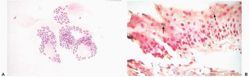
Figure 41-3 Microsporidia. A. Corneal scrapings from a 34-year-old man who was HIV-positive. The tiny organisms are present in the cytoplasm, surrounding a central clear area that is the nucleus. B. Biopsy of the limbal conjunctiva showing the Gram-positive organisms within the cytoplasm of the superficial epithelial cells. (Brown and Brenn stain.) (Courtesy of Dr. Pearl Rosenbaum, Montefiore Medical Center, Bronx, NY.)


Onchocerciasis (River Blindness)

Onchocerca volvulus is the largest of human filariae, transmitted by blackflies rather than mosquitoes (Ash and Spitz, 1945). It is the cause of severe dermatitis and river blindness, prevalent in Africa and parts of Central and South America, caused by numerous filariae accumulating in eye chambers, resulting in sclerosing keratitis. The disease is curable with appropriate drugs. To our knowledge, cytologic techniques have not been applied to the diagnosis of this very important disorder.


Echinococcosis

Sodhani et al (1996) described a case of echinococcosis affecting the eye. For description of this parasite, see Chapters 19 and 38.


Benign Tumors

Scrapings of benign tumors of the skin of the eyelids, such as xanthelasma, molluscum contagiosum (a viral disorder), and squamous papillomas may sometimes be of diagnostic advantage (Naib, 1981). In xanthelasma, lipid-filled cells may be observed. The cytologic presentation of molluscum contagiosum is discussed in Chapters 14 and 36. Squamous papilloma may show evidence of human papillomavirus infection in the form of squamous cells with abnormal nuclei and a large perinuclear clear zone, known as koilocytes (see Chap. 11 for extensive discussion of this entity). Other tumors occurring on the skin or surface of the eyelids are described in Chapter 36.

The sebaceous meibomian glands and the small serous glands located within the eyelids may be the site of benign tumors mimicking tumors of the salivary glands, notably pleomorphic adenomas (see Chap. 32). Leiomyomas of the eyelids have been described (Henkeind and Friedman, 1976).


Malignant Tumors


Squamous Carcinoma and Its Precursors

Malignant epithelial tumors of the conjunctiva and surface of the cornea are predominantly squamous carcinomas, which in fortuitous situations, may be diagnosed as preinvasive neoplastic lesions, such as carcinomas in situ (Fig. 41-4B,D). The term ocular surface squamous neoplasia (OSSN) has been proposed to include intraepithelial and invasive squamous lesions (Lee and Hirst, 1995; Nolan et al, 1997). The spectrum of abnormalities ranged from “simple dysplasia” to carcinoma in situ to invasive carcinoma. Subsequently, Nolan et al used the term “high-grade” lesions for carcinomas in situ and related lesions. The presence of human papillomavirus type 16, determined by PCR, was reported in a substantial proportion of premalignant and malignant lesions of the conjunctiva and cornea (McDonell et al, 1989). For further discussion of human papillomavirus, see Chapter 11. It has been reported that conjunctival intraepithelial neoplasia commonly occurs in patients with AIDS and may be a marker for this disease (Karp et al, 1996).

The clinical presentation of carcinoma in situ is often misleading. Slight thickening of the conjunctiva combined with increased vascularity may be mistaken for an inflammatory or degenerative lesion and treated as such by ophthalmologists. This was the experience of Dykstra and Dykstra (1969), who used cytologic techniques for the diagnosis of squamous carcinoma of the conjunctiva. In three of their eight cases, the lesion was still in situ and was not suspected clinically. Nolan et al (2001) used touch preparations (impression samples—see methods of sampling) of the corneae in 267 patients and compared the cytologic samples with biopsies. In 231 of these patients, the lesions were preinvasive, and in several of these patients, there was no evidence of clinical disease (Hirst et al, 1998; Nolan et al, 2001). These observations from Australia, where corneal carcinoma is common in elderly people as a consequence of exposure to ultraviolet light, indicate yet another use of cytologic techniques in the detection of an important form of cancer, particularly in patients with AIDS.

Cytology of squamous carcinoma in situ of the cornea and conjunctiva is identical to similar lesions of the uterine cervix or oral cavity (see Chaps. 11 and 21) and consists of small to moderately sized atypical, or frankly malignant, squamous cells with markedly enlarged, hyperchromatic nuclei of variable sizes (Fig. 41-4A). Nolan et al (1997) stressed that in many of the preinvasive squamous lesions and in some invasive squamous carcinomas, there was evidence of heavy keratinization and the cytologic presentation of these lesions was similar to the keratinizing variant of carcinoma of the uterine cervix (Fig. 41-4C). In some of the invasive squamous carcinomas, the smear patterns were those of poorly differentiated tumors with little or no keratin formation and large nuclei containing large nucleoli.

There is limited knowledge on the cytologic presentation of low-grade lesions and it may be assumed they resemble similar lesions of the uterine cervix (see below and Chap. 11).


Dysplasia after Exposure to Mustard Gas

Safaci et al (2001) reported on several cases of conjunctival dysplasia in soldiers exposed to mustard gas during the Iraq-Iran war. The cells illustrated in this paper were reminiscent of low-grade squamous precancerous lesions of the uterine cervix (see Chap. 11

Only gold members can continue reading. Log In or Register to continue

Stay updated, free articles. Join our Telegram channel

Jun 8, 2016 | Posted by in PATHOLOGY & LABORATORY MEDICINE | Comments Off on The Eyelids, Orbit, and Eye

Full access? Get Clinical Tree

Get Clinical Tree app for offline access