Chapter 5 The Ear
René Théophile Hyacinthe Laennec, 1821; letter to his cousin Menadec
St. John Baptiste de la Salle (1651–1719), The Rules of Christian Manners and Civility, I


A. External Ear
6 What is the auricle (or pinna)?
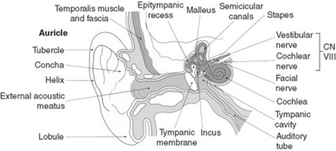
It is the part of the external ear that is outside the canal (Fig. 5-1). Made of cartilage and skin, it is highly flexible.
7 What are auricular bumps? What causes them?
 Darwin’s tubercle (Fig. 5-2): Benign and congenital nodule near the auricular apex (on the helix, at the junction of upper and middle thirds). Nontender and rarely bilateral, it was first described by the British sculptor Thomas Woolner, a founding member of the Pre-Raphaelite Brotherhood and a spare-time anatomist. Woolner depicted it in his statue of “Puck,” and Charles Darwin was so impressed that he named it the Woolnerian tip. It is an atavistic feature (i.e., a trait typical of our mammalian ancestors—more specifically, monkeys).
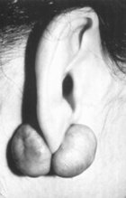
 Keloids (Fig. 5-3): Smooth and flesh-colored papule(s) on one or both sides of the earlobe. They indicate an exuberant and fibrotic response to injury.


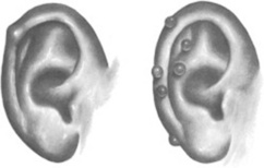
Figure 5-2 Darwin’s tubercle (left) and tophi (right).
(From Seidel HM, Ball JW, Daims JE, Benedict GW: Mosby’s Guide to Physical Examination, 3rd ed. St. Louis, Mosby, 1995.)


16 What is the value of pushing over the mastoid process?
Tenderness suggests suppurative mastoiditis—an ominous complication of ear infections.
19 What may induce vesicles in the auricle?
Not too many causes: (1) severe contact dermatitis (such as poison ivy); (2) varicella/zoster; and (3) Ramsay Hunt syndrome (painful and vesicular rash of the inferior portion of the auricle, due to herpetic infection of the geniculate ganglion and treated with acyclovir. See questions 48 and 49).
20 What are the causes of auricular red spots?


22 What is a tender and swollen auricle?
It is an uncommon but dramatic event. A diffusely swollen auricle is usually due to:
 Trauma: Easily identifiably by a history of recent altercation, especially if supported by other evidence of trauma, like a broken nose or a black eye. In fact, a “cauliflower” ear auricle is a time-honored occupational hazard of boxers, first portrayed in a beautiful Hellenistic statue of a resting fighter (Fig. 5-4). Unless evacuated, auricular hematomas heal with fibrosis and deformity and may even result in hearing loss. For instance, it has been suggested that Edison’s deafness was the result of having been picked up by the ears as a child. Still, there is no evidence that he had a cauliflower ear. President Johnson, on the other hand, contributed to our advance in veterinary medicine by demonstrating that cauliflower ears do not occur in dogs, especially beagles. In fact, he used to pick up his pooch by the ears and then toss him around in front of the press corps. LBJ, however, had no ear problems we know of, with the possible exception of selective deafness to war protesters in nearby Lafayette Park.


Stay updated, free articles. Join our Telegram channel

Full access? Get Clinical Tree


