Chapter 9 The Breast
A. Generalities
The clinical breast examination (CBE) is an effective screening tool for breast cancer; its accuracy depends on methodology and operator. Most of the research data stress palpation over inspection.
1 Why do a clinical breast examination (CBE)?
For either diagnosis of breast complaints (and primarily to rule out cancer) or for screening (to detect cancer in asymptomatic women). In primary care, CBEs are more screening than diagnostic (73% and 27%, respectively). They also get intertwined with medical litigation, since failure to detect breast cancer is a leading reason for malpractice claims, and primary care clinicians account for one half of all indemnities made.
2 Who should undergo a screening CBE? How frequently?
According to the American Cancer Society, CBEs should be carried out every 3 years in women between 20 and 39 years, and annually in women 40 and older. In this age group, the CBE can detect at least 50% of asymptomatic cancers and possibly contribute to a mortality reduction. A well-conducted exam cannot only detect potentially curable cancers, but also add to the yield of mammography, especially in women older than 70 (since fatty changes in their breast make lump detection easier).
3 What is the precision of CBE?
Hard to say, since CBEs are often carried out in nonstandardized fashion. Hence, their high interobserver variability. For example, 37 to 74 of 100 women screened by four different surgeons were found to have abnormal findings, and yet for only 25 of these women did all four surgeons agree on the findings. Agreement also varied by lesion: 13.5% for nipple discharge, 22.1% for dilated veins, 24.2% for “peau d’orange,” 59.4% for the lump per se, 61.5% for ulceration, and 68.1% for finding visibility.
4 What is the accuracy of CBE?
Hard to say, too, since to determine its screening accuracy the CBE should be compared to a standard criterion, which mammography cannot be, since cancers missed by mammograms can be found on CBE. Having said that, CBE sensitivity on pooled data is rather low (54%), but specificity is much stronger (94%). Overall, CBE has high false positive rates and even higher false negative rates. In silicone models, it has a sensitivity similar to that of population studies (40–71%), but a much lower specificity (41–77%).
5 What is the value of breast examination as compared to mammography?
CBE alone can detect 3–45% of cancers that screening mammography missed, with randomized clinical trials demonstrating reduced mortality rates in women screened by both techniques. Hence, CBE is an effective screening tool for breast cancer. Although unable to rule out disease when used alone, detection of certain abnormal findings by CBE can greatly increase the probability of breast cancer. Sensitivity of both professional and lay examiners can be improved by learning the correct examination method, and then practicing it on silicone breast models. Overall, for lesions of similar size, both examiner and patient factors can affect CBE’s accuracy.
7 What patient’s factors can adversely affect CBE’s accuracy?
Age. Denser breasts (typical of younger women) are harder to examine than fatty breasts (typical of older women), thus making lump detection more difficult. Larger breasts are also difficult, and so are fibrocystic breasts, which tend to be rather lumpy.
8 What is the bottom line for CBE modifiers?
Duration of exam, number of correct techniques, patient’s age, and the size/lumpiness of the breast may all affect CBE’s sensitivity. Size and hardness of the cancer can do it, too.
(1) Inspection
9 Which areas should be examined?
Breast tissue is contained in an imaginary pentagon, whose lateral border follows the midaxillary line, from the middle of the axilla down to the inframammary (or bra) line (fifth to sixth interspace); the lower border crosses along the inframammary fold toward the xiphoid process; the medial border ascends the midsternal line toward the suprasternal notch; and the upper border follows the clavicle before turning down toward the midaxilla. This pentagon can then be divided into four quadrants (Fig. 9-1). Most cancers originate in the upper outer quadrant of the breast, and also below the areola and nipple, two areas containing a large amount of glandular tissue. Always inspect first and palpate later.
10 What is the best way to inspect the breasts?
By examining the patient supine and then seated. Look for the breasts’ size, shape, and contour. Identify asymmetries, swelling, erythema, increase in venous pattern, and skin dimpling. Also search for nipple changes, including deviation, retraction, and inversion.
11 Which bedside maneuver can help to detect breast abnormalities on inspection?
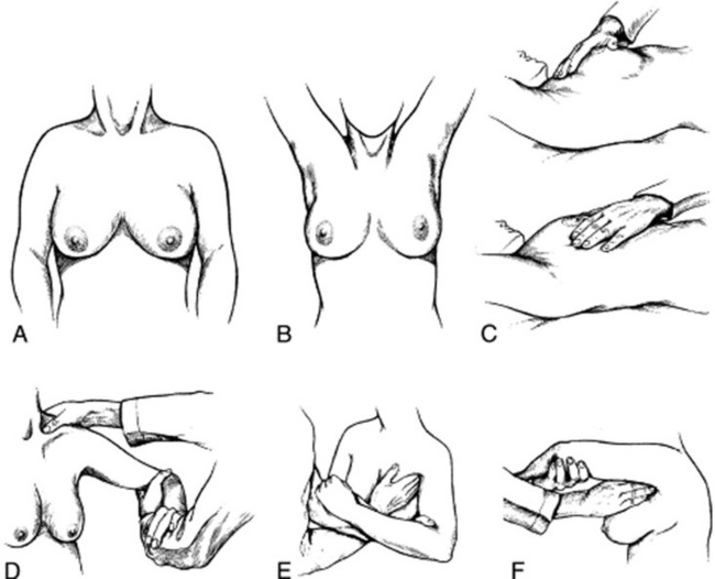
The most commonly taught and used maneuvers include a change in position of the patient’s arms and hands, first described by Haagensen (Fig. 9-2). To do so, ask the patient to carry out the following sequence:
1. >Rest the hands on the lap (to relax the pectoralis muscles).
2. Press them over the hips (to tense the pectoralis muscles and make dimpling and retraction more visible).
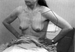
3. Raise them above the head, clasping them behind it (to also trigger skin dimpling, an important harbinger of cancer).
4. Lean forward (to allow the breasts to hang out pendulous from the chest).

Figure 9-2 Physical examination of the breast. A, Observation with patient sitting and arms resting at side. B, Observation with arms raised above the head. C, Systematic palpation with palm side of hand and fingers while patient is supine. D, Palpation of supraclavicular region of sitting patient with examiner supporting and elevating arm. E and F, Examination of axillae with volar surface of fingers by examiner standing on opposite side and totally supporting patient’s arm.
(From James EC, Corry RJ, Perry JF: Principles of Basic Surgical Practice. Philadelphia, Hanley & Belfus, 1987.)
Although these positions are commonly taught and practiced, they do take time. Moreover, the screening value of positioning (and even inspection) remains largely unproven. In a series of 296 breast cancers found on exam, 96% were discovered by palpation, 3% by visible nipple abnormalities, and only 1% by retraction alone. Yet, if the patient is symptomatic (or an abnormality is discovered during palpation), then careful inspection should definitely be carried out.
12 What are the most significant abnormalities that can be detected by inspection?
Noncongenital nipple asymmetry/deviation and retraction/inversion (both valuable clues to an underlying cancer). Nipple inversion may actually be normal, but only if long standing and correctable by manual pulling. Skin inspection also is important. Look for:
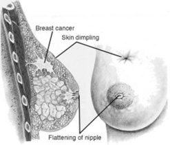
13 What is skin dimpling?
A slight depression or indentation in the breast’s surface (Fig. 9-3). This is an important clue to an underlying infiltrating carcinoma, causing fibrosis and retraction of the breast tissue. The same mechanism is responsible for nipple deviation.
14 What are the suspensory ligaments of the breast?
Suspensory (or Cooper’s) ligaments are thin, fibrous bands that run through the breast, attaching stroma to skin. Tension on these ligaments produces the characteristic dimpling that sometimes occurs over malignant masses.
15 What is peau d’orange?
It is French for orange peel—an apt description of the skin overlying an infiltrating cancer. It is due to lymphatic blockage by the tumor, resulting in localized lymphedema, with skin thickening and unusually large pores. It typically involves the lower aspect of the breasts.
16 What is Paget’s disease of the breast?
A malignant lesion of the areola and/or nipple, almost always associated with an in situ or invasive carcinoma. It is caused by extension of neoplastic cells from the lactiferous ducts into the epidermis, eventually causing it to become irritated. It presents as a scaly dermatitis of the nipple, with itching, crusting, and even erosion. Eventually, it involves the skin extensively, possibly without any palpable underlying mass. It may resemble other scaly and excoriative nipple diseases, such as eczema or the trauma of nursing.
17 Who was Paget?
“/>Sir James Paget (1814–1899) was a British surgeon who spent his entire career at the St. Bartholomew’s Hospital of London. A tall, brilliant, and charming man, he was so much enamored with clear and concise language to often quip, “To be brief is to be wise.” His fame grew to the point that Prime Minister Gladstone remarked that people are divided into two classes, “those who had, and those who had not heard of James Paget.” His name is linked to the first demonstration of trichinosis in humans (which he reported while still a medical student), osteitis deformans (described the same year in which he was made a baronet), the aforementioned breast disease, and a skin cancer of the apocrine glands presenting with the same cells as Paget’s disease of the breast.
(2) Palpation
18 What is the best way to palpate the breast?
One that uses (1) proper patient position; (2) awareness of breast boundaries; (3) adequate examination patterns; and (4) correct finger position, movement, and pressure. Duration of palpation is also crucial to success, with longer exams having overall a better yield. Note that a careful examination of an average-sized breast takes at least 3 minutes (6 minutes for both breasts), which is much more than the average 1.8 minutes physicians usually spend teaching breast self-examination (BSE) and examining both breasts.
19 Describe the proper patient position.
Since CBE requires flattening of the breast tissue against the chest, the patient must be supine. To further flatten the breast, one could try bedside maneuvers, especially in women with larger breasts. For example, to flatten the lateral part of the breast, you can ask the patient to roll onto her contralateral hip, rotate the shoulders back into a supine position, and place her ipsilateral hand on the forehead. Conversely, to flatten the medial part of the breast, you can ask the patient to lie flat on her back and raise her elbow until it is at the same level with her shoulder.

Stay updated, free articles. Join our Telegram channel

Full access? Get Clinical Tree

