The Bone Marrow


The Bone Marrow


Xiangdong Xu, M.D., Ph.D.

Anjum Hassan, M.D.


Acute lymphoblastic leukemia (ALL) is the most common malignancy in children and classically presents with pancytopenia, bleeding, and signs of anemia or infection. Characterized by an almost complete loss of normal hematopoietic elements, this disease tragically illustrates the fragility of the otherwise harmonically orchestrated “fluid-organ,” the bone marrow. Ultimately forming approximately 3% to 6% of the total body weight and reconstructing the peripheral blood throughout life, this organ undergoes a fascinating embryologic development. From midfetal development on and extending throughout life, the bone marrow is the site of origin of peripheral blood hematopoietic elements, specialized dendritic cells of monocyte-macrophage lineage, mast cells lymphocytes, natural killer (NK) cells, platelets, and osteoclasts (1). At this point, we know that the potency of some of the stem cells extends beyond this spectrum and through bone marrow mesenchymal stem cells; the bone marrow also contributes to endothelial cells, adipocytes, fibroblasts, osteoblasts, myofibroblasts, and reticular cells.


DEVELOPMENT

Mesenchymal-derived primitive erythroblasts in the yolk sac are the earliest signs of hematopoiesis in the embryo at a crown rump length of 95 mm (2). While the presence of lymphoid elements in the yolk sac is controversial, it has been shown that the aorta (aorta-gonad-mesonephros [AGM]) (3) and the placenta contribute in this earliest phase to the lymphomyeloid stem cell pool (4,5). The proposed candidates for hematopoietic stem cells (HSCs) in the AGM express CD34, CD45, CD117 (c-kit), and the transcription factor GATA-2 (6). The cells arising in the yolk sac show myeloid restriction (7). At weeks 10 to 24, the liver is the primary hematopoietic organ with production of red cells, granulocytes, and megakaryocytes in the primitive sinusoids. At this time, the spleen also contributes with approximately 20% to hematopoiesis. Slowly, the production within the bone marrow takes over, and at 4 to 5 months, it becomes the primary site of hematopoiesis. Typically by term, liver and spleen show minimal myelopoiesis. This switch is often referred to as embryo-to-fetal-to-adult-type hematopoieses (8). The development of the bone marrow continues in a topographically organized fashion. Hematopoiesis changes from the axial and radial skeleton (newborns) to the flat bones of the central skeleton by 12 to 16 years. Microscopically, the bone marrow is an inhomogeneous organ, which is often illustrated by higher cellularity within deeper areas of the medullary cavity than in subcortical zones. Due to the relatively short lifespan of peripheral blood elements, the production rates within the bone marrow are astronomic (9). The turnaround time of neutrophils (approximately 2 hours) requires the production of approximately 700,000 cells per second to maintain the normal value of 5000µL; exponentially higher values are needed in neutrophilia or sepsis, illustrating the dynamics of this system.

Significant age-related normal variations are seen in overall bone marrow cellularity with approximately 80% cellularity until 9 years, approximately 50% until 70 years, and less than 30% beyond. Relative proportions of various cell types also change. Hematopoiesis is a developmental continuum of HSCs and progenitor cells, which are very rare in normal bone marrow accounting for less than 0.001% of nucleated cells (Table 24-1). It is an exquisitely regulated, dynamic, and highly complicated system that involves complex interaction with diverse bone marrow microenvironment, coordinated expression of many genes, progressive loss of proliferative capacity, differentiation commitment, and maturation with specific biochemical, functional, and morphologic features (10,11).


BONE MARROW STRUCTURE

Encased and protected by cortical bone, traversed and supported by trabecular bone, the bone marrow consists of a highly organized thin-walled capillary network, venous sinuses, and surrounding extracellular matrix. The capillary-venous
sinuses, which result from bifurcations of the nutrient or medullary arteries, are the basic structural unit of the bone marrow (12). Within this histologic compartment, HSC and progenitor cells are exposed to the extracellular matrix that comprises the bone marrow microenvironment (Figure 24-1). The outer adventitial reticular cells (ARCs) add connective tissue elements and form the outer sinusoidal wall and synthesize collagen, laminin, fibronectin, and proteoglycans. All regulatory factors, adhesion molecules, and other proteins necessary for the regulation of hematopoiesis are contained within this matrix (1,13). Furthermore, the ARCs are phagocytic and can become lipocytes. As outlined before, the fat/hematopoietic ratio (“marrow cellularity”) is variable and a rough estimate can be calculated as cellularity = 100% — age (see below). Mitotically active cells are normally found around the supporting bone, typically paratrabecular and perivascular from where cells mature progressively into the medullary cavity. All newly formed mature hematopoietic cells are released into the bone marrow capillary-venous sinuses. Most cells pass through the sinus wall, but megakaryocytes reside adjacent to sinuses and extend pseudopodia directly into the vascular space (14,15). The capillary-venous sinuses coalesce into the venules and ultimately become veins that carry newly formed hematopoietic cells to the systemic circulation (12).


TABLE 24-1 GENERAL FEATURES OF THE BONE MARROW AND HEMATOPOIESIS


  • Microenvironment with regulatory factors for stem/progenitor cells and structural support via stromal framework and surrounding liquid matrix


  • Stem/progenitor cells localize to specific niches based on complementary adhesion molecule expression between hematopoietic cells, microenvironment, and stromal cells


  • Stem/progenitor cell proliferation and maturation under exquisite regulatory control; regulated “cross talk” between stromal cells and hematopoietic cells maintains steady state.


  • Stimulatory and suppressive factors within microenvironmental matrix; regulatory factors consist of CSFs, ILs, and inhibitory cytokines.


  • Stem cellsa are capable of self-renewal and multilineage differentiation.


  • Committed progenitor cellsa are destined to a specific lineage.


a Not morphologically distinct.


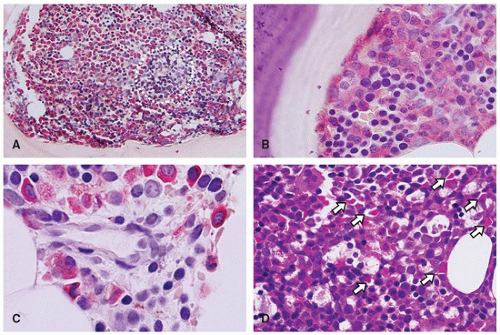
FIGURE 24-1 • Bone marrow microarchitecture. A: Bone marrow biopsy from a 1-day-old boy showing hematopoietic tissue that occupies approximately 90% of the marrow space. Only few regions of bone marrow fat are seen. The myeloid lineage is highlighted in red (Leder stain), and the perivascular region (circle) shows lack of myeloid cells. B: The paratrabecular region shows myeloid and erythroid precursors. C: Perivascular distribution of precursors in a bone marrow biopsy from an 18-year-old girl; note the delicate reticulum and extracellular matrix derived from ARCs. D: Highly cellular (>90%) bone marrow biopsy in a preterm girl shows numerous capillaries (arrows) interspersed between the hematopoietic cells and extracellular matrix.


STEM CELLS AND PROGENITOR CELLS

HSC can be defined by their ability to regenerate long-term multilineage hematopoiesis in myeloablated recipients. Although not morphologically recognizable, stem cells can be detected by either functional features (the simultaneous capability of sustained self-renewal and multilineage differentiation potential) or immunophenotype (CD34+, Thyr-1+, c-kit+, CD38, cytokine receptor, and adhesion molecule expression) (1,16) (Figure 24-2). HSCs are estimated to constitute 1 in 104 nucleated marrow cells. In contrast, progenitor cells are progressed stem cells with lineage commitment. The process of lineage commitment is incompletely understood; however, the resulting committed stem/progenitor cells are also morphologically unrecognizable but immunophenotypically defined by CD34, c-kit, and CD38 expression (16,17). Further, maturation is characterized by the acquisition of morphologic and immunophenotypic properties of the corresponding hematopoietic lineages. Both proliferation and lineage maturation are regulated by the synergistic
stimulatory activities of colony-stimulating factors (CSFs) and interleukins (ILs), whereas antagonistic effects are driven by inhibitory factors that include tumor necrosis factor (18,19). It is known that mature hematopoietic elements play a role in the regulation of lineage production and in maintaining steady-state hematopoiesis. In addition to the complicated molecular pathways, endocrine, paracrine, mesenchymal, and autonomic nervous system regulations have been implicated for homeostasis (18,19).


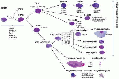
FIGURE 24-2 • Selected aspects of hematopoiesis. See text for details. CLP, committed lymphoid progenitor; CMP, committed myeloid progenitor (e.g., CFU-S: colony-forming unit—spleen); GEMM, granulo-erythro-megakaryo-monocytic; GM, granulo-monocytic (= myelomonocytic); HPC, hematopoietic progenitor committed; HSC, hematopoietic stem cell; Im-B, immature B-lymphocyte; PC, plasma cell; PSC, peripheral stem cell.


HEMATOPOIETIC LINEAGES

A detailed discussion of all hematopoietic lineages is beyond the scope of this chapter; however, some selected lineages are described below.


Granulopoiesis

The process of granulocytic maturation is characterized by a progressive nuclear segmentation, simultaneous decrease in the nuclear-to-cytoplasmic (N/C) ratio, as well as acquisition and increase of primary and later secondary cytoplasmic granules. The earliest morphologically recognizable cell in the granulocytic lineage is the myeloblast (20 µm; N/C ratio >85%); the subsequent arbitrary stages of this continuous maturation process include promyelocytes (the largest granulocytic cell), myelocytes, metamyelocytes, band neutrophils, and segmented neutrophils (Figure 24-3). The last maturation stage with a proliferative potential is a myelocyte. The key regulatory factors involved in granulopoiesis are granulocyte-macrophage colony-stimulating factor (GM-CSF), granulocyte colony-stimulating factor (G-CSF), and interleukin-3 (IL-3) (20). G-CSF is an 813 amino acid membrane protein that functions by binding to its specific cell surface receptor (G-CSFr) and activates cytoplasmic tyrosine kinases (21). Granulopoiesis is also under the control of retinoic acid receptors (RAR), which bind to all-trans retinoic acid (ATRA) and 9-cis-retinoic acid (21). The combination of four otherwise non-myeloid-restricted transcription factors is unique to the granulocyte lineage: C/EBPa (restricted to CD34+/CD33+ myeloid cells), PU.1 (Ets family member), CBF (AML1), and c-Myb (22,23,24,25). Other transcription factors (e.g., WT-1, Rb, and Hox) have also been implicated in granulopoiesis (25). Granulopoiesis occurs predominantly in paratrabecular and perivascular regions within the bone marrow (13). Thus, in normal bone marrow biopsy sections, immature granulocytic precursors selectively localize to the paratrabecular and, less conspicuously, the perivascular regions. This distribution may be altered after cytokine treatment, after chemotherapy, as well as after bone marrow transplantation (see below). Normal localization can be highlighted by immunoperoxidase staining for myeloperoxidase (MPO). Metamyelocytes, bands,
and neutrophils comprise the largest maturation storage compartment of the bone marrow, which can be released into the peripheral blood in response to multiple host-mediated challenges.


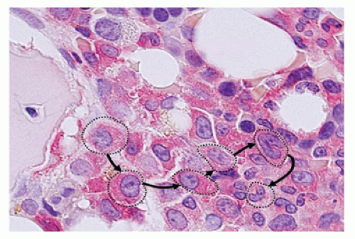
FIGURE 24-3 • Granulopoiesis. Immature granulocytic precursors (Leder positive) localize to the paratrabecular regions. Subsequent arbitrary stages are indicated (circles) and maturation progresses to, for example, band neutrophils.
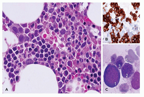

Erythropoiesis

The earliest morphologically recognizable cell in the erythroid lineage is the erythroblast (normoblast). The subsequent maturation has been arbitrarily divided into the basophilic normoblast, polychromatophilic normoblast, orthochromic normoblast, reticulocyte, and mature erythrocyte stages (Figure 24-4). The maturational process is characterized by progressive nuclear condensation with ultimate extrusion of the pyknotic nucleus at the end of the orthochromic normoblastic stage, which results in the young erythrocyte (reticulocyte). Simultaneously, the cytoplasm gradually changes from a deeply basophilic, organelle-rich substance to one that consists almost entirely of hemoglobin. In addition to the general growth factors (GM-CSF, IL-3, and IL-11), the primary growth factor responsible for red blood cell (RBC) production is erythropoietin (EPO), a 30.4-kDa glycoprotein that induces proliferation and maturation of committed erythroid progenitor cells by binding to its specific cell receptor (R-EPO), which inhibits apoptosis and thereby regulates the rate of red cell production (26,27). EPO does not cross the placenta, and therefore, the fetus primarily controls erythropoiesis (27). Although erythroid and megakaryocytic lineages share several transcription factors such as GATA-1 and NF-E2 (28,29), specific growth factors act selectively and allow committed cells to differentiate and proliferate. Erythropoiesis occurs in small colonies (erythroblast islands), and even though related to vascular structures, they appear randomly dispersed throughout the hematopoietic cavity. They are neither paratrabecular nor perivascular in distribution (30). Erythroid architecture can be highlighted by immunohistochemistry utilizing E-cadherin, hemoglobin A, or glycophorin (Figure 24-4).


FIGURE 24-4A: Erythropoiesis occurs in small colonies (erythroblast islands) related to vascular structures. B: Glycophorin A; marker of erythroid differentiation. C: Subsequent stages of erythroid differentiation.


Megakaryopoiesis

Megakaryocytes are the largest nucleated cell (50 to 150 µm) in the bone marrow. Unlike the maturation of the other lineages, megakaryocyte maturation from the blast to the mature cell stage is not associated with mitotic divisions. Megakaryocyte differentiation occurs via endomitosis, resulting in increasing nuclear lobulations without cell division (31), controlled via thrombopoietin (TPO) (32,33). The earliest megakaryocyte precursor identified in cell culture studies is the promegakaryoblast. Subsequent maturational stages have been arbitrarily designated as megakaryoblast, basophilic megakaryocyte, granular megakaryocyte, and platelet-producing megakaryocyte. The maturational sequence is characterized by a progressive increase in the overall size, an increase in nuclear lobulations (n = 8, 16 or 32), without nucleoli, and the development of demarcation membranes and multiple types of (purple-red or pink) cytoplasmic granules. Megakaryocyte production is regulated by a variety of factors, including multilineage growth factors such as GM-CSF, stem cell factor, IL-3, IL-6, and lineageselective factors such as IL-11 and TPO (32,33,34). TPO binds to c-Mpl and acts in synergy with other cytokines (see above, EPO, IFN-α, IFN-β) (35). Even though megakaryocytes appear randomly distributed in biopsy sections, they are localized selectively to the parasinusoidal regions within the bone marrow microanatomy. Megakaryocytes project pseudopodia into the vascular space, and proplatelets are directly released into the blood stream by this mechanism.


Monopoiesis and Dendritic Cell Development

Monocytes, the largest leukocyte (12 to 20 µm), are derived from the same precursor cells that give rise to neutrophils. Macrophage colony-stimulating factor (M-CSF) is instrumental in influencing the progenitor cells to differentiate into monocyte-macrophages (36). Gradual nuclear folding
and the acquisition of cytoplasmic granules characterize the stages of maturation, designated as monoblast, promonocyte, and mature monocyte. Although characteristically, monocytes have fewer and smaller granules than neutrophils, neither monoblasts nor promonocytes are generally recognizable in normal bone marrow. Monocytes circulate in the blood and subsequently migrate to solid tissues and become macrophages or various types of immune accessory cells. Due to this accessory role and evidence that these cells play an integrated, multifaceted role in humoral and cellular immunity beyond simple phagocytosis, the former designation mononuclear phagocyte system (37) has been replaced. Foucar and Foucar (38) proposed the alternative name mononuclear phagocyte and immunoregulatory effector (M-PIRE) system as a more accurate descriptor. The M-PIRE system includes monocytes, macrophages, multiple dendritic cells (e.g., Langerhans and dendritic reticulum cells), and their bone marrow precursors. Some evidence suggests a common cell of origin (39). Because the constituent cells show unique immunophenotypic and functional properties, the M-PIRE designation remains controversial. Regardless of the name, both macrophages (histiocytes) and dendritic cells are inconspicuous normal constituents of virtually all organ systems, and mature cells of monocyte-macrophage lineage also remain as a major constituent of the bone marrow microenvironment.


Lymphopoiesis

T and B lymphocytes are derived from the same HSCs that give rise to all hematopoietic elements. Regulatory molecules known to influence B-cell proliferation, differentiation, and function include IL-1, IL-2, IL-4, IL-10, adhesion molecules, and IFN-γ. In comparison, regulatory factors of T-cell development and function include IL-1 through IL-9 (40). The bone marrow microenvironment serves as the “bursal equivalent” in humans and is the primary site of postnatal B-cell development, whereas T-cell precursors migrate from the marrow to the thymus for maturation and differentiation. Antigenetically mature T and B cells can proliferate in response to a variety of cytokines.

The stages of maturation of both B and T lymphocytes are generally defined by the surface antigen profile rather than by morphologic features (Figure 24-2). The earliest immunologically recognizable B cells express nuclear terminal deoxynucleotidyl transferase (TdT), surface CD34, CD79a, and HLA-DR; CD10 expression is variable but common (41,42). Further maturation is characterized by the acquisition of cytoplasmic mu heavy chain and, later, surface immunoglobulin. B-cell precursors are generally infrequent in normal bone marrow, although these immature cells are much more prominent in specimens from infants and young children. When they are abundant, the term hematogones has been applied to immature lymphocytes (see below).

T-cell maturation is characterized by the presence of cytoplasmic and, later, surface CD3 together with the expression of many other antigens associated with T cells (43). Terminal maturation is defined by the development of either a helper (CD4+) or a cytotoxic (CD8+) suppressor T cell.

Although the terms lymphoblast and prolymphocyte have been applied to developing lymphoid cells and are utilized in leukemia classification, the distinction is not easy in normal bone marrow specimens. Lymphocytes migrate from blood to specific tissue sites throughout the body, selectively homing to B- or T-cell regions of lymph node, spleen, and thymus, and to widespread extranodal regions. T lymphocytes are characteristically long-lived and periodically recirculate.


Development of Natural Killer Cells

NK cells are morphologically indistinguishable from CD8+ cytotoxic T cells, both of which are large granular lymphocytes. NK cells were initially defined by a functional activity, that is, major histocompatibility complex (MHC)-nonrestricted cytotoxicity (44). These cells were subsequently found to perform many other functions (43,44,45). Evidence suggests a common T/NK progenitor cell, and the thymus may be an additional site of NK-cell maturation.

On immunophenotype analysis, NK cells are defined by the expression of adhesion molecules such as CD56, CD57, and CD16. However, the expression of these adhesion molecules is not restricted to NK cells. The fact that true NK cells lack surface CD3 and CD8 expression facilitates their distinction from cytotoxic suppressor T cells.

Cells with NK activities (both cytotoxic suppressor T cells and true NK cells) are concentrated within the large granular lymphocyte population of peripheral blood. The mature cells have round nuclei, condensed chromatin, inconspicuous nucleoli, and moderate amount of pale blue cytoplasm that contains a small number of coarse, azurophilic granules. The granules contain cytolytic perforin and associated granule proteases (e.g., granzyme) essential for their cytolytic activity.


NORMAL HEMATOPOIETIC PARAMETERS

The peripheral blood and bone marrow profiles are characterized by prominent age-related physiologic variations (Table 24-2 and Table 24-3). As previously outlined, bone marrow cellularity decreases with age (46) and is classically best evaluated on biopsy sections or imprints. Particle sections are the next best choice, and aspirate smears may be difficult to evaluate; however, section imprints and aspiration smears are all reported as equally reliable (47). While earlier references specified 100% cellularity at birth, more recent studies show that bone marrow cellularity is somewhat lower than previously estimated (48); therefore, the percentage should be taken as a representative figure. The distribution of erythroid and lymphoid elements also varies by age, whereas the proportion of bone marrow devoted to granulopoiesis is

generally stable. A dramatic decline in erythroid elements parallels the drop in EPO levels that occurs after birth in full-term neonates (49). Erythropoiesis returns to normal steady-state levels following resolution of this so-called physiologic anemia of infancy. Likewise, dramatic age-related variations occur in the proportion of bone marrow lymphoid cells, with up to 40% lymphocytes in bone marrow specimens of very young children and infants (50). The proportion of lymphocytes decreases in bone marrow specimens, and B-cell production in general declines with age (51).


TABLE 24-2 NORMAL VALUES FOR BONE MARROW AND DIFFERENTIAL CELL COUNTS


Parameter (Unit)


Cord Blood


Week 1


Week 4


1 Year


Child


Adult


Hemoglobin (g/dL)


16.5


17


14


12


13.5


M: 16 F: 14


Hematocrit (%)


53


54


43


37


40


M: 47 F: 41


RBC (×106µL)


5.3


5


4


4.6


4.6


M: 5.2 F: 4.6


MCV (fL)


115


100


98


80


84


90


MCHC (g/dL)


32


33


33


34


34


34


Reticulocytes (% of RBC)


3-7


0-1


0


0-1


0-1


0-1


Nucleated RBC (per 100 WBC)


500


0


0


0


0


0


WBC (×109/L)


20


12


10


10


7


6


Absolute neutrophil count (×109/L)


13


5


4


4


3


3


Absolute lymphocyte count (×109/L)


5


5


6


6-8


4


3


Platelet count (×109/L)


290


250


250


250


250


250


Cell Type


Normal Range (%)


Cell Type


Normal Range (%)


Myeloblasts


0-3


Basophils and precursors


0-1


Promyelocytes


2-8


Monocytes


0-1


Myelocytes


10-13


Erythroblasts


0-2


Metamyelocytes


10-15


Other erythroid elements


10-25


Band/neutrophils


25-40


Lymphocytes


10-35


Eosinophils and precursors


1-3


Plasma cells


0-1


TABLE 24-3 HEMATOLOGIC PROFILE DURING THE FIRST MONTH OF LIFE AND IN YOUNG INFANTS


I. Term infants to 1 month


  • Hgb and Hct drop from 16.5 g/dL and 53% at birth to 14 g/dL and 43% at 1 mo of age, respectively.


  • MCV declines from 115 fL at birth to about 98 fL at 1 mo.


  • Reticulocyte count drops from 5% to7% at birth to ˜0% at 1 mo.


  • Nucleated RBCs are present at birth but disappear in the first week of life.


  • Marked leukocytosis with neutrophilia is normal at birth and lymphocytes predominate by 1 mo.


II. Preterm infants


  • Lower Hgb and Hct levels at birth than term neonates


  • Higher MCV, more nucleated RBCs, and higher reticulocyte counts compared with term neonates


  • More rapid and pronounced physiologic nadir


  • Lower leukocyte counts than term neonates


III. Young infants


  • Neonatal assessment complex because of dramatic physiologic variations in conjunction with potential maternal, familial, obstetric, and other fetal and neonatal factors


  • Maternal factors: infections, medications, obstetrical complications, and underlying illnesses


  • For example, maternal and paternal incompatibility for RBC antigens can result in hemolysis (hemolytic disease of the newborn).


  • Numerous constitutional hereditary disorders of hematopoietic cell production and survival including:


    • Diamond-Blackfan anemia (red cell aplasia)


    • Thalassemias (hemoglobinopathy)


    • Congenital neutropenia (granulocyte aplasia)


  • Thrombocytopenia with absent radii (megakaryocyte aplasia)


  • Constitutional disorders can manifest at birth or in early infancy.


  • Fetomaternal hemorrhage or internal hemorrhage can produce neonatal anemia.


  • Other causes include various congenital malformations and congenital neoplasms.


Hct, hematocrit; Hgb, hemoglobin; MCV, mean corpuscular volume.


TABLE 24-4 INDICATIONS FOR BONE MARROW EXAMINATION IN CHILDREN


  • Peripheral blood abnormality (undetermined after regular workup)


  • Evaluation of possible constitutional hematopoietic disorder


  • Evaluation for leukemia, myelodysplasia, myeloproliferative/myelodysplastic disorders, and myeloproliferative neoplasms


  • Evaluation for fever of unknown origin, storage diseases, and unexplained splenomegaly


  • Staging and management of patients with certain types of neoplasms (e.g., Hodgkin and non-Hodgkin lymphoma, various other solid tumors)


  • Evaluation of patient with atypical but nondiagnostic lymphoreticular process in other sites


  • Evaluation of patient who does not follow predicted course of initial diagnosis (e.g., patient with presumed idiopathic thrombocytopenic purpura who does not respond to therapy)


  • Ongoing monitoring of response to therapy in patients with a variety of hematologic and lymphoreticular disorders


  • Bone marrow assessment prior to autologous bone marrow transplantation


TABLE 24-5 SPECIALIZED TECHNIQUES IN BONE MARROW EXAMINATIONS


Technique


Specimen Required


Indications


Culture


Aspirate, sterile


Workup for infection


Cytochemical stains


Air-dried aspirate smears


Lineage identification of immature cells


Immunohistochemical stains


FFPE tissues


Numerous antibodies available to assess for lymphoid, myeloid, erythroid, and megakaryocytic antigens as well as to determine lineage of metastatic processes Selected antibodies to assess immaturity (e.g., CD34, TdT) also available


Immunophenotyping (by flow cytometry)


Aspirate, sterile


Useful in determining immunophenotypic profile of wide variety of neoplastic disorders (e.g., leukemias and lymphomas) as well as benign infiltrates (e.g., hematogones)


Cytogenetics


Aspirate, sterile


Yield prognostic and diagnostic information in acute leukemias, myeloid neoplasms, and lymphoma Essential in the evaluation of acute leukemias


Fluorescence in situ hybridization


Air-dried smears, cell culture smears, or FFPE tissues


Assess for specific cytogenetic abnormality if probe available Useful in minimal residual disease assessment


Molecular analysis


FFPE tissues (PCR)


Useful in determining B- and T-cell clonality as well as gene rearrangements and other genetic aberrations


Aspirate, sterile (other methods)


Useful in detecting gene amplifications in metastatic neuroblastoma


FFPE: formalin-fixed, paraffin-embedded


Age-related variations in peripheral blood values are well delineated (Table 24-4), and the most dramatic changes are found in erythrocyte, neutrophil, and lymphocyte parameters (52).


HEMATOLOGIC PROFILE OF THE NEONATE

The 1st month of life is characterized by remarkable physiologic changes in erythrocyte and white blood cell (WBC) parameters (Table 24-5), and these parameters vary between full-term and preterm neonates (52). In the full-term neonates, the hematocrit, mean corpuscular volume (MCV), RBC, and WBC counts are higher than those in later life. The neonatal period is also the only time when circulating erythroid precursors are physiologic. The nucleated RBCs are cleared rapidly from the blood and do not normally persist
beyond the first 3 to 4 days of life (53). In healthy neonates, the relative hypoxia in utero is reversed at birth, so that a marked, transient, abrupt decline in erythropoiesis (so-called physiologic anemia of infancy) occurs. These physiologic changes are exaggerated in preterm infants (49).

The neonate assessment for a hematologic disorder is uniquely challenging because of the complex interplay between possible maternal, familial, and obstetric factors in conjunction with the normal and dramatic physiologic variations (53), all of which must be considered in the workup of any hematologic aberration.


EXAMINATION OF THE BONE MARROW IN CHILDREN

Indications for bone marrow examination in children are listed in Table 24-4. The decision to examine the bone marrow is made on an individual basis by correlating laboratory and hematologic findings with the clinical history. While the posterior iliac crest is the preferred site for the evaluation in older children, aspirates and even biopsy specimens can be obtained from the tibia in young infants (54). Before performing a bone marrow examination, careful consideration must be given to the types of specimens necessary for optimal evaluation of the most likely differential diagnosis (Table 24-5). Except for cultures, as a general rule, all specialized studies should be delayed until the bone marrow aspirate smears have been reviewed as adequate. Flow cytometry is one of the routine techniques for immunophenotyping and aids in determining the lineage and maturation of neoplastic infiltrates. Cytogenetic evaluation provides essential diagnostic and prognostic information not only in acute leukemias but also in other myeloid disorders and nonhematopoietic malignancies. Other ancillary techniques are also useful to assess for minimal residual disease in patients with leukemias/lymphomas and to evaluate metastatic processes (Table 24-5).


TABLE 24-6 CONSTITUTIONAL HEMATOLOGIC DISORDERS


Fanconi anemia


DNA repair defect (autosomal/X-linked recessive) with increased incidence of AML Aplastic anemia in >90%, prominent neonatal cytopenia, pancytopenia by midchildhood Gradual development of single and multilineage aplasia Associated congenital anomalies of bone, skin, kidney; mental retardation


15 genes (A-P) identified—most common:


FANCA (16q24.3; exon 43) 60%-70%


FANCC (9q22.3; exon 14) ˜14%


FANCG (9p13; exon 14) ˜10%


Dyskeratosis congenita


DNA repair defect, unable to maintain telomere complex (uncharacterized genetic subtype in 50%) Gradual development of pancytopenia and aplastic anemia (˜80%) Initial hypercellularity common Associated with congenital anomalies of skin, nails, mucosa; frequent mental retardation


Four genes identified:


X-linked recessive (˜30%)


DKC1/dyskerin (Xq28; exon 15)


Autosomal dominant (10%)


TERC (3q26; exon1)


TERT (5p15; exon16)


Autosomal recessive (˜1%)


NOP10 (15q14; exon 2)


TERT (5p15; exon 16)


Diamond-Blackfan anemiaa


90% cases are sporadic. Inherited forms are <10%, due to autosomal inheritance and haplodeficiency. Likely intrinsic progenitor cell defect Constitutional red cell aplasia (rare erythroblasts present) Some patients develop marrow failure.


Associated with congenital anomalies, especially skeletal (30%-40%)


Nine genes identified: (RPS19, RPL5, RPL11, RPL35A, RPS24, RPS17, RPS7, RPS10, and RPS26) 50%-60% cases carry at least one of these mutants.


Autosomal dominant


RPS19 (19q13.2), 25%, 129 distinct mutations


RPL5 (1p22.1), 6.6%, 39 mutations


RPS10 (6p21.31), 6.4%, 3 mutations


Congenital dyserythropoietic/idiopathic aplastic anemiaa


Erythroid hyperplasia/aplasia


Associated with distinctive bone marrow abnormalities including multinucleation, nuclear bridging, and megaloblastic changes/bone marrow failure


Chromosomal instability and increased incidence of malignancy (repair defect)


Heterozygous mutations in TERC and TERT are risk factors for some cases.


Shwachman-Diamond syndromeb


Constitutional neutropenia with frequent development of aplasia (˜20%)


Associated with congenital anomalies including exocrine pancreas insufficiency


One gene identified:


Autosomal recessive (˜90%)


SBDS (7q11; exon 5)


Thrombocytopenia with absent radiic (TAR)


Constitutional thrombocytopenic disorder with reduced megakaryocytes and bone anomalies


Compound inheritance (biallelic) of a low-frequency noncoding SNP and a rare null mutation in RBM8A (55)


RBM8A (1q21)


Congenital amegakaryocytic thrombocytopeniac


Isolated thrombocytopenia with decreased/no megakaryocytes


50% patients develop aplastic anemia at the age of 5.


Can evolve into MDS


Genetically heterogeneous; one autosomal recessive subtype characterized


C-MPL (1p34.2)


Lysosomal enzyme defects/storage disorders (multiple types):


Over 40 genetic disorders (˜1 in 7000 live births) with mostly secondary hematologic manifestations


Accumulation of substrate protein within histiocytes/macrophages


Increased bone marrow histiocytes with distinctive morphology


Classification into six groups:


Lipid storage disorders (Gaucher, Niemann-Pick) Gangliosidosis (Tay-Sachs disease) Leukodystrophies (ADL, MLD, Krabbe, Refsum, Pelizaeus-Merzbacher) Mucopolysaccharidosis (Hunter syndrome, Hurler disease) Glycoprotein storage disorders (mucolipidosis, pseudoHurler) Mucolipidoses (ML type I-IV; sialidosis)


a Considered a constitutional erythrocyte disorder; this group also includes hemoglobinopathies, membrane defects, and enzyme defects; for example, thalassemias, sickle cell disorders, hereditary spherocytosis, and pyruvate kinase deficiency (not discussed here).


b Considered a constitutional granulocyte disorder; this group also includes Kostmann agranulocytosis syndrome, cyclic neutropenia, and Chediak-Higashi syndrome (see Table 24-5).


c Considered a constitutional megakaryocytic disorder.


ALD, adrenal leukodystrophy; MLD, metachromatic leukodystrophy.


Inherited Bone Marrow Failure Syndromes and Constitutional Disorders

Bone marrow biopsies for constitutional/inherited hematologic disorders may be encountered in clinical practice. The different entities represent a heterogeneous group of diseases and involve individual lineage with defects of, for example, erythroid, megakaryocytic, and/or histiocytic elements (Table 24-6). Many of these disorders (e.g., thrombocytopenia with absent radii; see below) are evident at birth or shortly thereafter, whereas the multilineage abnormalities that characterize the constitutional aplastic anemias usually develop gradually, sometimes not until adulthood (62,63). Another interesting pattern is that these hematologic disorders are frequently associated with a variety of abnormalities in other organ systems (Table 24-6), while the bone marrow picture is largely one of single lineage aplasia or multilineage failure without distinctive morphologic aberrations (62). Exceptions include marked dyserythropoiesis in congenital dyserythropoietic anemia and erythroid hyperplasia in various constitutional erythrocyte survival disorders (64,65).

The related group of storage diseases typically occurs as a consequence of lysosomal enzyme defects, affecting mainly histiocytes. Various tissues throughout the body could be involved, and the affected cells exhibit distinctive morphologic abnormalities caused by the accumulation of substrate proteins. Although not a primary hematologic disorder, the accumulation of abnormal histiocytes in the bone marrow produces secondary hematologic effects (62) (see Chapter 5).


Aplastic Anemia in Children

Aplastic anemia in children can be separated into constitutional/inherited versus acquired (66). This heterogeneous group of disorders, characterized by bone marrow failure with/without somatic abnormalities, typically presents with bone marrow failure in childhood. Eventually, severe trilineage hypoplasia develops; however, despite the name (aplastic), initial presentation is often trilineage hyperplasia, megaloblastic changes, or single lineage aplasia. It is noteworthy that some cases may not present until adulthood, highlighting the importance not only for pediatric pathologists. Since cloning of the first aplastic anemia-related gene in 1992 [Fanconi anemia (FA)-gene], considerable advances in the syndromic entities have been made (62). It is clear that approximately 20% of bone marrow failure syndromes in children are inherited and approximately 10% represent secondary causes. The latter includes radiation, chemicals and drugs (typically busulfan, chloramphenicol, nonsteroids), viruses (e.g., hepatitis), and immunologic causes (e.g., systemic lupus erythematosus). The classic diepoxybutane/mitomycin C-induced chromosome fragility testing in cytogenetics has been complemented by targeted molecular approaches (67). The former test assayed the underlying constitutional DNA repair defect in FA, which represents the most common genetic aplastic anemia (Table 24-6). Our current understanding of the molecular mechanisms underlying this group of diseases is convergence in the DNA repair-FBRCA pathway (62). The diseases affect telomere complex maintenance in dyskeratosis congenita-related genes (e.g., DKC1/dyskerin, TERC, TERT, NOP10), ribosome biogenesis in Shwachman-Diamond syndrome (SBDS) and Diamond-Blackfan anemia genes (nine genes including RPS19 and RPL5) (68,69), or TPO receptor (C-MPL) in congenital amegakaryocytic thrombocytopenia (62,67). Despite the availability of mutational information and mode of inheritance (Table 24-6), the majority (approximately 70%) of “classical” bone marrow failures are “idiopathic” or uncharacterized, and therefore, the main/primary pathogenesis remains unknown (62,70). The peak incidence for secondary and idiopathic aplastic anemia in children is 3 to 5 years of age, and the morphologic features in the bone marrow are generally severely reduced or absent hematopoiesis.


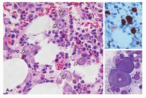
FIGURE 24-5 • Parvovirus. A,B: Intranuclear viral inclusions in a patient with parvo B19-induced red cell aplasia (arrows) and corresponding parvovirus immunohistochemistry. C: Morphology of intranuclear inclusion on smear (Wright-Giemsa stain).


Benign Erythroid Disorders in Children

Nonneoplastic erythroid disorders (also known as pure red cell aplasia) consist primarily of congenital and acquired anemias (Table 24-6) (71). The congenital form is induced by intrauterine damage to early erythroid precursors (65). Although the uncommon familial or tumor-associated polycythemia/erythrocytosis can be seen in the neonatal period, the most common neonatal polycythemia is physiologic, resulting from intrauterine hypoxia. The prevalence of specific types of anemia varies by patient age and ethnicity. In neonates, anemias secondary to blood loss predominate, followed by immune and nonimmune hemolytic processes. Anemias secondary to either maturation or proliferation defects are uncommon in infants and include constitutional red cell aplasia and congenital dyserythropoietic anemias (65).

Depending on the ethnic composition in a given practice area, constitutional erythrocyte survival disorders, including hemoglobinopathies and erythrocyte membrane disorders (72), could be relatively common causes of anemia in infants. However, bone marrow examination is generally not required for diagnosis.

A relatively common diagnostic challenge in bone marrow biopsies in children is the classification of red cell aplasia. The three primary causes of red cell aplasia in young children are Diamond-Blackfan anemia, transient erythroblastopenia of childhood, and red cell aplasia secondary to parvovirus infection (73). The latter (also-called acquired pure red cell aplasia) is typically transient and self-limited. If a variety of clinical, laboratory, hematologic, and bone marrow morphologic findings are integrated, the types of red cell aplasia in young children can generally be distinguished. In all types of constitutional and acquired red cell aplasia, the bone marrow is characterized by a profound decrease in maturing erythroid elements, although usually a variable number of erythroblasts are apparent. In addition, distinctive intranuclear inclusions within the residual enlarged erythroblasts are the hallmark of parvovirus infection, but these may not be readily apparent in all cases, and immunohistochemistry can be helpful (Figure 24-5). Consequently, acute parvovirus infection should always be excluded by serologic or molecular studies in cases of red cell aplasia, even when the
morphologic features of parvovirus infection are lacking. Another distinctive bone marrow finding, increased hematogones (74,75), may accompany any type of red cell aplasia in children, especially in very young patients.

In older infants and children, the most common cause of anemia is iron deficiency; other causes of anemia in this age group include chronic diseases, such as HIV-1 infection, and red cell aplasia. The frequency of constitutional hemolytic anemias varies by ethnicity. Other nutritional anemias, including vitamin B12 and folate deficiency, occur in both constitutional and acquired forms in children, but the incidence is low. For many of these types of anemia, the diagnosis can be established by integrating clinical and laboratory parameters, and bone marrow examination may not be required; however, bone marrow evaluation is necessary for diagnosis in most patients with red cell aplasia, pancytopenia, and suspected congenital dyserythropoietic anemia (76,77).

Finally, drugs and chemicals are associated with the development of pure red cell aplasia (78). Common examples include ampicillin, azathioprine, carbamazepine, cephalothin, chlormadinone, cotrimoxazole, D-penicillamine, erythromycin, estrogens, furosemide, gold, indomethacin, rifampicin, and valproic acid (79).


TABLE 24-7 SELECTED CONSTITUTIONAL HEMATOLOGIC DISORDERS INVOLVING GRANULOCYTES


Immunodeficiency Disorder


Mode of Inheritance and Predominant Morphologic Findings


Clinical Observations


Phagocytic/motility/adhesion defects in granulocytes


Chronic granulomatous disease (56)


X-linked or autosomal recessive. Abscess and granulomas common; normal leukocyte morphology


Defective microbial killing by phagocytic cells; any site can be involved, most documented are GI lesions and lungs; incomplete response to infections


Leukocyte adhesion defect (57)


LAD-1: Autosomal recessive, mutations in ITGB2 (a subunit of CD18). LAD-2: mutation in a fucose transporter on chromosome 11; rolling defects LAD-3: mutation in FERMT3 gene; defects in integrin activation Distinct lack of neutrophils at sites of infection despite peripheral neutrophilia and myeloid hyperplasia in bone marrow; normal morphology


Delayed wound healing, delayed attachment of umbilical cord, recurrent infections


Chediak-Higashi syndrome (58)


Autosomal recessive; mutations in CHS1/LYST gene; functionally defective neutrophils with giant cytoplasmic granules


Recurrent pyogenic infections, partial oculocutaneous albinism, progressive neuropathy


Cyclic neutropenia (59)


Autosomal dominant or sporadic; mutations of ELA2 gene; absence of granulocytic precursors in neutropenic phase; normal morphology


Cyclic hematopoiesis with periods of neutropenia lasting from 9 to 21 days followed by neutrophilia. Increased infections correspond to neutropenic cycle


Kostmann syndrome (60,61)


Autosomal recessive or sporadic; severe neutropenia with sustained myeloid aplasia in the bone marrow


Recurrent bacterial infections; myelodysplasia and AML may be seen in those treated with G-CSF therapy.

Only gold members can continue reading. Log In or Register to continue

Stay updated, free articles. Join our Telegram channel

Sep 23, 2016 | Posted by in PATHOLOGY & LABORATORY MEDICINE | Comments Off on The Bone Marrow

Full access? Get Clinical Tree

Get Clinical Tree app for offline access