Rare cases may transform to syringocystadenocarcinoma papilliferum
Rarely, basal cell carcinoma may develop within SCP
Microscopic
• Endophytic/exophytic adnexal tumors
Invaginations that communicate with epidermal surface
Invaginations have papillary architecture
• Papillary structures lined by glandular epithelium with double layer
Papillae communicate with duct-like structures in deeper aspects
Basal layer is flattened to cuboidal
Luminal layer usually columnar
• Characteristic stroma
Fibrovascular connective tissue within papillae
Numerous plasma cells admixed with some lymphocytes
Top Differential Diagnoses
• Hidradenoma papilliferum
Lacks connection with epidermis
Lacks plasma cells
• Tubular apocrine adenoma
Lacks epidermal attachment
• SCP/malignant syringocystadenoma
Architectural complexity
Cytologic atypia
Mitotic activity
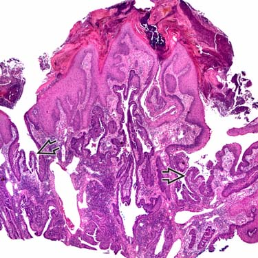
Low-Power Image of Syringocystadenoma Papilliferum Syringocystadenoma papilliferum has an endo/exophytic growth pattern with invaginations into the underlying dermis that have a papillary architecture .
Syringocystadenoma Papilliferum With Transition From Squamous to Glandular Epithelium The superficial aspects of the imaginations are lined by squamous epithelium that transitions to glandular epithelium .
Syringocystadenoma Papilliferum: High Magnification of Glandular Epithelium The glandular component is composed of 2 cell layers with a basal myoepithelial layer and a columnar glandular layer .
High Magnification of Apocrine Glandular Epithelium and Plasma Cells The glandular epithelium in this case shows evidence of apocrine differentiation , with focal apical snouts and secretions. The stroma contains numerous plasma cells .
TERMINOLOGY
Abbreviations
• Syringocystadenoma papilliferum (SCP)
Definitions
• Benign adnexal tumor with endophytic and exophytic growth pattern
ETIOLOGY/PATHOGENESIS
Cytogenetics
• Subset of tumors show loss of heterozygosity for PTCH1 (PTCH) &/or CDKN2A (P16), suggesting role for loss of these tumor suppressor genes in some cases
CLINICAL ISSUES
Epidemiology
• Age
~ 1/2 present at birth or childhood
Presentation
• Scalp most common location, followed by face
• Solitary gray to dark brown papillomatous lesion
• Rarely multiple
• SCP seen in 5-19% of nevus sebaceus
2nd most common tumor arising in nevus sebaceus after trichoblastoma
• Rarely associated with other neoplasms (e.g., tricholemmoma, apocrine hidrocystoma)
Treatment
• Surgical approaches
Simple excision is curative
Prognosis
• Benign
Rare cases may have associated basal cell carcinoma
Only gold members can continue reading. Log In or Register to continue


 .
.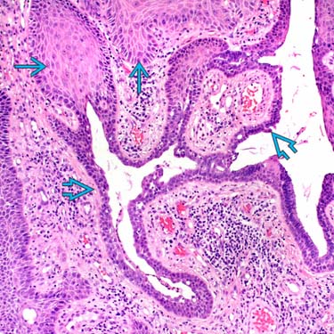
 that transitions to glandular epithelium
  .
.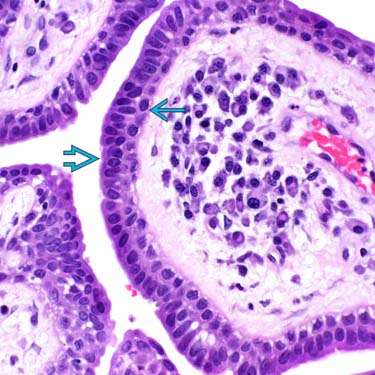
 and a columnar glandular layer
  .
.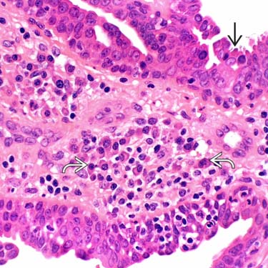
 , with focal apical snouts and secretions. The stroma contains numerous plasma cells
  .
.


