
OVERVIEW
OLFACTORY NERVE (CN I)
TABLE 9.1. Cranial Nerves: Attachment to Central Nervous System, General Functions, and Distribution
TABLE 9.2. Summary of Cranial Nerves
TABLE 9.3. Cranial Parasympathetic Ganglia: Location, Parasympathetic and Sympathetic Roots, and Main Distribution 1060
OPTIC NERVE (CN II)
OCULOMOTOR NERVE (CN III)
TROCHLEAR NERVE (CN IV)
TRIGEMINAL NERVE (CN V)
Ophthalmic Nerve (CN V1)
Maxillary Nerve (CN V2)
Mandibular Nerve (CN V3)
TABLE 9.4. Summary of Divisions of Trigeminal Nerve (CN V)
ABDUCENT NERVE (CN VI)
FACIAL NERVE (CN VII)
Somatic (Branchial) Motor
Visceral (Parasympathetic) Motor
Somatic (General) Sensory
Special Sensory (Taste)
VESTIBULOCOCHLEAR NERVE (CN VIII)
GLOSSOPHARYNGEAL NERVE (CN IX)
Somatic (Branchial) Motor
Visceral (Parasympathetic) Motor
Somatic (General) Sensory
Special Sensory (Taste)
VAGUS NERVE (CN X)
SPINAL ACCESSORY NERVE (CN XI)
HYPOGLOSSAL NERVE (CN XII)
TABLE 9.5. Summary of Vagus Nerve (CN X)
BLUE BOX: Cranial Nerves. Cranial Nerve Injuries. Olfactory Nerve. Anosmia–Loss of Smell; Olfactory Hallucinations. Optic Nerve. Demyelinating Diseases and Optic Nerves; Optic Neuritis; Visual Field Defects. Oculomotor Nerve. Injury to Oculomotor Nerve; Compression of Oculomotor Nerve; Aneurysm of Posterior Cerebral or Superior Cerebellar Artery. Trochlear Nerve. Trigeminal Nerve. Injury to Trigeminal Nerve; Dental Anesthesia. Abducent Nerve. Facial Nerve. Vestibulocochlear Nerve. Injuries to Vestibulocochlear Nerve; Deafness; Acoustic Neuroma; Trauma and Vertigo. Glossopharyngeal Nerve. Lesions of Glossopharyngeal Nerve; Glossopharyngeal Neuralgia. Vagus Nerve. Spinal Accessory Nerve. Hypoglossal Nerve
TABLE 9.6. Summary of Cranial Nerve Lesions
The regional aspects of the cranial nerves are described in the preceding chapters, especially those for the head and neck. This chapter summarizes all of the cranial nerves, largely in figures and tables. Figures 9.1–9.3 and Tables 9.1 and 9.2 summarize specific cranial nerves. Figure 9.4 and Table 9.3 summarize the cranial parasympathetic ganglia, their location, sympathetic and parasympathetic roots, and main distribution.

FIGURE 9.1. Summary of cranial nerves.

FIGURE 9.2. Cranial nerves in relation to internal aspect of cranial base. The tentorium cerebelli has been removed and the venous sinuses have been opened on the right side. The dural roof of the trigeminal cave has been removed on the left side and CN V1, CN III, and CN IV have been dissected from the lateral wall of the cavernous sinus.
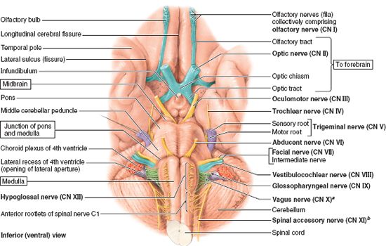
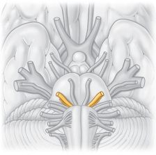
FIGURE 9.3. Superficial origins of cranial nerves from brain and spinal cord (except for CN IV, which arises from the posterior aspect of the midbrain). aThe traditional “cranial root of the accessory nerve” is considered here as part of the vagus nerve. bThe spinal accessory nerve as listed here refers to only the traditional “spinal root of the accessory nerve.”

FIGURE 9.4. Summary of cranial parasympathetic ganglia.
TABLE 9.1. CRANIAL NERVES: ATTACHMENT TO CENTRAL NERVOUS SYSTEM, GENERAL FUNCTIONS, AND DISTRIBUTION
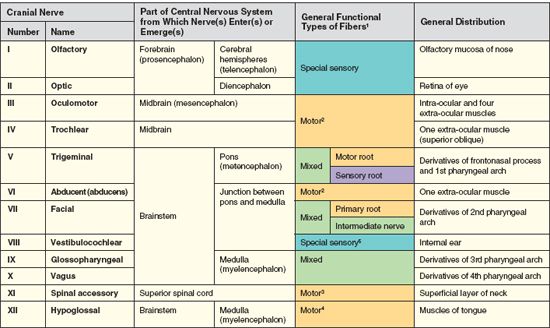
1Note that the colors in this column match those of the nerves in Figure 9.3.
2The presence and function of proprioceptive afferent fibers to the extra-ocular muscles is controversial.
3Cranial nerve XI is purely motor as it leaves the CNS, but gains pain and proprioceptive fibers from the cervical plexus in the lateral cervical region (posterior triangle) of the neck.
4Cranial nerve XII is purely motor as it leaves the CNS; pathways for proprioception associated with the tongue are unknown and may involve the lingual and glossopharyngeal nerves, and cervical spinal nerves that communicate with CN XII.
5The cochlear part of CN VIII, traditionally considered “purely sensory,” actually conveys some efferent fibers that appear to modulate sensory sensitivity.
TABLE 9.2. SUMMARY OF CRANIAL NERVES


TABLE 9.3. CRANIAL PARASYMPATHETIC GANGLIA: LOCATION, PARASYMPATHETIC AND SYMPATHETIC ROOTS, AND MAIN DISTRIBUTION

TABLE 9.4. SUMMARY OF DIVISIONS OF TRIGEMINAL NERVE (CN V)

OVERVIEW
Like spinal nerves, cranial nerves are bundles of sensory or motor fibers that innervate muscles or glands, carry impulses from sensory receptors, or have a combination of motor and sensory fibers. They are called cranial nerves because they emerge through foramina or fissures in the cranium and are covered by tubular sheaths derived from the cranial meninges. There are 12 pairs of cranial nerves, which are numbered I–XII, from rostral to caudal (Figs. 9.1–9.3). Their names reflect their general distribution or function.
Cranial nerves carry one or more of the following five main functional components (Fig. 9.3, Table 9.1).
• Motor (efferent) fibers.
1. Motor fibers to voluntary (striated) muscle. These include the somatic motor (general somatic efferent) axons. On the basis of the embryologic/phylogenetic derivation of certain muscles of the head and neck,1 some motor fibers conveyed by cranial nerves to striated muscle have traditionally been classified as “special visceral.” When appropriate, these fibers are designated somatic (branchial) motor, referring to the muscle tissue derived from the pharyngeal arches in the embryo (e.g., muscles of mastication).
2. Motor fibers involved in innervating involuntary (smooth) muscles or glands. These include visceral motor (general visceral efferent) axons that constitute the cranial outflow of the parasympathetic division of the autonomic nervous system (ANS). The presynaptic (preganglionic) fibers emerge from the brain and synapse outside the central nervous system (CNS) in a parasympathetic ganglion. The postsynaptic (postganglionic) fibers continue to innervate smooth muscles and glands (e.g., the sphincter pupillae and lacrimal gland).
• Sensory (afferent) fibers.
3. Fibers transmitting general sensation (e.g., touch, pressure, heat, cold, etc.) from the skin and mucous membranes. These include somatic sensory (general somatic afferent) fibers, mainly carried by CN V, but also by CN VII, CN IX, and CN X.
4. Fibers conveying sensation from the viscera. These include visceral sensory (general visceral afferent) fibers conveying information from the carotid body and sinus (see Fig. 8.17), pharynx, larynx, trachea, bronchi, lungs, heart, and gastrointestinal tract.
5. Fibers transmitting unique sensations. These include special sensory fibers conveying taste and smell (special visceral afferent fibers) and those serving the special senses of vision, hearing, and balance (special somatic afferent fibers).
Some cranial nerves are purely sensory, others are considered purely motor, and several are mixed. CN III, CN IV, CN VI, CN XI, CN XII, and the motor root of CN V are considered to be pure motor nerves that appear to have evolved from primordial anterior roots. However, a small number of sensory fibers for proprioception (nonvisual perception of movement and position) are also present in these nerves, the cell bodies of which are probably located in the mesencephalic nucleus of CN V. The sensory root of CN V is purely a somatic (general) sensory nerve. Four cranial nerves (CN III, CN VII, CN IX, and CN X) contain presynaptic parasympathetic (visceral motor) axons as they emerge from the brainstem. CN V, CN VII, CN IX, and CN X are mixed nerves with both somatic (branchial) motor and somatic (general) sensory components, and each nerve supplies derivatives of a different pharyngeal arch.
The fibers of cranial nerves connect centrally to cranial nerve nuclei—groups of neurons in which sensory or afferent fibers terminate and from which motor or efferent fibers originate (Fig. 9.5, see p. 1061). Except for CN I and CN II, which involve extensions of the forebrain, the nuclei of the cranial nerves are located in the brainstem. Nuclei of similar functional components (e.g., somatic or visceral motor, or somatic or visceral sensory) are generally aligned into functional columns in the brainstem.

FIGURE 9.5. Cranial nerve nuclei
OLFACTORY NERVE (CN I)
Function: Special sensory (special visceral afferent)—that is, the special sense of smell. “Olfaction is the sensation of odors that results from the detection of odorous substances aerosolized in the environment” (Simpson, 2006).
The cell bodies of olfactory receptor neurons are located in the olfactory organ (the olfactory part of the nasal mucosa or olfactory area), which is located in the roof of the nasal cavity, and along the nasal septum and medial wall of the superior nasal concha (Fig. 9.6, see p. 1062). Olfactory receptor neurons are both receptors and conductors. The apical surfaces of the neurons possess fine olfactory cilia, bathed by a film of watery mucus secreted by the olfactory glands of the epithelium. The olfactory cilia are stimulated by molecules of an odiferous gas dissolved in the fluid.

FIGURE 9.6. Olfactory system. A. This sagittal section through the nasal cavity shows the relationship of the olfactory mucosa to the olfactory bulb. B. The bodies of the olfactory receptor neurons are in the olfactory epithelium. These bundles of axons are collectively called the olfactory nerve (CN I).
The basal surfaces of the bipolar olfactory receptor neurons of the nasal cavity of one side give rise to central processes that are collected into approximately 20 olfactory nerves (L. fila olfactoria), constituting the right or left olfactory nerve (CN I). They pass through tiny foramina in the cribriform plate of the ethmoid bone, surrounded by sleeves of dura mater and arachnoid mater, and enter the olfactory bulb in the anterior cranial fossa (Figs. 9.2 and 9.3). The olfactory bulb lies in contact with the inferior or orbital surface of the frontal lobe of the cerebral hemisphere. The olfactory nerve fibers synapse with mitral cells in the olfactory bulb. The axons of these secondary neurons form the olfactory tract. The olfactory bulbs and tracts are anterior extensions of the forebrain.
Each olfactory tract divides into lateral and medial olfactory striae (distinct fiber bands). The lateral olfactory stria terminates in the piriform cortex of the anterior part of the temporal lobe, and the medial olfactory stria projects through the anterior commissure to contralateral olfactory structures. The olfactory nerves are the only cranial nerves to enter the cerebrum directly.
The Bottom Line
OLFACTORY NERVE
♦The olfactory nerves (CN I) have sensory fibers concerned with the special sense of smell. ♦ The olfactory receptor neurons are in the olfactory epithelium (olfactory mucosa) in the roof of the nasal cavity. ♦ The central processes of the olfactory receptor neurons ascend through foramina in the cribriform plate of the ethmoid bone to reach the olfactory bulbs in the anterior cranial fossa. These nerves synapse on neurons in the bulbs, and the processes of these neurons follow the olfactory tracts to the primary and associated areas of the cerebral cortex.
OPTIC NERVE (CN II)
Function: Special sensory (special somatic afferent)—that is, the special sense of vision.
Although officially nerves by convention, the optic nerves (CN II) develop in a completely different manner from the other cranial nerves. The structures involved in receiving and transmitting optical stimuli (the optical fibers and neural retina, together with the pigmented epithelium of the eyeball) develop as evaginations of the diencephalon. The optic nerves are paired, anterior extensions of the forebrain (diencephalon) and are therefore actually CNS fiber tracts formed by axons of retinal ganglion cells (Moore et al., 2012). In other words, they are third-order neurons, with their cell bodies located in the retina (Fig. 9.7B).

FIGURE 9.7. Visual system. A. The origin, course, and distribution of the visual pathway are shown. The axons of retinal ganglionic neurons convey visual information to the lateral geniculate body of the diencephalon (thalamus) through the optic nerve (CN II) and optic tract. Fibers from the lateral geniculate body project to the visual cortices of the occipital lobes. The axons of the ganglion cells of the nasal halves of the retinas cross in the optic chiasm; those from the temporal halves do not cross. B. The visual pathway begins with photoreceptor cells (rods and cones) in the retina. The responses of the photoreceptors are transmitted by bipolar cells (neurons with two processes) to ganglion cells in the ganglion cell layer of the retina. The central processes of this third-order neuron are the fibers conducted by the optic nerves.
The optic nerves are surrounded by extensions of the cranial meninges and subarachnoid space, which is filled with cerebrospinal fluid (CSF). The meninges extend all the way to the eyeball. The central artery and vein of the retina traverse the meningeal layers and course in the anterior part of the optic nerve. Cranial nerve II begins where the unmyelinated axons of retinal ganglion cells pierce the sclera (the opaque part of the external fibrous coat of the eyeball) and become myelinated, deep to the optic disc.
The nerve passes posteromedially in the orbit, exiting through the optic canal to enter the middle cranial fossa, where it forms the optic chiasm (Fig. 9.7A). Here, fibers from the nasal (medial) half of each retina decussate in the chiasm and join uncrossed fibers from the temporal (lateral) half of the retina to form the optic tract.
The partial crossing of optic nerve fibers in the chiasm is a requirement for binocular vision, allowing depth-of-field perception (three-dimensional vision). Thus fibers from the right halves of both retinas form the right optic tract. The decussation of nerve fibers in the chiasm results in the right optic tract conveying impulses from the left visual field and vice versa. The visual field is what is seen by a person who has both eyes wide open and who is looking straight ahead. Most fibers in the optic tracts terminate in the lateral geniculate bodies of the thalamus. From these nuclei, axons are relayed to the visual cortices of the occipital lobes of the brain.
The Bottom Line
OPTIC NERVE
♦The optic nerves (CN II) have sensory fibers concerned with the special sense of vision. ♦ The optic nerve fibers arise from ganglion cells in the retina. ♦ The nerve fibers exit the orbit via the optic canals; fibers from the nasal half of the retina cross to the contralateral side at the optic chiasm. ♦ The nerve fibers then pass via the optic tracts to the geniculate bodies of the thalamus, where they synapse on neurons whose processes form the optic radiations to the primary visual cortex of the occipital lobe.
OCULOMOTOR NERVE (CN III)
Functions: Somatic motor (general somatic efferent) and visceral motor (general visceral efferent–parasympathetic).
Nuclei: There are two oculomotor nuclei, each serving one of the functional components of the nerve. The somatic motor nucleus of the oculomotor nerve is in the midbrain (Fig. 9.5). The visceral motor (parasympathetic) accessory (Edinger-Westphal) nucleus of the oculomotor nerve lies dorsal to the rostral two thirds of the somatic motor nucleus (Haines, 2006).
The oculomotor nerve (CN III) provides the following (Fig. 9.8):
• Motor to the striated muscle of four of the six extra-ocular muscles (superior, medial, and inferior recti and inferior oblique) and superior eyelid (L. levator palpebrae superioris); hence the nerve’s name.
• Parasympathetic through the ciliary ganglion to the smooth muscle of the sphincter pupillae, which causes constriction of the pupil and ciliary muscle, which produces accommodation (allowing the lens to become more rounded) for near vision.

FIGURE 9.8. Distribution of oculomotor (CN III), trochlear (CN IV), and abducent (CN VI) nerves. CN IV supplies the superior oblique, CN VI supplies the lateral rectus, and CN III supplies five striated extra-ocular muscles (levator palpebrae superioris, superior rectus, medial rectus, inferior rectus, and inferior oblique) and two intra-ocular muscles (ciliary muscle and sphincter pupillae muscle—not shown; see Chapter 7, p. 896).
CN III is the chief motor nerve to the ocular and extra-ocular muscles. It emerges from the midbrain, pierces the dura mater lateral to the sellar diaphragm roofing over the hypophysis, and then runs through the roof and lateral wall of the cavernous sinus. CN III leaves the cranial cavity and enters the orbit through the superior orbital fissure. Within this fissure, CN III divides into a superior division (which supplies the superior rectus and levator palpebrae superioris) and an inferior division (which supplies the inferior and medial rectus and inferior oblique). The inferior division also carries presynaptic parasympathetic (visceral efferent) fibers to the ciliary ganglion, where they synapse (Fig. 9.4; Table 9.3). Postsynaptic fibers from this ganglion pass to the eyeball in the short ciliary nerves to innervate the ciliary body and sphincter pupillae (see Chapter 7, p. 896).
The Bottom Line
OCULOMOTOR NERVE
♦The oculomotor nerves (CN III) send somatic motor fibers to all extra-ocular muscles, except the superior oblique and lateral rectus. ♦ These nerves also send presynaptic parasympathetic fibers to the ciliary ganglion for innervation of the ciliary body and sphincter pupillae. ♦ These nerves originate from the brainstem, emerging medial to the cerebral peduncles, and run in the lateral wall of the cavernous sinus. ♦ These nerves enter the orbit through the superior orbital fissures and divide into superior and inferior branches.
TROCHLEAR NERVE (CN IV)
Functions: Somatic motor (general somatic efferent) to one extra-ocular muscle (superior oblique).
Nucleus: The nucleus of the trochlear nerve is located in the midbrain, immediately caudal to the oculomotor nucleus (Fig. 9.5).
The trochlear nerve (CN IV) is the smallest cranial nerve. It emerges from the posterior (dorsal) surface of the midbrain (the only cranial nerve to do so), passing anteriorly around the brainstem. It has the longest intracranial (subarachnoid) course of the cranial nerves. The trochlear nerve pierces the dura mater at the margin of the tentorium cerebelli, and passes anteriorly in the lateral wall of the cavernous sinus (Fig. 9.8). CN IV then continues through the superior orbital fissure into the orbit, where it supplies the superior oblique—the only extra-ocular muscle that uses a pulley, or trochlea, to redirect its line of action (hence the nerve’s name).
The Bottom Line
TROCHLEAR NERVE
♦The trochlear nerves (CN IV) supply somatic motor fibers to the superior oblique muscles, which abduct, depress, and medially rotate the pupil. ♦ The trochlear nerves emerge from the posterior aspect of the brainstem. ♦ The nerves run a long intracranial course, passing around the brainstem to enter the dura mater in the free edge of the tentorium cerebelli close to the posterior clinoid process. ♦ The nerves then run in the lateral wall of the cavernous sinus, entering the orbit via the superior orbital fissures.
TRIGEMINAL NERVE (CN V)
Functions: Somatic (general) sensory and somatic (branchial) motor to derivatives of the 1st pharyngeal arch.
Nuclei: There are four trigeminal nuclei (Fig. 9.5)—one motor (motor nucleus of trigeminal nerve), and three sensory (mesencephalic, principal sensory and spinal nuclei of trigeminal nerve).
The trigeminal nerve (CN V) is the largest cranial nerve (if the atypical optic nerve is excluded). It emerges from the lateral aspect of the pons by a large sensory root and a small motor root (Fig. 9.3). The roots of CN V are comparable to the posterior and anterior roots of spinal nerves. CN V is the principal somatic (general) sensory nerve for the head (face, teeth, mouth, nasal cavity, and dura mater of the cranial cavity). The large sensory root of CN V is composed mainly of the central processes of the pseudounipolar neurons that make up the sensory trigeminal ganglion (Fig. 9.9). The ganglion is flattened and crescent shaped (hence its unofficial name, semilunar ganglion), and is housed within a dural recess (trigeminal cave) lateral to the cavernous sinus.

FIGURE 9.9. Distribution of trigeminal nerve (CN V). A. Cutaneous (sensory) zones are innervated by the three divisions of the trigeminal nerve. B. Each cranial nerve division supplies skin and mucous membranes and sends a branch to the dura of the anterior and middle cranial fossae. Each division is associated with one or two parasympathetic ganglia and delivers the postsynaptic parasympathetic fibers from that ganglion: CN V1 for the ciliary ganglion, CN V2 for the pterygopalatine ganglion, and CN V3 for the submandibular and otic ganglia. C. This open-book view shows the innervation of the lateral wall and septum of the nasal cavity and palate. CN V1 supplies the anterosuperior portions of the cavity, and CN V2, the postero-inferior portions and the palate.
The peripheral processes of the ganglionic neurons form three nerves or divisions: ophthalmic nerve (CN V1), maxillary nerve (CN V2), and sensory component of the mandibular nerve (CN V3). Maps of the zones of cutaneous innervation by the three divisions resemble the dermatome maps for cutaneous innervation by spinal nerves (Fig. 9.9A). Unlike spinal nerve dermatomes, however, there is little overlap in innervation by the divisions; lesions of a single nerve result in clearly demarcated areas of numbness.
The fibers of the motor root of CN V pass inferior to the trigeminal ganglion along the floor of the trigeminal cave, bypassing the ganglion (just as the anterior roots of spinal nerves bypass the spinal sensory ganglia). They are distributed exclusively via the mandibular nerve (CN V3), blending with the sensory fibers as the nerve traverses the foramen ovale in the cranium. Branches pass to the muscles of mastication, mylohyoid, anterior belly of the digastric, tensor veli palatini, and tensor tympani, which are derived from the 1st pharyngeal arch.
Although CN V conveys no presynaptic parasympathetic fibers from the CNS, all four parasympathetic ganglia are associated with the divisions of CN V. Postsynaptic parasympathetic fibers from the ganglia join branches of CN V and are carried to their destinations along with the CN V sensory and motor fibers (Fig. 9.9; Table 9.3).
Ophthalmic Nerve (CN V1)
In contrast to the other two CN V divisions, CN V1 is not a branchial nerve (i.e., it does not supply pharyngeal arch derivatives). It serves structures derived from the paraxial mesoderm of the embryonic frontonasal process. The ophthalmic nerve’s association with the other CN V divisions is a secondary occurrence. The somatic (general) sensory fibers of CN V1 are distributed to skin and mucous membranes and conjunctiva of the front of the head and nose (Fig. 9.9).
Testing CN V1: The integrity of this division is tested by checking the corneal reflex—touching the cornea, which is also supplied by CN V1, with a wisp of cotton will evoke a reflexive blink if the nerve is functional (Table 9.4).
Maxillary Nerve (CN V2)
CN V2 innervates derivatives of the maxillary prominence of the 1st pharyngeal arch. Exiting the cranial cavity via the foramen rotundum, its somatic (general) sensory fibers are generally distributed to skin and mucous membranes associated with the upper jaw. The pterygopalatine (parasympathetic) ganglion is associated with this division of CN V, involved in innervating the lacrimal gland and glands of the nose and palate.
Mandibular Nerve (CN V3)
CN V3 innervates derivatives of the mandibular prominence of the 1st pharyngeal arch. CN V3 is the only division of CN V to convey somatic (branchial) motor fibers, distributed to the striated muscle derived from mandibular prominence mesoderm, primarily the muscles of mastication. Two parasympathetic ganglia, the otic and submandibular, are associated with this division of CN V; both are concerned with the innervation of salivary glands.
Tables 9.1 and 9.2 provide a general summary of CN V. Table 9.4 summarizes the branches of the three divisions.
The Bottom Line
TRIGEMINAL NERVE
♦The trigeminal nerve (CN V) supplies somatic motor fibers to the muscles of mastication, mylohyoid, anterior belly of the digastric, tensor tympani, and tensor veli palatini muscles. ♦ It also distributes postsynaptic parasympathetic fibers of the head to their destinations. ♦ CN V is sensory to the dura of the anterior and middle cranial fossae, skin of the face, teeth, gingiva, mucous membrane of the nasal cavity, paranasal sinuses, and mouth. ♦ CN V originates from the lateral surface of the pons by two roots: motor and sensory. ♦ These roots cross the medial part of the crest of the petrous part of the temporal bone and enter the trigeminal cave of the dura mater lateral to the body of the sphenoid and cavernous sinus. ♦ The sensory root leads to the trigeminal ganglion; the motor root runs parallel to the sensory root, then bypasses the ganglion and becomes part of the mandibular nerve (CN V3).
Stay updated, free articles. Join our Telegram channel

Full access? Get Clinical Tree


