Has granular layer, rather than eosinophilic cuticle of steatocystoma


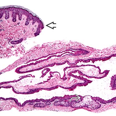
Steatocystomas present as thin-walled dermal cysts. Note the normal epidermis in the upper left corner
 and sebaceous glands in the cyst wall
  .
.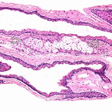
The cyst is lined by a thin squamous epithelial lining that has an eosinophilic cuticle and is associated with sebaceous glands
 .
.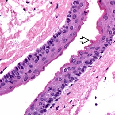
The key diagnostic feature is the
 eosinophilic cuticle lining the cyst wall. It has an undulating crenulated appearance.
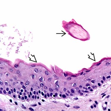
The eosinophilic cuticle
 with its undulating, crenulated appearance is a key diagnostic feature. This cyst also has a fragment of a vellus hair shaft
  , a feature sometimes seen in steatocystomas.
CLINICAL ISSUES
Epidemiology


Site
• May occur at any site, but trunk is most common location for steatocystoma multiplex


Stay updated, free articles. Join our Telegram channel
Full access? Get Clinical Tree
