pN2a: Metastasis in single ipsilateral lymph node, > 3 cm to < 6 cm in greatest dimension


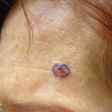
This is a small invasive squamous cell carcinoma (SCC) arising on the forehead of an adult female. The tumor is less than 2 cm in greatest dimension and did not have any high-grade features histologically, consistent with a T1 tumor.
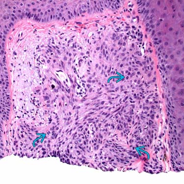
This is an example of a small, superficially invasive, well- to moderately differentiated SCC (T1). Note the enlarged, hyperchromatic staining nuclei with focal spindling and scattered mitoses
 .
.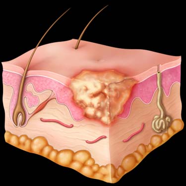
Squamous carcinoma in situ (Bowen disease) shows growth limited to the epidermis (Tis).
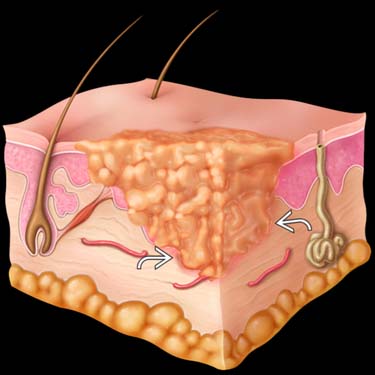
Invasive SCC shows superficial invasion into the dermis
 . This is a tumor ≤ 2 cm in greatest dimension with < 2 high-risk features (T1).
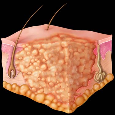
A tumor > 2 cm in greatest dimension ± 1 additional high-risk feature or any size with ≥ 2 high-risk features is staged as T2.
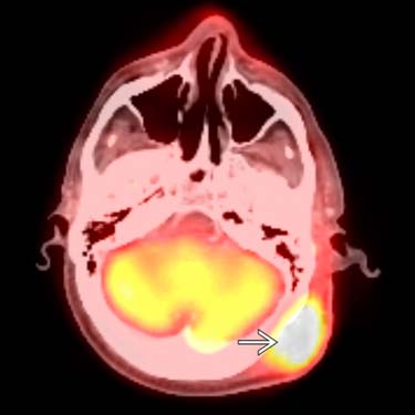
Axial fused PET/CT scan shows an increase in size and metabolic activity of a left scalp mass
 ; excision confirmed an invasive SCC of > 2 cm in greatest dimension (T2).
Stay updated, free articles. Join our Telegram channel

Full access? Get Clinical Tree


