Clinical Photograph of SCCis on Forearm This is an example of squamous cell carcinoma in situ (SCCis)/Bowen disease presenting as an erythematous plaque on the forearm of an elderly male patient who had a history of multiple skin cancers.
SCCis (Bowen Disease) Bowen disease (SCCis) is characterized by a proliferation of atypical intraepidermal keratinocytes filling the entire epidermis, including the granular layer . Many of the cells show cytoplasmic clearing .
SCCis at High Magnification High magnification of Bowen disease shows prominent cytologic atypia, an atypical mitotic figure in the midepidermis, and apoptotic cells .
Verrucoid SCCis Verrucoid SCCis shows hypergranulosis, mild papillomatosis, and superficial koilocytic-appearing cells, suggesting an HPV etiology. Note the scattered mitotic figures and enlarged, bizarre-appearing, hyperchromatic-staining nuclei .
TERMINOLOGY
Abbreviations
• Squamous cell carcinoma in situ (SCCis)
Synonyms
• Bowen disease
• Squamous intraepithelial neoplasia
Definitions
• Full-thickness intraepidermal atypia of squamous keratinocytes, often with numerous mitotic figures and apoptotic cells
ETIOLOGY/PATHOGENESIS
Sun Exposure
• Chronic UV radiation strongly implicated in SCCis
Immunosuppression
• Increased risk of developing SCCis in immunosuppressed patients, especially organ transplant recipients
HPV Infection
• Some cases of SCCis are related to HPV infection, particularly in anogenital sites
SCCis also may arise in verrucae and condylomata
CLINICAL ISSUES
Site
• Most common on head and neck region, other sun-exposed sites
Presentation
• Scaly patch or plaque lesion
• Ulceration and hemorrhage may be present
Treatment
• Surgical approaches
Complete surgical excision is standard and definitive therapy
Mohs surgery often performed for facial lesions to minimize amount of tissue taken
Electrodesiccation and curettage may also be used
• Drugs
Topical therapy with immunomodulators, including imiquimod or 5-fluorouracil, may be used
– Patients with extensive lesions or poor surgical candidates
Prognosis
• Excellent in most cases
• Small risk for invasive squamous cell carcinoma
Greater risk in patients with immunosuppression or numerous lesions
MACROSCOPIC
General Features
• Broad, superficial lesion with epidermal thickening and overlying scale
MICROSCOPIC
Histologic Features
• Atypical intraepidermal proliferation of squamous cells extending into upper levels of epidermis
Basilar keratinocytes are often spared, leading to so-called eyeliner sign
• Overlying parakeratosis often diffusely present, without skip areas over adnexal structures
• Follicular epithelial involvement is typically seen
• Cells are usually markedly enlarged and atypical-appearing, with nuclear hyperchromasia and enlarged nucleoli
Only gold members can continue reading. Log In or Register to continue
 of an elderly male patient who had a history of multiple skin cancers.
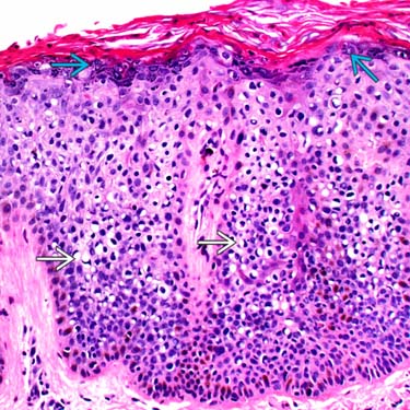
 . Many of the cells show cytoplasmic clearing
  .
.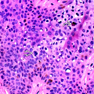
 in the midepidermis, and apoptotic cells
  .
.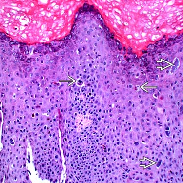
 and enlarged, bizarre-appearing, hyperchromatic-staining nuclei
  .
.


