Squamous Carcinoma of the Uterine Cervix and Its Precursors
NATURAL HISTORY, EPIDEMIOLOGY, ETIOLOGY, AND PATHOGENESIS
Carcinoma of the uterine cervix and its precursors belong to the best studied forms of human cancer. In this chapter, only cancers and precancerous lesions with the origin in, or characteristics of, squamous epithelium will be discussed. The term squamous carcinoma has been in general use to describe these lesions. The alternate term epidermoid carcinoma will be used to describe lesions with limited formation of keratin. Adenocarcinomas and related lesions are discussed in Chapter 12.
It has been repeatedly documented that invasive carcinoma of the uterine cervix, regardless of type, develops from precursor lesions or abnormal surface epithelium, which, in its classic form, is known as carcinoma in situ (International Stage O). The precursor lesions do not produce any specific alterations of the cervix visible to the naked eye. Therefore, before the introduction of cervicovaginal cytology and colposcopy, these lesions were a rarity and their discovery was incidental in biopsies of the cervix and in hysterectomy specimens. Since the introduction of mass screening by smears, and with accumulated experience, it has been shown that these lesions are quite common. The investigations of the precursor lesions is facilitated by the accessibility of the cervix to clinical examination and inspection by the colposcope and the ease of cytologic and histologic sampling that could be subjected, not only to microscopic scrutiny, but also to cytogenetic and molecular biologic analysis. Although considerable progress has been made in the understanding of the natural history of these lesions, there are still many areas of ignorance requiring further clarification.
The assumption of the prevention programs of cancer of the uterine cervix is that the precursor lesions may be identified in cervicovaginal preparations and eradicated, thus preventing the occurrence of invasive cancer. The success of these programs has been confirmed because, over the past half century, the rate of invasive cancer of the uterine cervix has been reduced by about 70% in the United States and other developed countries (summaries in Koss, 1989; Cannistra and Niloff, 1996). In developing countries, however, cancer of the cervix remains a common disease with a high mortality rate.
The first part of the chapter is devoted to epidemiology, etiology, pathogenesis, and natural history of precursor lesions and squamous cancer of the uterine cervix. The cytology and histopathology of these lesions are discussed in Part 2.
HISTORICAL PERSPECTIVE
The identification of invasive carcinoma of the uterine cervix as a distinct disease, different from other tumors of the uterus, was significantly enhanced with the introduction of uterine biopsies by Ruge and Veit in 1877. The histologic features of invasive squamous cancer were well known toward the end of the 19th century and were illustrated in a number of textbooks, such as that by Amann, published in 1897. In fact, Amann also recognized the component cells of squamous carcinoma (Fig. 11-1) but neither he nor his contemporaries addressed the issue of the origin of invasive cancer. The credit for this contribution goes to W. Schauenstein, a gynecologist from Graz, Austria, who published, in 1908, a remarkable paper pointing out the striking similarity between the histologic patterns of cancerous surface
epithelium (Krebsbelag in the original German) and superficially infiltrating squamous cancer of the cervix. He expressed the opinion that the abnormal surface epithelium deserved the name of cancer because it was the source of origin of infiltrating carcinoma (Fig. 11-2).
epithelium (Krebsbelag in the original German) and superficially infiltrating squamous cancer of the cervix. He expressed the opinion that the abnormal surface epithelium deserved the name of cancer because it was the source of origin of infiltrating carcinoma (Fig. 11-2).
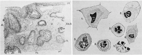 Figure 11-1 Facsimile of drawings of a cervical carcinoma and cancer cells, derived from Amann’s book on gynecologic histology, which appeared in 1897. The tissue lesion that was diagnosed as “carcinoma of cervix originating from squamous epithelium” would undoubtedly be classified today as a carcinoma in situ with extension to endocervical glands. Note the remarkably accurate drawings of “pyknotic cancer cells.” (JF Bergman, publisher, Wiesbaden, Germany.) |
Pronai in 1909 and Rubin in 1910 supported Schauenstein’s observations by additional examples. The matter was also dealt with in considerable detail in a large book by Schottländer and Kermauner, published in 1912, which contains a detailed analysis of several hundred cases of uterine cancer. In reference to cancer of the uterine cervix, Schottländer and Kermauner coined the term carcinoma in situ to describe the cancerous epithelium on the surface of the uterine cervix and considered this lesion to be malignant. Although, in the American literature, the term “carcinoma in situ” is often attributed to the pathologist A.C. Broders of the Mayo Clinic, who published a paper on this topic in 1932, he was not the first person to use this term. Numerous synonyms, such as preinvasive carcinoma, intraepithelial carcinoma, precancerous epithelium, Bowen’s disease of the cervix, and squamous or epidermoid carcinoma without evidence of invasion, have been used intermittently in the literature for many years to describe and discuss this lesion. The critical issue of whether such epithelial abnormalities may be recognized as cancerous in the absence of an invasive component was the subject of numerous controversies in the first decades of the 20th century, first addressed by Rubin in 1910. In the 1920s and 1930s, two German gynecologic pathologists, Walter Schiller and Robert Meyer (both of whom escaped to the United States to avoid Hitler’s racial laws) wrote extensively on the subject of interpretation of cervical biopsies and concluded that precancerous intraepithelial lesions were indeed precursors of invasive cervical cancer and could be so identified under the microscope. Still, because the behavior of the precancerous lesions has been shown to be unpredictable and not necessarily leading to invasive cancer, the controversy was not put to rest. With the onset of the 21st century, there are few observers who use the term “carcinoma in situ.” Most of them favor other terms, such as dysplasia, cervical intraepithelial neoplasia (CIN), and squamous intraepithelial lesions (SIL) of low (LGSIL) and high-grade (HGSIL), to be defined and discussed below.
Stay updated, free articles. Join our Telegram channel

Full access? Get Clinical Tree


