Spiradenoma
Steven D. Billings, MD
Key Facts
Clinical Issues
Upper 1/2 of body most commonly involved
Dermal mass/nodule, < 2 cm in size
Often tender or painful lesion
Microscopic Pathology
Basophilic tumor lobules/nodules in dermis
Tumor may be partially encapsulated
Biphasic appearance with 2 cell types
Small cells with scant cytoplasm and small hyperchromatic nuclei; typically at periphery of tumor lobules
Larger cells with eosinophilic cytoplasm and oval, vesicular nuclei; typically in centers of tumor lobules
Focal to diffuse duct lumen formation
Tumor lobules associated with vascularized stroma, hemorrhage may be present
Top Differential Diagnoses
Cylindroma
Significant overlap with spiradenoma, and may have combined tumors
Lobules in cylindroma are typically smaller and have a “jigsaw puzzle” pattern
Spiradenocarcinoma (malignant spiradenoma)
Associated with precursor spiradenoma, usually with abrupt transition
Merkel cell carcinoma
More cytologic atypia and high mitotic rate
Basal cell carcinoma
Peripheral palisading with tumor-stroma retraction
Lymphoid infiltrate (pseudolymphoma)
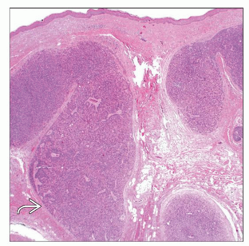 Spiradenoma is characterized by circumscribed basophilic tumor nodules or lobules in the dermis. The tumor lobules often have at least a partial fibrous capsule  . . |
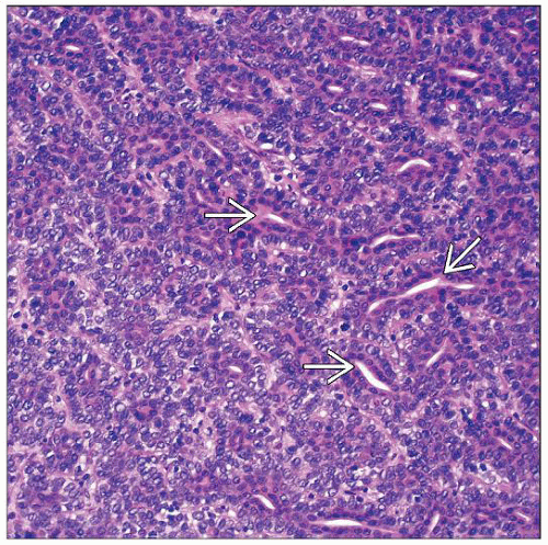 The tumor is composed of a biphasic population of smaller basaloid cells and larger pale cells. Duct lumen formation  is present and may be focal or relatively prominent, as in this case. is present and may be focal or relatively prominent, as in this case. |
TERMINOLOGY
Synonyms
“Eccrine” spiradenoma
Definitions
Benign adnexal tumor composed of nodules of basaloid cells with ductal differentiation
May show evidence of apocrine differentiation rather than eccrine differentiation
ETIOLOGY/PATHOGENESIS
Genetic Syndrome
Familial cases associated with Brooke-Spiegler syndrome
Also known as familial cylindromatosis or turban tumor syndrome
Autosomal dominant
Multiple cylindromas, but can also have spiradenomas and trichoepitheliomas
CLINICAL ISSUES
Epidemiology
Age
Most common in young adults, but can present at any age
Site
Upper 1/2 of body most commonly involved
> 75% present on ventral surface
Presentation
Dermal mass/nodular lesion
Often tender or painful
May have bluish color
Usually solitary, but may be multiple
Multiple lesions may be part of Brooke-Spiegler syndrome, and associated with multiple cylindromas and trichoepitheliomas
Treatment
Surgical approaches
Complete surgical excision is curative
Prognosis
Benign, but local recurrence may occur
Very rare malignant transformation
MACROSCOPIC FEATURES
General Features
Dermal-based, bluish nodule
Size
Typically small, < 1-2 cm
MICROSCOPIC PATHOLOGY
Histologic Features
Basophilic tumor nodules in dermis
Tumor lobules may be partially encapsulated
Biphasic appearance with 2 cell types
Small cells with scant cytoplasm and small hyperchromatic nuclei
Small cells are typically at periphery of tumor lobules
Larger cells with eosinophilic cytoplasm and oval, vesicular nuclei ± a distinct nucleolus
Stay updated, free articles. Join our Telegram channel

Full access? Get Clinical Tree


