CT Scan of Soft Tissue Chondroma 
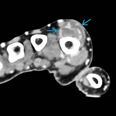
Soft tissue chondroma typically occurs in the digits of the hands and feet. It presents as a painless, well-demarcated mineralized mass, as demonstrated in this CT of a dorsal index finger lesion. Note the peripheral distribution of mineralization .
Lobular Architecture of Chondroma 
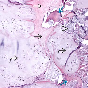
Soft tissue chondroma typically has a lobular architecture with islands of hyaline cartilage separated by fibrous bands . Matrix calcification and ossification are common.
Cytological Features of Chondroma 
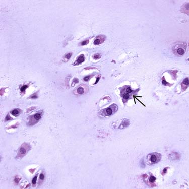
High-power micrograph illustrates typical cytological features of soft tissue chondroma. The chondrocytes are often arranged in clusters, situated in lacunar spaces within a pale blue hyaline matrix, and have uniform round nuclei and abundant eosinophilic cytoplasm. Rare binucleated cells can be seen . Mitoses are rare.
Calcified Hyaline Cartilage 
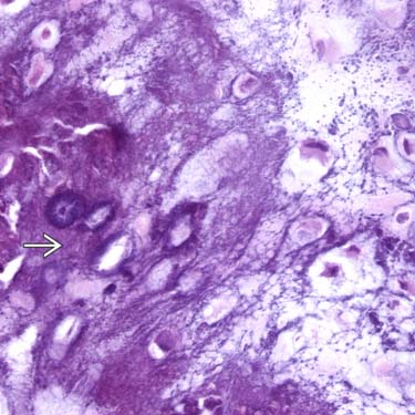
Areas of very dense calcification are common, illustrated by heavy basophilic staining of the cartilage matrix.
TERMINOLOGY
Synonyms
• Extraskeletal chondroma
• Chondroma of soft parts
• Fibrochondroma
• Osteochondroma
• Myxochondroma
• Chondroblastoma-like chondroma
Definitions
• Benign hyaline cartilage neoplasm of soft tissue with predilection for hands and feet
ETIOLOGY/PATHOGENESIS
Neoplasm With Genetic Abnormalities
• Rearrangement of 12q13-15
• Trisomy 5
• Aberrations of chromosome 11
• HMGA2 abnormalities
CLINICAL ISSUES
Epidemiology
• Incidence
Uncommon
– Exact incidence unknown
• Age
Median: 4th decade
– Range: Infancy to 9th decade
• Sex
F = M
Presentation
• Painless mass
• Most common in hands and feet (60-95%)
Especially in fingers (40-50%)
• Rare sites
Proximal extremities, trunk, head and neck, oral cavity, ear, upper aerodigestive tract, dura/extradural, skin, fallopian tube
Treatment
• Simple surgical excision
Prognosis
• Low recurrence rate (15-20%)
Recurrences controlled by reexcision
• No reports of malignant degeneration
IMAGING
General Features
• Best diagnostic clue
Small, well-demarcated lesion
Mineralized soft tissue mass
Acral extremity
• Location
Hands and feet
Often in vicinity of joint or tendon
No intraarticular or subperiosteal localization by definition
• Morphology
Most are calcified or ossified
Sometimes erode and deform underlying bone
MACROSCOPIC
General Features
• Well demarcated and bosselated
• Spherical or oval
• Rubbery or hard
• Sometimes soft, friable, gelatinous, or cystic
Size
• Median: 1.6 cm
Range: 0.3-6.5 cm
MICROSCOPIC
Histologic Features
• Well-circumscribed and lobulated subcutaneous tumor
• Mostly composed of mature hyaline cartilage
• Chondrocytes located in lacunae
Arranged diffusely or in small clusters
Some have enlarged nuclei and moderate pleomorphism
Very low mitotic rate
• Variable amounts of calcification
Granular stippled calcification that surrounds chondrocytes in lace-like pattern
Only gold members can continue reading. Log In or Register to continue