1 Skull and Face Fractures
Anatomy of the Skull and Facial Skeleton
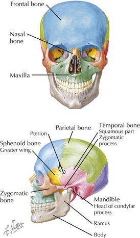
Skull and Facial Bones
• Viscerocranium (facial skeleton): maxilla, nasal, lacrimal, zygomatic, vomer, palatine, mandible bones
• Most of the bones of the skull are flat (type), with inner and outer “tables” (layers) of compact (cortical) bone surrounding trabecular bone and marrow space (diploë).
• Emissary veins connect diploic spaces with cerebral veins/sinuses (intracranial) and scalp and superficial veins: potential route for intracranial spread of infection.
Scalp Layers
• Aponeurosis of occipitofrontalis muscle, with lateral attachments of temporoparietalis and posterior auricular muscles (collectively the epicranius)
• Loose areolar tissue: allows aponeurosis movement; danger space for infections owing to emissary vein drainage into diploic spaces of cranium
Neurovascular Supply
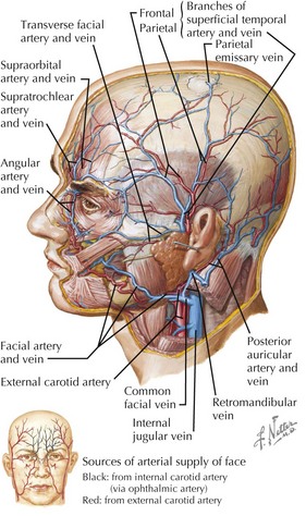
Arteries of Face and Cranium
External Carotid (Proximal to Distal)
• Maxillary: deep auricular, anterior tympanic, deep temporal, middle meningeal, inferior alveolar, posterior alveolar, infraorbital branches; to deep face
Other
• Vertebral: basilar, pontine, posterior and inferior cerebellar, posterior cerebral, posterior communicating branches
• Facial: face richly perfused, with anastomoses across midline, anterior to posterior, and between intra- and extracranial branches
Stay updated, free articles. Join our Telegram channel

Full access? Get Clinical Tree


