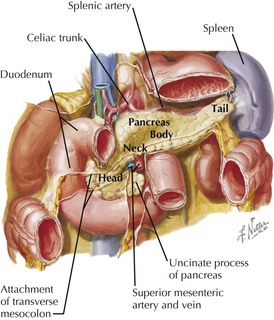
14 Pancreatic Diseases Anatomy of the Pancreas Pancreas in Situ Parts and Landmarks • Head (includes uncinate process), neck, body, tail • Uncinate process lies behind superior mesenteric artery and anterior to aorta. • Neck overlies superior mesenteric artery and vein and portal vein. • Development Two endodermal gland buds of caudal foregut merge to form pancreas. Buds rotate with foregut. Dorsal bud forms body and tail. Ventral bud makes head; uncinate process rotates behind superior mesenteric artery. Original mesentery fuses with posterior peritoneum, and pancreas becomes retroperitoneal. Location and Locale of the Pancreas • Retroperitoneal and posterior to stomach: typically nonpalpable on physical examination • Neck of pancreas overlies L1 and L2 vertebral bodies in the transpyloric plane. • Head is to the right of and inferior to transpyloric plane. • Body and tail are to the left and above transpyloric plane. Duct System Main Pancreatic Duct • Begins in tail, runs medially into head • Turns inferiorly, closely related to bile duct • Ducts unite to form hepatopancreatic ampulla (of Vater). • Ampulla empties into descending duodenum at the major duodenal papilla. • Smooth muscle sphincter of pancreatic duct around terminal portion • Smooth muscle sphincter lies around terminal bile duct. • Hepatopancreatic sphincter (of Oddi) around hepatopancreatic ampulla Accessory Pancreatic Duct (Variable) • Can open into duodenum at minor duodenal papilla • Accessory duct more often joins main duct (~60%). • If main duct is small, and there is no juncture, accessory duct can carry majority of secretion. Functional Anatomy • Tubuloacinar gland structure with a variety of cell types, including intermingled islets of Langerhans • Parasympathetic and sympathetic nerves are distributed to islets and acini. • Cells’ secretions are controlled by endocrine and autonomic nervous activities. Exocrine Functions • Mediated by secretin and cholecystokinin formed by duodenal and jejunal epithelium • Acinar cells secrete amylase, lipase, trypsinogen, chymotrypsinogen, carboxypeptidase, and Cl−. • Ductal cells secrete HCO3−. • Some secretomotor input comes from vagal parasympathetic fibers. Endocrine Functions • Alpha cells secrete glucagon. • Beta cells (central islets) secrete insulin. Only gold members can continue reading. Log In or Register to continue Share this: Share on X (Opens in new window) X Share on Facebook (Opens in new window) Facebook Like this:Like Loading… Related Related posts: Uterus and Adnexal Diseases Prostate Diseases Pelvic Fractures Liver Diseases Stay updated, free articles. Join our Telegram channel Join Tags: Netters Surgical Anatomy Review PRN Aug 12, 2016 | Posted by admin in ANATOMY | Comments Off on Pancreatic Diseases Full access? Get Clinical Tree
14 Pancreatic Diseases Anatomy of the Pancreas Pancreas in Situ Parts and Landmarks • Head (includes uncinate process), neck, body, tail • Uncinate process lies behind superior mesenteric artery and anterior to aorta. • Neck overlies superior mesenteric artery and vein and portal vein. • Development Two endodermal gland buds of caudal foregut merge to form pancreas. Buds rotate with foregut. Dorsal bud forms body and tail. Ventral bud makes head; uncinate process rotates behind superior mesenteric artery. Original mesentery fuses with posterior peritoneum, and pancreas becomes retroperitoneal. Location and Locale of the Pancreas • Retroperitoneal and posterior to stomach: typically nonpalpable on physical examination • Neck of pancreas overlies L1 and L2 vertebral bodies in the transpyloric plane. • Head is to the right of and inferior to transpyloric plane. • Body and tail are to the left and above transpyloric plane. Duct System Main Pancreatic Duct • Begins in tail, runs medially into head • Turns inferiorly, closely related to bile duct • Ducts unite to form hepatopancreatic ampulla (of Vater). • Ampulla empties into descending duodenum at the major duodenal papilla. • Smooth muscle sphincter of pancreatic duct around terminal portion • Smooth muscle sphincter lies around terminal bile duct. • Hepatopancreatic sphincter (of Oddi) around hepatopancreatic ampulla Accessory Pancreatic Duct (Variable) • Can open into duodenum at minor duodenal papilla • Accessory duct more often joins main duct (~60%). • If main duct is small, and there is no juncture, accessory duct can carry majority of secretion. Functional Anatomy • Tubuloacinar gland structure with a variety of cell types, including intermingled islets of Langerhans • Parasympathetic and sympathetic nerves are distributed to islets and acini. • Cells’ secretions are controlled by endocrine and autonomic nervous activities. Exocrine Functions • Mediated by secretin and cholecystokinin formed by duodenal and jejunal epithelium • Acinar cells secrete amylase, lipase, trypsinogen, chymotrypsinogen, carboxypeptidase, and Cl−. • Ductal cells secrete HCO3−. • Some secretomotor input comes from vagal parasympathetic fibers. Endocrine Functions • Alpha cells secrete glucagon. • Beta cells (central islets) secrete insulin. Only gold members can continue reading. Log In or Register to continue Share this: Share on X (Opens in new window) X Share on Facebook (Opens in new window) Facebook Like this:Like Loading… Related Related posts: Uterus and Adnexal Diseases Prostate Diseases Pelvic Fractures Liver Diseases Stay updated, free articles. Join our Telegram channel Join Tags: Netters Surgical Anatomy Review PRN Aug 12, 2016 | Posted by admin in ANATOMY | Comments Off on Pancreatic Diseases Full access? Get Clinical Tree