Chapter 3 Skin, nails and hair
SYMPTOMS OF SKIN DISEASE
The history should evaluate possible precipitating factors and determine whether the skin problem is localised or a manifestation of systemic illness.
EXAMINATION OF THE SKIN, NAILS AND HAIR
Examining the skin
Examination relies almost entirely on careful inspection.
ABNORMAL SKIN COLOUR
Iron overload (haemosiderosis and haemochromatosis) causes the skin to turn a slate-grey colour. Addison’s disease is characterised by darkening of the skin, occurring first in the skin creases of the palms and soles, scars and other skin creases. The mucosa of the mouth and gums also becomes pigmented. Striking pigmentation arises after bilateral adrenalectomy for adrenal hyperplasia (Nelson’s syndrome).
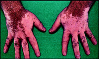
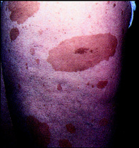
Common localised abnormalities of skin pigmentation include vitiligo (Fig. 3.1) and café au lait spots (Fig. 3.2). When examining a patient, you may notice an erythematous flush in the necklace area which is caused by anxiety. Purpura is the term used for red-purplish lesions of the skin caused by seepage of blood from skin blood vessels. These lesions do not blanch with pressure. If the lesions are small (<5 mm) they are called petechiae, whereas larger lesions are purpura.
LOCALISED SKIN LESIONS
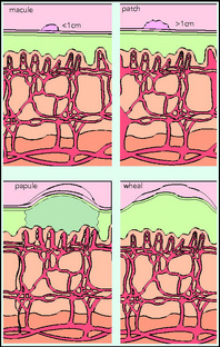
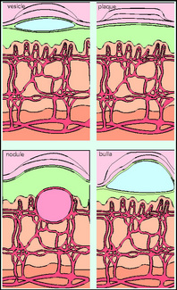
Decide whether the lesion is flat, nodular or fluid-filled. Flat circumscribed changes in colour are termed macules if less than 1 cm, or patches if more than 1 cm. If the lesion is raised and can be palpated, assess whether the mass is a papule, plaque, nodule, tumour or wheal. If a circumscribed elevated lesion is fluctuant and fluid-filled, describe whether it is a vesicle, bulla or pustule (Fig. 3.3).
COMMON SKIN LESIONS
Rosacea
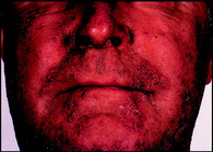
This facial rash usually presents in the fourth decade. Papules and pustules erupt on the forehead, cheeks, bridge of the nose and the chin. The erythematous background highlights the rash (Fig. 3.4). Eye involvement is characterised by grittiness, conjunctivitis and even corneal ulceration.
Drug reactions
Drug reactions may occur within minutes or hours of taking the medication but there may also be delays of up to 2 weeks for the reaction to manifest. It is important to recognise different expressions of drug sensitivity.
Erythema nodosum
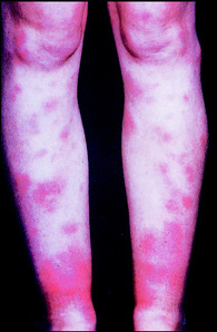
Symmetrical in distribution, the acute crops of painful, tender, raised red nodules usually affect the extensor surfaces, especially the shins but also the thighs and upper arms (Fig. 3.5). Over 7–10 days, lesions change colour from bright red through shades of purple to a yellowish area of discoloration. Erythema nodosum is caused by vasculitis, and most commonly associated with sulphonamides, oral contraceptives and barbiturates.

 Questions to ask: Skin history
 Differential diagnosis: Systemic diseases causing pruritus
 Questions to ask: Hair history
 Differential diagnosis: Hirsutism
 Questions to ask: Hirsutism
 Differential diagnosis: Skin lesions associated with drug sensitivity
 Differential diagnosis: Erythema nodosum

