• Complete excision is curative but not necessary given benign nature of lesions
Microscopic
• Well-circumscribed vascular proliferation
• Vessels are thin-walled and closely packed, with little intervening stroma
• Lining cells are small endothelial cells with nuclear hyperchromasia
• Pseudopapillary pattern may be seen (due to tangential sectioning)
• Mitotic figures typically not seen
• Thrombosis may occur and be associated with intravascular papillary endothelial hyperplasia (Masson tumor/change)
• Calcifications rarely seen
Top Differential Diagnoses
• Arteriovenous hemangioma (malformation)
Usually occurs on lips, perioral skin, or nose of older adult males
• Venous malformation (cavernous hemangioma)
Occurs in children, usually present at birth
• Cherry angioma
Much more common, small superficial lesions occurring in adults
• Glomeruloid hemangioma
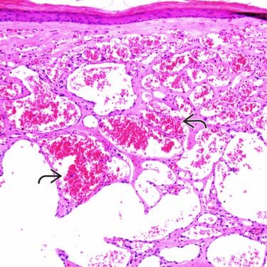
Sinusoidal Hemangioma in Superficial Dermis Low-magnification histologic examination shows the superficial portion of a cutaneous sinusoidal hemangioma (SH) involving the papillary and reticular dermis with large, dilated vascular spaces, some of which are filled with red blood cells .
Sinusoidal Hemangioma With Dilated Vascular Spaces Higher magnification shows sinusoidal vascular spaces with large areas of thrombosis , a finding often seen in SH.
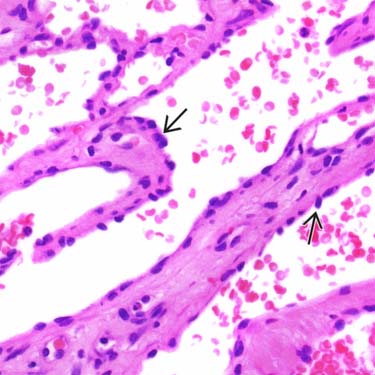
Sinusoidal Hemangioma With Thrombosis Another example of an SH shows hemorrhage and thrombosis with adjacent areas of papillary endothelial hyperplasia (Masson change) .
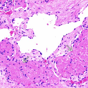
Sinusoidal Hemangioma: Cytologic Features High-magnification examination demonstrates the cytologic features of the endothelial cells, which show small, uniform-appearing round to oval nuclei with hyperchromasia .
TERMINOLOGY
Abbreviations
• Sinusoidal hemangioma
Synonyms
• Cavernous hemangioma (variant)
Definitions
• Acquired vascular lesion in adults; features similar to cavernous hemangioma/venous malformation
ETIOLOGY/PATHOGENESIS
Unknown
• May represent reactive vascular proliferation rather than true neoplastic process
Only gold members can continue reading. Log In or Register to continue


 .
.
 , a finding often seen in SH.
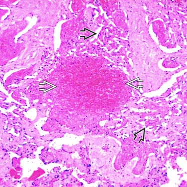
 with adjacent areas of papillary endothelial hyperplasia (Masson change)
  .
.
 .
.


