Cartilage Destruction by Melanoma The nasal septum cartilage is being destroyed by the infiltrative neoplasm. The tumor forms a thick, sheet-like distribution. No pattern of growth can be seen at this magnification, although ulceration is present.
Junctional Melanoma Confirms Primary Melanoma Neoplastic, atypical junctional melanocytes are noted within the respiratory epithelium, arranged in pagetoid spread . The tumor cells are also present within the stroma. This change helps to confirm a primary tumor.
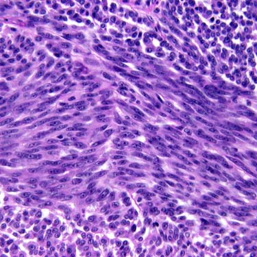
Spindled Cells in STMMM The neoplastic cells are arranged in a short fascicular architecture, comprised of spindled cells. Nucleoli are quite prominent.
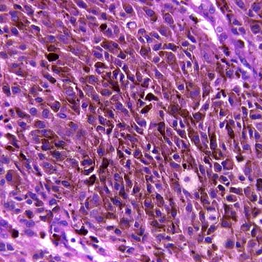
Pigmented STMMM Hematoxylin and eosin shows a spindled to polygonal population of highly atypical, pigmented neoplastic cells. These changes are characteristic for a pigmented melanoma. The pigment must be within the atypical cells (rather than histiocytes) to qualify as a pigmented melanoma.
TERMINOLOGY
Definitions
• Neural crest-derived neoplasms originating from melanocytes and demonstrating melanocytic differentiation
ETIOLOGY/PATHOGENESIS
Environmental Exposure
• Formalin, possibly radiation, &/or UV exposure
CLINICAL ISSUES
Epidemiology
• Incidence
Rare
– Represents < 1% of all melanomas
– < 5% of all sinonasal tract neoplasms
– 15-20% of all skin melanomas occur in head and neck
– Sinonasal tract and nasopharynx mucosal malignant melanoma (STMMM) represent < 4% of all head and neck melanomas
• Age
Wide range, usually in 5th-8th decades
• Sex
Equal gender distribution
• Ethnicity
Increased incidence in Japanese patients
Site
• ~ 15-20% of melanomas arise in head and neck
80% are cutaneous in origin
Ocular origin account for majority of remaining malignant mucosal melanoma (MMM)
Sinonasal tract is next most common site
• Anterior nasal septum > maxillary sinus
Presentation
• Nasal obstruction
• Epistaxis or nasal discharge
Melanorrhea: Black-flecked (melanin) discharge
• Polyp
• Pain is uncommon
Treatment
• Options, risks, complications
Metastatic melanoma to sinonasal tract can develop but is vanishingly rare
Breslow thickness and Clark level are not used in sinonasal tract
• Surgical approaches
Wide local excision is treatment of choice
• Radiation
Adjuvant postoperative radiation therapy may improve locoregional control but does not affect survival
Prognosis
• Poor overall
• 5-year survival: 17-47%
5-year disease-free survival: < 20%
• Recurrences are common
• Poor prognosis associated with
Obstruction as presenting symptom; nasopharynx or mixed site of involvement; tumor ≥ 3 cm; undifferentiated histology; high mitotic count; recurrence; stage of tumor
• Tyrosine kinase inhibitors may work when protooncogene KIT mutations are detected
• Matrix metalloproteinases (MMPs) (proteolytic enzymes required for extracellular matrix degradation) expression may be associated with patient outcome
Decreased MMP2 expression associated with greater overall survival
Positive MMP14 expression associated with poor survival
IMAGING
Radiographic Findings
• Usually identifies extent of tumor and bone invasion
• PET tends to show posterior nasal cavity and sinus tumors better than anterior nasal tumors
• Locoregional and metastatic disease can be detected
MACROSCOPIC
General Features
• Most are polypoid
• White to gray, brown, or black
• Surface ulceration/erosion is common
Size
• Range: Up to 6 cm; mean: 2-3 cm
MICROSCOPIC
Histologic Features
• Protean histology, mimic of many other primary tumor types
• Junctional activity and epidermal migration (pagetoid spread) help to confirm primary tumor
• Surface ulceration is common, obscuring in situ component
• Bone or soft tissue invasion is common
• Many patterns of growth
Peritheliomatous: Distinctive and unique for STMMM

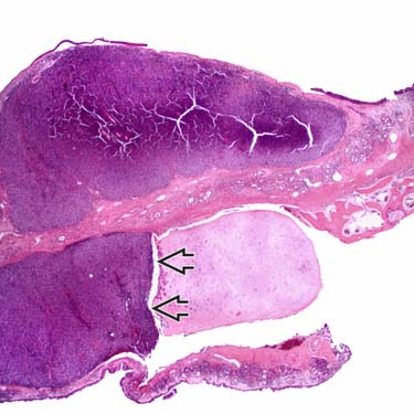
 is being destroyed by the infiltrative neoplasm. The tumor forms a thick, sheet-like distribution. No pattern of growth can be seen at this magnification, although ulceration is present.
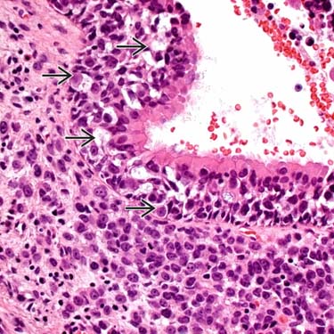
 . The tumor cells are also present within the stroma. This change helps to confirm a primary tumor.