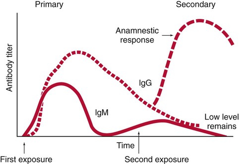
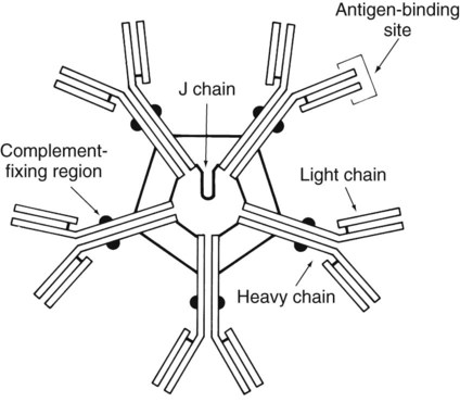
Serologic Diagnosis of Infectious Diseases


Serologic Diagnosis of Infectious Diseases


Immunochemical methods are used as diagnostic tools for serodiagnosis of infectious disease. An understanding of how these methods have been adapted for this purpose requires a basic working knowledge of the components and functions of the immune system. Immunology is the study of the components and functions of the immune system. The immune system is the body’s defense mechanism against invading “foreign” antigens. One of the functions of the immune system is distinguishing “self” from “nonself” (i.e., the proteins or antigens from foreign substances). (Chapter 3 presents a more in-depth discussion of the host’s response to foreign substances.) This chapter is intended to provide a brief overview and review of immunology. The complexity and detail required to fully understand immunology and serology are beyond the scope of this text.


Features of the Immune Response


The host, or patient, has physical barriers, such as intact skin and ciliated epithelial cells, and chemical barriers, such as oils produced by the sebaceous glands and lysozyme found in tears and saliva, to prevent infections by foreign organisms. In addition, natural (innate) immunity, which is not specific, activates chemotaxis, the process by which phagocytes are recruited to a site of invasion and engulf organisms entering the host. Acquired active immunity is the specific response of the host to an infecting organism.


The human specific immune responses are simplistically divided into the following two categories: cell-mediated and antibody-mediated.


Cell-mediated immune responses are carried out by special lymphocytes of the T-cell (thymus derived) class. T cells proliferate and differentiate into various effector T cells, including cytotoxic and helper cells. Cytotoxic T lymphocytes specifically attack and kill microorganisms or host cells damaged or infected by pathogens. Helper cells promote the maturation of B cells by producing activator cytokines that induce the B cells to produce antibodies and attach to and kill invading organisms. Although diagnosis of certain diseases may be aided by measuring the cell-mediated immune response to the pathogen, such tests entail skin tests performed by physicians or in vitro cell function assays performed by specially trained immunologists. These tests are usually not within the repertoire of clinical microbiology laboratories.


Antibody-mediated immune responses are produced by specific proteins generated by lymphocytes of the B-cell (bone marrow derived) class. Because these proteins exhibit immunologic function and fold into a globular structure in the active state, they are also referred to as immunoglobulins. Antibodies are either secreted into the blood or lymphatic fluid (and sometimes other body fluids) by activated B lymphocytes (plasma cells), or they remain attached to the surface of the lymphocyte or other cells. Because the cells involved in this category of immune response primarily circulate in the blood, this type of immunity is also called humoral immunity. For purposes of determining whether a patient’s body has produced an antibody against a particular infectious agent, the serum (or occasionally the plasma) is examined for the presence of the antibody. The study of the diagnosis of disease by measuring antibody levels in serum is referred to as serology.


Characteristics of Antibodies


Immunocompetent humans are able to produce antibodies specifically directed against almost all the antigens with which they may come into contact throughout their lifetimes and that the body recognizes as “foreign.” Antigens may be part of the physical structure of a pathogen or a chemical produced and released by the pathogen, such as an exotoxin. One pathogen may contain or produce many different antigens that the host recognizes as foreign. Infection with one agent may cause the production of a number of different antibodies. In addition, some antigenic determinants on a pathogen may not be available for recognition by the host until the pathogen has undergone a physical change. For example, until a pathogenic bacterium has been digested by a human polymorphonuclear (PMN) leukocyte, certain antigens deep in the cell wall are not detected by the host immune system. Once the bacterium has been broken down, these new antigens are released and the specific antibodies can be produced. For this reason, a patient may produce different antibodies at different times during the course of a single disease. The immune response to an antigen also matures with continued exposure, and the antibodies produced become more specific and more avid (able to bind more tightly).


Antibodies function by (1) attaching to the surface of pathogens and making the pathogens more amenable to ingestion by phagocytic cells (opsonizing antibodies); (2) binding to and blocking surface receptors for host cells (neutralizing antibodies); or (3) attaching to the surface of pathogens and contributing to their destruction by the lytic action of complement (complement-fixing antibodies). Routine diagnostic serologic methods are used to measure primarily two antibody classes, IgM and IgG; however, antibodies are categorized into five classes: immunoglobulin G (IgG), immunoglobulin M (IgM), immunoglobulin A (IgA), immunoglobulin D (IgD), and immunoglobulin E (IgE). IgA, also referred to as secretory antibody, is the predominant class of antibody in saliva, tears, and intestinal secretions. IgD is attached to the surface of B cells and is involved in immune regulations. IgE levels increase as a result of infections caused by several parasites or in response to allergic reactions.


The basic structure of an antibody molecule comprises two mirror images, each composed of two identical protein chains (Figure 10-1). At the terminal ends are the antigen binding sites, or variable regions, which specifically attach to the antigen against which the antibody was produced. Depending on the specificity of the antibody, antigens of some similarity, but not total identity, to the inducing antigen may also be bound; this is called a cross reaction. The complement binding site is found in the center of the molecule in a structure similar for all antibodies of the same class and is referred to as the constant region. IgM is produced as a first response to many antigens, although the levels remain high transiently. Thus, the presence of IgM usually indicates recent or active exposure to an antigen or infection. IgG, on the other hand, may persist long after an infection has run its course.


The IgM antibody type (Figure 10-2) consists of five identical proteins (pentamer), with the basic antibody structures linked at the bases with 10 antigen binding sites on the molecule. IgG consists of one basic antibody molecule (monomer) that has two binding sites (see Figure 10-1). The differences in the size and conformation between these two classes of immunoglobulins result in differences in activities and functions.


Features of the Humoral Immune Response Useful in Diagnostic Testing


Immunocompetent individuals produce both IgM and IgG antibodies in response to most pathogens. In most cases, IgM is produced by a patient after the first exposure to a pathogen and is no longer detectable within a relatively short period. For serologic diagnostic purposes, it is important to note that IgM is unable to cross the placenta. Therefore, any IgM detected in the serum of a newborn must have been produced by the infant and indicates an infection in utero. The larger number of binding sites on IgM molecules provides for more rapid clearance of the offending pathogen, even though each individual antigen binding site may not be the most efficient for binding to the antigen. Over time, the cells producing IgM switch to production of IgG. IgG is the highest circulating antibody in the human body.


IgG is often more specific for the antigen (i.e., it has higher avidity). IgG has two antigen binding sites, but it can also bind complement. Complement is a complex series of serum proteins that is involved in modulating several functions of the immune system, including cytotoxic cell death, chemotaxis, and opsonization. When IgG is bound to an antigen, the base of the molecule (Fc portion) is exposed in the environment. Structures on this Fc portion attract and bind the cell membranes of phagocytes, increasing the chances of engulfment and destruction of the pathogen by the host cells. A second exposure to the same pathogen induces a faster and greater IgG response and a much lesser IgM response. Several B lymphocytes retain memory of the pathogen, allowing a more rapid response and a higher level of antibody production than the primary exposure or response. This enhanced response is called the anamnestic response. B-cell memory is not perfect. Occasional clones of memory cells can be stimulated through interaction with an antigen that is similar but not identical to the original antigen. Therefore, the anamnestic response may be polyclonal and nonspecific. For example, reinfection with cytomegalovirus may stimulate memory B cells to produce antibody against Epstein-Barr virus (another herpes family virus), which the host encountered previously, in addition to antibody against cytomegalovirus. The relative humoral responses are diagrammatically represented in Figure 10-3.


Interpretation of Serologic Tests


In serology, a change in antibody titer is a central concept for the diagnosis and monitoring of disease progression. The titer of antibody is the reciprocal of the highest dilution of the patient’s serum in which the antibody is still detectable. Patients with large amounts of antibody have high titers, because antibody is still detectable at very high dilutions of serum. Serum for antibody levels should be drawn during the acute phase of the disease (when it is first discovered or suspected) and again during convalescence (usually at least 2 weeks later). These specimens are called acute and convalescent sera. For some infections, such as legionnaires’ disease and hepatitis, titers may not rise until months after the acute infection, or they may never rise. Therefore, changes in titer must be carefully correlated with the patient’s signs and symptoms of the specific disease or suspected infectious agent.


Patients with intact humoral immunity develop increasing amounts of antibody to a pathogen over several weeks. If it is the patient’s first exposure to the pathogenic organism and the specimen has been obtained early enough, no or very low titers of antibody are detected at the onset of disease. In the case of a second exposure, the patient’s serum usually contains measurable antibody during the initial phase of the disease, and the antibody level quickly increases as a result of the anamnestic response. For most pathogens, an increase in the patient’s titer of two doubling dilutions (e.g., from a positive result of 1 : 8 to a positive result of 1 : 32) is considered to be diagnostic of current infection. This is defined as a fourfold rise in titer.


For many infections, accurate results used for diagnosis are achieved when acute and convalescent sera are tested concurrently in the same test system. Variables inherent in the procedures and laboratory error can cause a difference of one doubling (or twofold) dilution in the results obtained from a same sample tested concurrently in different laboratories. Unfortunately, a certain proportion of infected patients never demonstrate a rise in titer, necessitating the use of other diagnostic tests. Because the delay inherent in testing paired acute and convalescent sera results in diagnostic information arriving too late to affect the initial therapy, increasing numbers of early (IgM) serologic testing assays are being commercially evaluated. Moreover, it is sometimes more realistic to see a fourfold fall in titer between acute and convalescent sera when samples are tested concurrently in the same system. This is a result of the sera being collected late in the course of an infection, when antibodies have already begun to decrease.


Serodiagnosis of Infectious Diseases


With most diseases, a spectrum of responses may be seen in infected humans, such that a person may develop antibody from a subclinical infection or after colonization by an agent without actually having symptoms of the disease. In these cases, the presence of antibody in a single serum specimen or a similar titer of antibody in paired sera may merely indicate past contact with the agent and cannot be used to accurately diagnose recent disease. Therefore, in the vast majority of serologic procedures for diagnosis of recent infection, testing of both acute and convalescent sera is the method of choice. Except for detecting the presence of IgM, testing of a single serum may be recommended in certain cases. Mycoplasma pneumoniae and viral influenza B infections are examples in which high titers may indicate recent infection. IgM levels may be diagnostic if the infecting or disease-causing agent is extremely rare, such as rabies or exposure to botulism toxin, and people without disease or prior immunization would have no chance of developing an immune response.


The prevalence of antibody to an etiologic agent of disease in the population correlates with the number of people who have come into contact with the agent, not the number who actually develop disease. For most diseases, only a small proportion of infected individuals actually develop symptoms; others develop protective antibodies without experiencing signs and symptoms of the disease. In a number of circumstances, serum is tested to determine whether a patient is immune; that is, whether the patient has antibody to a particular agent either in response to a past infection or to immunization. These tests can be performed with a single serum sample. The results of the tests must be correlated with the actual immune status of individual patients to determine the level of detectable antibody present, in order to determine whether the individual has developed a true immunity to infection or a secondary reinfection. For example, sensitive tests can detect the presence of very tiny amounts of antibody to the rubella virus. Certain people, however, may still be susceptible to infection with the rubella virus with such small amounts of circulating antibody, and a higher level of antibody may be required to ensure protection from disease.


Alternatively, depending on the etiologic agent, even low levels of antibody may protect a patient from pathologic effects of disease and not prevent a second reinfection. For example, a person previously immunized with killed poliovirus vaccine who becomes infected with pathogenic poliovirus experiences multiplication of the virus in the gut and virus entry into the circulation. Damage to the central nervous system is blocked by humoral antibody in the circulation. As more sensitive testing methods are developed and these types of problems become more common, microbiologists must work closely with clinicians to develop guidelines for interpreting serologic test results in relation to the immune status of individual patients. Moreover, patients may respond to an antigenic stimulus by producing cross-reacting antibodies. These antibodies are nonspecific and may cause misinterpretation of serologic tests.


Table 10-1 provides a brief list of representative serologic tests available for immunodiagnosis of infectious diseases, the specimen required, interpretation of positive and negative test results, and examples of applications of each technique. Because serologic assays are rapidly evolving, this table is not intended to be all-inclusive.


TABLE 10-1


Noninclusive Overview of Tests Available for Serodiagnosis of Infectious Diseases


Test Sera Needed Interpretation Application
IgM Single, acute (collected at onset of illness) Newborn, positive: in utero (congenital) infection
Adult, positive: primary or current infection
Adult, negative: no infection or past infection
Newborn: STORCH* agents; other organisms
Adults: any infectious agent
IgG Acute and convalescent (collected 2-6 weeks after onset) Positive: fourfold rise or fall in titer between acute and convalescent sera tested at the same time in the same test system
Negative: no current infection or past infection, or patient is immunocompromised and cannot mount a humoral antibody response, or convalescent specimen collected before increase in IgG (Lyme disease, Legionella sp.)
Any infectious agent
IgG Single specimen collected between onset and convalescence Adult, positive: adult evidence of infection at some unknown time except in certain cases in which a single high titer is diagnostic (rabies, Legionella, Ehrlichia spp).
Newborn, positive: maternal antibodies that crossed the placenta
Newborn, negative: patient has not been exposed to microorganism or patient has a congenital or acquired immune deficiency or specimen collected before increase in IgG (Lyme disease or Legionella sp.)
Any infectious agent
Immune status evaluation Single specimen collected at any time Positive: previous exposure
Negative: no exposure
Rubella testing for women of childbearing age, syphilis testing may be required in some states to obtain a marriage license, cytomegalovirus testing for transplant donor and recipient

Stay updated, free articles. Join our Telegram channel

Aug 25, 2016 | Posted by in MICROBIOLOGY | Comments Off on Serologic Diagnosis of Infectious Diseases

Full access? Get Clinical Tree

Get Clinical Tree app for offline access