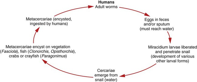
1. List the clinically significant trematodes capable of infecting the liver and lungs. 2. Describe the general life cycle of the liver and lung flukes and identify the infective stage for humans. 3. Describe the diagnostic methods used to identify the liver and lung flukes including the microscopic differentiation of eggs and serologic methods. 4. Describe the pathogenesis of the liver and lung flukes including location and associated disease manifestations. 5. List the drug of choice for infections with liver and lung flukes. 6. Describe the transmission of the liver and lung flukes and discuss how infection may be prevented. The life cycle of the liver flukes is very similar to that of the intestinal flukes. The adult worms produce eggs in the biliary ducts that are then excreted from the body in the feces. The free-swimming miracidium is released from the egg in freshwater and enters the snail host where it develops into a redia and then a cercariae, which leaves the snail and enters the water (Figure 57-1). The cercariae of Clonorchis and Opisthorchis are ingested by a second intermediate host, a freshwater fish. The cercariae then encyst and develop into the metacercariae within the intermediate host. The metacercaria is the infective stage for humans. When infected freshwater fish are eaten raw or undercooked, the metacercariae will excyst in the duodenum and then travel to the bile duct where they mature. The cercariae of Fasciola encyst on freshwater vegetation, such as watercress and water chestnuts, and develop into metacercariae. When the infected vegetation is eaten raw, the metacercariae will excyst in the duodenum and then travel to the bile duct and mature. Figure 57-2 depicts the general life cycles of the liver and lung flukes. Identification of the liver flukes is primarily made by recovery of the eggs in feces using a sedimentation method and a wet mount with or without iodine staining. Table 57-1 shows some diagnostic characteristics of the liver and lung flukes. TABLE 57-1 Characteristics of Liver and Lung Trematodes
Liver and Lung Trematodes
The Liver Flukes
General Characteristics
Epidemiology and Life Cycle
Laboratory Diagnosis
Trematode
Adult Location
Food Source
Size of Egg
Description of Egg
Fasciola hepatica
Bile ducts
Freshwater vegetation
130-150 µm × 70-90 µm
Operculated, brownish-yellow, unembryonated
Clonorchis sinensis
Bile ducts
Freshwater fish
28-34 µm × 14-18 µm
Operculated with shoulders, opposite end knob, yellow-brown, embryonated
Opisthorchis viverrini
Bile ducts
Freshwater fish
19-29 µm × 12-17 µm
Operculated with shoulders, opposite end knob, yellow-brown, embryonated
Paragonimus westermani
Lungs
Freshwater crabs or crayfish
80-120 µm × 45-60 µm
Operculated with shoulders, thick shelled, brownish-yellow, unembryonated
Paragonimus mexicanus
Lungs
Freshwater crabs
40 µm × 80 µm
Operculated with shoulders, thick shelled, brownish-yellow, unembryonated ![]()
Stay updated, free articles. Join our Telegram channel

Full access? Get Clinical Tree


Liver and Lung Trematodes