Selenium1


Selenium1


Roger A. Sunde


Selenium first attracted biologic interest in the 1930s when it was found to cause poisoning of livestock that grazed in areas with high-selenium soils (1). In 1957, Schwarz and Foltz (2) reported that small amounts of selenium prevented liver necrosis in vitamin E-deficient rats, a finding indicating that selenium was an essential nutrient and not only a toxin. Soon thereafter, deficiencies of selenium and vitamin E were shown to be involved in several economically important nutritional diseases of cattle, sheep, swine, and poultry (1). The first demonstration of a biochemical function for selenium in animals came in 1973, when the element was discovered to be a constituent of the enzyme glutathione peroxidase (GPX) (3).

The importance of selenium in human nutrition was documented in 1979, when Chinese scientists reported that selenium supplementation prevented the development of a cardiomyopathy known as Keshan disease in children living in low-selenium areas (4), and New Zealand workers reported a clinical response to selenium in a selenium-depleted patient (5). Information about the role of selenium in human nutrition increased rapidly in the 1980s, and a recommended dietary allowance (RDA) for selenium was established in 1989 (6) and revised in 2000 (7). Dietary recommendations from the World Health Organization (WHO) were issued in 1996 (8). Molecular and genetic studies, in humans, animals, and other organisms, are providing extensive new information on selenoproteins and the molecular biology of selenium.


CHEMICAL FORMS

Most selenium in biologic systems is present in proteins as a constituent of amino acids. Reflecting the similarity of selenium chemistry to that of sulfur, those amino acids are usually selenocysteine and selenomethionine. Selenocysteine (Fig. 14.1) is incorporated into the peptide backbone of selenoproteins, contains selenium in the selenol form, and is often referred to as the twenty-first amino acid. The standard amino acid symbols for selenocysteine are Sec (three letters) or U (one letter). Selenol has chemical properties that are distinct from the thiol in cysteine, and selenocysteine almost always performs catalytic functions in proteins. Selenomethionine, conversely, contains selenium bound covalently to two carbon atoms
and is considerably less reactive than selenocysteine. It is not known to have a biochemical function distinct from that of methionine.


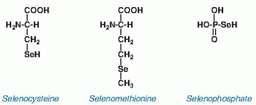
Fig. 14.1. Key selenium-containing molecules found in animals. Selenocysteine is the biologically active form of the element found in selenoproteins. Its selenol is largely ionized at physiologic pH and is a stronger nucleophile than the thiol of cysteine. These chemical properties contribute to its catalytic function in selenoenzymes. Selenomethionine contains selenium covalently bound to two carbon atoms. Thus, its selenium is shielded and is not as chemically active as the selenium in selenocysteine. Selenomethionine appears to be distributed nonspecifically in the methionine pool. Selenophosphate, the product of selenophosphate kinase, is the activated form of selenium used for synthesis of selenocysteine.

A selenoprotein contains stoichiometric amounts of selenium. Selenocysteine is the form of the element in all the animal selenoproteins identified so far and in nearly all the bacterial selenoproteins. The presence of selenium in an unidentified form (not selenocysteine) that is coordinated with molybdenum in nicotinic acid hydroxylase from Clostridium barkeri (9) indicates that selenoproteins containing forms of the element other than selenocysteine exist in nature. In addition, some prokaryotes synthesize a modified selenouridine base found in the anticodons of a just a few tRNA species (10).

Numerous proteins contain selenium as selenomethionine in nonstoichiometric amounts and are referred to as selenium-containing proteins. This designation has low utility because virtually all proteins that contain methionine contain selenomethionine in proportion to the relative abundance of these two amino acids in the organism. This is because enzymes that attach methionine to the methionine-tRNA for incorporation into protein or that metabolize methionine cannot distinguish selenomethionine from methionine.

Selenium enters the food chain through plants that incorporate it into compounds that usually contain sulfur. The result is that plant selenium is in the form of selenomethionine and, to a lesser extent, selenocysteine and other analogs of sulfur amino acids. Fungi and higher plants do not have selenoproteins or the selenocysteine incorporation machinery necessary for synthesis of selenoproteins (11), and they do not appear to require selenium for their existence.

Some plants express an enzyme that methylates free selenocysteine, thus producing selenium-methylselenocysteine (12). This is a detoxification product, and it cannot be incorporated into protein. It accumulates to high concentrations and can be responsible for selenium poisoning in animals that eat these plants.

Selenophosphate (see Fig. 14.1) is an important intermediate compound in selenium metabolism. It is produced by selenophosphate synthetase and serves as the selenium donor for the production of selenocysteine destined for incorporation into selenoproteins (13).

Methylated forms of selenium are produced as excretory metabolites and rapidly appear in the urine and breath (14, 15). This includes a methylated selenosugar, 1β-methylseleno-N-acetyl-D-galactosamine, which is synthesized in liver and is the major selenium species in urine with usual dietary intakes of selenium (15). Additional small molecule forms of the element have been detected in blood plasma, but their identities have not been established (16).


DIETARY CONSIDERATIONS


Food Sources

The richest food sources of selenium are organ meats and seafoods (0.4 to 1.5 µg/g fresh weight)2, followed by muscle meats (0.1 to 0.4), cereals and grains (<0.1 to >0.8), dairy products (<0.1 to 0.3), and fruits and vegetables (<0.1) (14). The wide variation in the selenium content of cereals and grains occurs because plants contain variable amounts, depending on how much soil selenium is available for uptake. For example, the selenium content of corn collected in China ranged from 0.005 to 8.1 µg/g, and the selenium content of the British diet declined from 65 to 31 µg/day after Britain switched its source of wheat from North America to Europe (14). Foods from animal sources vary somewhat in selenium content, but the degree of variation is less than in plants because of the homeostatic control of selenium metabolism in animals. The US Department of Agriculture National Nutrient Database for Standard Reference provides analytic or inferred values for the selenium content of hundreds of food items (17). Drinking water generally contributes negligible selenium to the overall intake, except in some localized highly seleniferous areas (14).

The Dietary Intake Data from the third National Health and Nutrition Survey (NHANES III) Total Diet Study found that median daily selenium intake was 149 and 98 µg for adult (19 to 50 years) men and women, respectively, between 1988 and 1994 (7). Lower daily selenium intakes, 30 µg or less, have been reported in countries with selenium-poor soils, such as New Zealand (14). Extremely low dietary selenium intakes from 3 to 22 µg/day have been reported in areas of China affected by Keshan disease. Conversely, very high dietary intakes (≤6690 µg/day) have been observed in a region of China with endemic human selenosis. The food in this area had been grown on soil contaminated with selenium leached from highly seleniferous coal fly ash (14).


Bioavailability

Only a few studies have determined the nutritional bioavailability of selenium in foods consumed by humans. A commonly used experimental procedure to estimate selenium availability has been to follow increases in hepatic GPX activity after feeding various food sources of selenium to rodents previously depleted of selenium. On this basis, selenium fed as mushrooms, tuna, and wheat was 5%, 57%, and 83% as available to rats, respectively, as sodium selenite (14). A human bioavailability trial performed in Finland with men of moderately low selenium status showed significant differences among various forms of selenium tested (e.g., selenate, wheat, yeast), depending on the criterion of availability used (increase in platelet GPX activity, elevation of plasma or red blood cell selenium content, retention of selenium) (14). A 16-week study of US subjects supplemented with 200 to 600 µg selenium/day as selenite, selenomethionine, or selenized yeast showed no effect of any of these supplements on plasma GPX or selenoprotein P (SEPP1) in this already selenium-adequate population, nor any increase with selenite in plasma selenium, but showed rapid increases in plasma selenium with high levels of selenomethionine or selenized yeast (18). These studies point out the need to consider the form of supplemental selenium, biologically active biomarkers and their function versus tissue selenium levels, short-term versus long-term changes in biomarkers, and repletion of deficient subjects versus maintenance of status of selenium-adequate subjects when interpreting the results of such studies.


Nutrient-Nutrient Interrelationships

Because GPXs and most other selenoproteins are potential oxidoreductases (see later), selenium is likely to interact with other nutrients that affect the antioxidant-prooxidant balance of the cell. Selenium also protects against the toxicity of mercury, cadmium, and silver, and a physiologic role for selenium in counteracting heavy metal pollutants has been proposed (14). A novel compound, called selenoneine, has selenium bound to the imidazole ring of modified histidine and is the major form of selenium in liver and blood of tuna (19). The low bioavailability of the selenium in tuna may result from this form or from complexation with mercury (14), but this issue needs further investigation.


METABOLISM

Selenium enters the body in several forms (Fig. 14.2). The two major forms are selenomethionine, derived ultimately from plants, and selenocysteine, mainly from animal selenoproteins. Selenomethionine is present in blood and tissues as methionine-containing proteins. This selenium in selenomethionine is made available for specific use when the amino acid is catabolized by the transsulfuration pathway (see also the chapter on proteins and amino acids) in liver or kidney. The selenium then enters the regulated selenium metabolic pool and can be incorporated into selenoproteins, transported to other organs, or excreted.


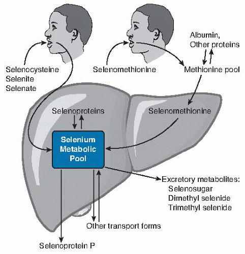
Fig. 14.2. Relationship of dietary form of selenium with tissue forms of the element. Ingested selenocysteine and inorganic selenium forms selenite and selenate enter the selenium metabolic pool directly (more detail in Fig. 14.3) directly. Selenomethionine enters the methionine pool and is incorporated into methionine-containing proteins throughout the body. When selenomethionine is metabolized to selenocysteine by the transsulfuration pathway, it enters the selenium metabolic pool. In liver, the selenium metabolic pool produces hepatic selenoproteins and selenoprotein P (SEPP1) for export. Homeostasis of selenium is maintained by the production of excretory metabolites and transport forms of selenium.

Figure 14.3 is an outline of selenium metabolism in a typical cell. Free selenocysteine, whether derived from catabolism of intracellular or extracellular selenoproteins, is degraded by selenocysteine β-lyase. The resulting selenide can enter the anabolic pathway by conversion to selenophosphate, can be converted to an excretion form, or can be modified for transport out of the cell. This metabolism of selenide is the likely point of homeostatic regulation of selenium in the cell, but the mechanism of this regulation has not been determined.


Absorption

Absorption appears to play no role in the homeostatic regulation of selenium. Virtually complete absorption occurs when the element is supplied as selenomethionine and, presumably, as selenocysteine. Absorption of selenite and selenate is greater than 50% but can vary significantly. Thus, selenium absorption is usually in the range of 50% to 100% and is not affected by selenium nutritional status (14).


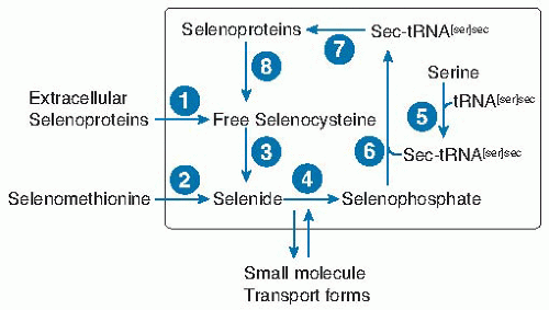
Fig. 14.3. Selenium metabolic pool in a typical nonliver or kidney cell. Selenium enters the cell as selenocysteine from extracellular selenoproteins (probably mainly selenoprotein P) (1) or breakdown of selenomethionine (2) or from unidentified small molecule forms. Free selenocysteine is produced by catabolism of (8) cellular selenoproteins or (1) extracellular selenoproteins. Free selenocysteine does not accumulate because it is metabolized by (3) selenocysteine β-lyase. The resulting selenide is transformed into selenophosphate by (4) selenophosphate synthetase, by using the cosubstrate adenosine triphosphate. The amino acid serine is acylated (5) to the tRNA[ser]sec to form ser-tRNA[ser]sec. In bacteria, selenophosphate is a substrate for selenocysteine synthetase (6), which forms sec-tRNA[ser]sec directly, whereas in eukaryotes a kinase in an additional step phosphorylates serine on the ser-tRNA[ser]sec, which then with selenophosphate is the substrate for selenocysteine synthetase (6). sec-tRNA[ser]sec donates selenocysteine to the growing peptide chain in the synthesis of selenoproteins (7) (see Fig. 14.4B).


Transport

Two selenoproteins, SEPP1 and extracellular GPX (GPX3), have been identified in plasma, and both contain the element as selenocysteine. Work using mice with targeted deletion of the SEPP1 gene has shown that this protein is involved in supplying selenium to the brain and testes, results in marked decreases in testes selenium even in mice supplemented with normally adequate levels of selenium (20, 21), and results in sperm defects indistinguishable from those elicited by dietary selenium deficiency (22, 23). Deletion of SEPP1 also results in neurologic dysfunction in the form of motor incoordination that can be prevented but not reversed by supernutritional selenium supplementation (21, 24). Supernutritional selenium supplementation largely restores selenium levels in brain but not testes (20). A SEPP1-specific receptor, the apolipoprotein E receptor (ApoER2), is expressed in brain and testes, and deletion of ApoER2 in mice results in marked reduction in brain and testes selenium concentration and in neurologic dysfunction and sperm defects that are identical to those present in SEPP1-deletion mice or in selenium-deficient mice and rats (25, 26). Finally, transgenic restoration of SEPP1 expression in liver alone is fully sufficient to restore selenium uptake by the brain and testes and to prevent the neurologic and fragile sperm phenotypes (23). These studies illustrate how molecular and genetic research is leading to detailed understanding of the targeted selenium delivery to testes and brain.

In addition to the ApoER2 receptor, the megalin receptor mediates SEPP1 uptake by the kidney and thus provides a second targeting mechanism for selenium (27). In SEPP1 knockout mice, the ability of higher levels of dietary selenium to restore brain selenium levels (20), or to provide selenium for development in fetuses and nursing pups (28), indicates that other, perhaps low-molecularweight, forms may provide additional transport functions.


Incorporation into Protein

Mammalian selenoproteins contain selenocysteine in their primary structure. The mechanism by which selenocysteine, the twenty-first amino acid, is synthesized and then incorporated into selenoproteins is complex (see Fig. 14.3, steps 4 to 7). Serine provides the carbon skeleton for selenocysteine (29), and inorganic selenium at the selenide level provides the selenium (30). The serine is acylated to tRNA[ser]sec, a unique tRNA with the anticodon for UGA, by regular seryl tRNA ligases. The serine while bound to the ser-tRNA[ser]sec is then converted to selenocysteine, as described in Figure 14.3 (31).

Figure 14.4A represents a typical selenoprotein mRNA. A selenoprotein mRNA requires a UGA in the open reading frame to code for insertion of selenocysteine and a stem-loop structure in the 3′ untranslated region (3′ UTR). This stem-loop structure is known as a selenocysteine insertion sequence (SECIS) element. Absence of the SECIS element or modification of its essential features causes the UGA to function instead as a termination codon (32). In eukaryotes, the 3′ UTR serves as a tether that allows the SECIS to loop back to interact with the UGA codon and facilitate selenocysteine incorporation. The SECIS in prokaryotes is adjacent to the UGA codon (33).

In eukaryotes, two selenium-specific protein factors facilitate selenocysteine insertion at the UGA (see Fig. 14.4B). One factor, selenocysteine binding protein 2 (SBP2), binds to the SECIS element (34). The other, elongation factor for selenocysteine (EFsec), binds to the sec-tRNA[ser]sec (35, 36). These two proteins bind to each other to form a complex that delivers the sec-tRNA[ser]sec to the ribosome for incorporation of selenocysteine into the growing polypeptide chain. It is becoming clear that several additional factors with roles in the usual ribosomal complex for peptide synthesis are also important for incorporation of selenocysteine into the peptide backbone of selenoproteins (37, 38).


Excretion

Homeostasis of selenium in the body is achieved through regulation of excretion. As dietary intake increases from the deficient range into the adequate range, urinary excretion of the element increases and accounts for maintenance of homeostasis. At very high intakes, volatile forms of selenium are exhaled, and the breath becomes a significant route of excretion. No evidence indicates that fecal selenium is regulated. Thus, under physiologic conditions,
urinary excretion is the primary means whereby body selenium is regulated (14).


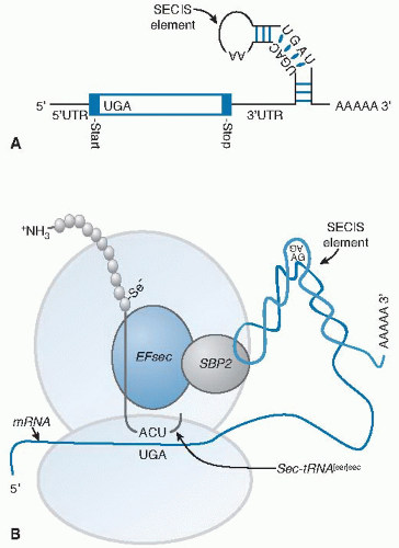
Fig. 14.4. Selenoprotein synthesis. A. The mRNA of a selenoprotein has a UGA in the open reading frame specifying selenocysteine (Sec) incorporation and a specialized stem-loop structure, known as an Sec insertion sequence (SECIS) element, in the 3′ untranslated (3′UTR) region. B. Two trans-acting proteins, SBP2 (SECIS-binding protein 2) and EFsec (elongation factor for selenocysteine), facilitate the recognition of the sec-tRNA[ser]sec by the UGA. SBP2 binds to the SECIS element and interacts with EFsec that has the sec-tRNA[ser]sec attached. This complex delivers the sec-tRNA[ser]sec to the ribosome for incorporation of Sec into the growing polypeptide chain (see Fig. 14.3, step 7).

Most excretory metabolites of selenium appear to be methylated forms produced in the liver or kidneys. Under deficient to adequate conditions, a large fraction of urinary selenium is present as a methylated selenosugar (15), and a much smaller percentage is trimethylselenonium ion. Selenium in the breath is largely dimethylselenide, especially with high levels of selenium intake. The biochemical mechanisms that regulate formation of these metabolites are not known (14).


BIOCHEMICAL FUNCTIONS

Twenty-five selenoprotein genes have been identified in the human genome by bioinformatics methods (Table 14.1) (39). The selenoproteins that result from expression of these genes are responsible for the biochemical function of selenium. Selenium is present in most of these proteins as part of a Sec-Cys pair (or Sec-serine or Sec-threonine), a finding strongly suggesting roles for these proteins as oxidoreductases (40). In addition, several of these proteins appear to be localized in the endoplasmic reticulum (41), thereby suggesting roles in protein folding or regulation of protein degradation. Nearly half of the selenoproteins, however, have not been characterized sufficiently to identify their activities. Some of the better-known selenoproteins are discussed briefly to show the breadth of selenium biochemical function.


TABLE 14.1 SELENOPROTEINS IN HUMANS


GROUP/NAME


SYMBOL


Selenoproteins involved in thiol redox reactions


Glutathione peroxidases


Cellular glutathione peroxidase


GPX1


Gastrointestinal glutathione peroxidase


GPX2


Extracellular glutathione peroxidase


GPX3


Phospholipid hydroperoxide glutathione peroxidase


GPX4


Odorant metabolizing glutathione peroxidase


GPX6


Thioredoxin reductases


Cytosolic thioredoxin reductase


TXNRD1


Thioredoxin/glutathione reductase


TXNRD2


Mitochondrial thioredoxin reductase


TXNRD3


Other U-C motif redox selenoproteins


Methionine-R-sulfoxide reductase


SELR


Selenoprotein 15 (ER residenta)


SEP15


Selenoprotein H (may regulate glutathione metabolism)


SELH


Selenoprotein M (ER resident)


SELM


Selenoprotein O (largest mammalian selenoprotein)


SELO


Selenoprotein T (ER resident)


SELT


Selenoprotein V (related to Sepw1; expressed in testes)


SELV


Membrane selenoproteins


Selenoprotein I (may be phosphotransferase)


SELI


Selenoprotein K (ER resident)


SELK


Selenoprotein S (ER resident)


SELS


Selenoproteins involved in thyroid


Hormone Metabolism


Type I iodothyronine deiodinase


DIO1


Type II iodothyronine deiodinase (ER resident)


DIO2


Type III iodothyronine deiodinase


DIO3


Muscle selenoproteins


Selenoprotein W (binds glutathione)


SEPW1


Selenoprotein N (ER resident)


SEPN1


Selenocysteine synthesis selenoproteins


Selenophosphate synthetase-2


SEPHS2


Transport selenoproteins


Selenoprotein P


SEPP1


a Selenoproteins that appear to be localized in the endoplasmic reticulum (ER) (41).


Selected data from Kryukov GV, Castellano S, Novoselov SV et al. Characterization of mammalian selenoproteomes. Science 2003;300:1439-43, with permission.

Only gold members can continue reading. Log In or Register to continue

Stay updated, free articles. Join our Telegram channel

Jul 27, 2016 | Posted by in PUBLIC HEALTH AND EPIDEMIOLOGY | Comments Off on Selenium1

Full access? Get Clinical Tree

Get Clinical Tree app for offline access