Patient Story
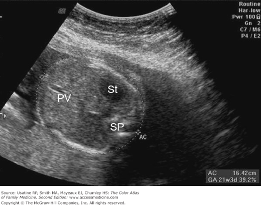
A 23-year-old pregnant gravida 2, para 1 woman is being seen for ultrasound because of her uncertain dates. Her best recollection of her last period gave her an estimated gestational age (EGA) of 19 weeks. Her vital signs are normal and her fundal height is 20 cm. Figures 78-1, 78-2, and 78-3 are still images taken from her ultrasound examination demonstrating an EGA of 21 weeks and 6 days by measurement of the baby’s biparietal diameter (BPD), head circumference (HC), abdominal circumference (AC), and femur length (FL). All four measurements allow the computer to calculate an estimated fetal weight of 431 g. The pregnancy proceeded without complications and the patient delivered a healthy boy at 403/7 weeks based on the ultrasound calculated estimated date of delivery. No interventions were needed for postdates because of the ultrasound calculations earlier in the pregnancy.
Figure 78-1
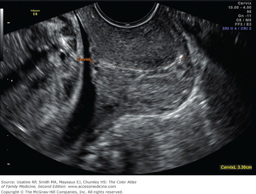
Transvaginal cervical length of a 24-week uterus. The hyperechoic line identifies the glandular tissue of the cervix and correct positioning to include the entire cervix. Measurement is from the internal os traced out to the external os. The fetal head is visible in this shot but not necessary for the measurement. (Courtesy of Danielle Cooper, MD.)
Figure 78-2
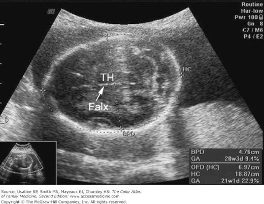
Ultrasound examination of a 21-week EGA fetus showing measurement of the baby’s biparietal diameter (BPD) and head circumference (HC). Note the presence of the falx cerebri and the thalamus (TH) to ensure the measurement is at the right anatomic level. (Courtesy of E.J. Mayeaux, Jr., MD.)
Introduction
The ideal time to perform an obstetric ultrasound is between 18 and 20 weeks’ gestation. The fetus has developed all of its organ systems and is large enough to visualize these in detail. If resources are limited or patient costs are an issue, the second trimester ultrasound provides the most bang for the buck.
Indications for Ultrasound
- Second and third trimester ultrasound examination can be used to determine fetal number and presentation, and for documentation of fetal cardiac activity, placental location, and amniotic fluid volume.
- Second trimester is a good time to perform ultrasound for fetal assessment for gestational age and weight, and it is also an integral part of performing diagnostic amniocentesis.
- If only a single ultrasound can be performed on a patient, optimal timing is 18 to 20 weeks. Fetal anatomy is best seen during this interval. If fetal abnormalities are detected, a more detailed examination (level 2 ultrasound) by a specialized sonographer is indicated.
- Ultrasound examination is indicated to establish the number of fetuses when a multiple gestation pregnancy is suspected. Risk factors for a multiple gestation pregnancy include assisted reproductive technology, family history of twins (or higher multiples), and uterine size larger than that expected by menstrual dating. The number of fetuses can be best established by obtaining an image that includes a cross-section of all fetal poles that have distinct cardiac activity within a single frame.
- The cervix and lower uterine segment may be imaged in the second trimester to look for funneling (membranes protruding into the cervical canal), a short cervix that is a significant risk factor for preterm labor.
The placental location, appearance and relationship to the internal cervical os should be recorded.1


