Patient Story
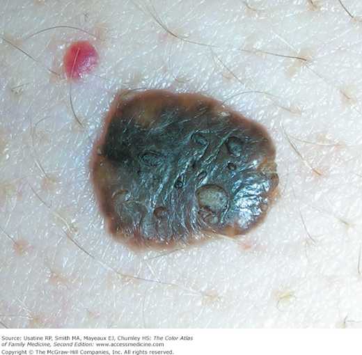
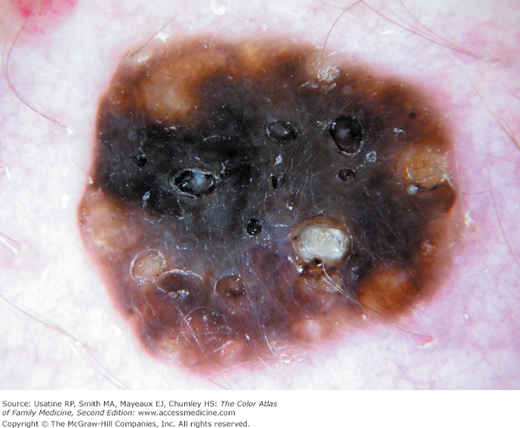
An elderly woman noted a growth of a lesion on her chest (Figure 158-1). She was afraid that it might be melanoma. Her family physician recognized the typical features of a seborrheic keratosis (SK) (stuck-on with visible horn cysts) and attempted to reassure her. Dermoscopy was performed and the features were so typical of an SK; the physician was able to convince the patient to not have a biopsy (Figure 158-2). The black comedonal-like openings and white milia-like cysts are typical of an SK and can be seen with the naked eye and magnified with a dermatoscope.
Introduction
Epidemiology
- Most common benign tumor in older individuals; frequency increases with age.
- In an older study of individuals older than age 64 years in North Carolina, 88% had at least one SK. Ten or more SKs were found in 61% of the black men and women, 38% of the white women, and 54% of the white men in the study.1
- In an Australian study preformed in 2 general practices, 23.5% (40 out of 170) of individuals between ages 15 and 30 years had at least 1 SK; prevalence and size increased with age.2
- Approximately half of cases of multiple SKs occur within families, with an autosomal dominant mode of inheritance.3
Etiology and Pathophysiology
- In pigmented SKs, the proliferating keratinocytes secrete melanocyte-stimulating cytokines triggering activation of neighboring melanocytes.3
- A high frequency of mutations have been found in certain types of SKs in the gene encoding the tyrosine kinase receptor fibroblast growth factor receptor 3 (FGFR3).3 One study found that FGFR3 and transcription factor forkhead box N1 (FOXN1) were highly expressed in SKs but close to undetectable in squamous cell skin cancer.4 This may represent a positive regulatory loop between FGFR3 and FOXN1 that underlies a benign versus malignant skin tumor phenotype.
- Reticulated SKs, usually found on sun-exposed skin, may develop from solar lentigines.3
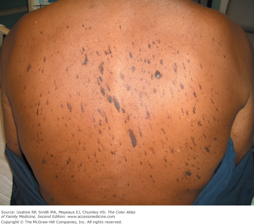
- Multiple eruptive seborrheic keratoses (the sign of Leser-Trélat) can be associated with internal malignancy (most often adenocarcinoma of the GI tract) (Figure 158-3),5 although this association has been questioned.6
- An eruption of seborrheic keratoses may develop after an inflammatory dermatosis such as severe sunburn or eczema.3 There is also a report of exacerbation of a SK by topical fluorouracil.7
Diagnosis
- Typically oval or round brown plaques with adherent greasy scale (Figure 158-4).
- Color ranges from black to tan (Figures 158-4 to 158-6).
- Most often have a velvety to finely verrucous surface and appear to be “stuck on.”
- Some are so verrucous they can appear to be warty (Figure 158-6).
- Lesions may be large (up to 35 × 15 cm), pigmented, and have irregular borders (Figure 158-5).
- Lesions can also be flat (Figure 158-7).
- Many lesions show keratotic plugging of the surface (Figures 158-1 and 158-2).
- May have surface cracks and associated horn cysts (keratin-filled cystic structures). The dermoscopy terms for the horn cysts are comedo-like openings and milia-like cysts (Figures 158-1 and 158-2; see Appendix C, Dermoscopy).
- Occasionally, lesions become irritated and can itch, grow, and bleed; secondary infection may occur.
- Variants of SKs include:
- Dermatosis papulosa nigra—Consists of multiple brown-black dome-shaped, smooth papules found on the face in young and middle-age persons of color, predominantly African Americans (Figures 158-8 and 158-9

Stay updated, free articles. Join our Telegram channel

Full access? Get Clinical Tree


- Dermatosis papulosa nigra—Consists of multiple brown-black dome-shaped, smooth papules found on the face in young and middle-age persons of color, predominantly African Americans (Figures 158-8 and 158-9
