Sebocytes in lobules, rimmed by more than 1 layer of basaloid cells
Often connecting directly to epidermal surface
• Ectopic sebaceous glands in other sites (e.g., nipple)
Often connect directly to epidermal surface
• Phymatous rosacea
Prominent sebaceous glands, edema, and fibrosis on different sites
Clinical history is key
• Sebaceous trichofolliculoma
Mature, central hair follicle, often producing hair shaft
Smaller, baby hair follicles and sebaceous glands radiating away from central follicle
• Folliculosebaceous cystic hamartoma
Central dilated infundibulum surrounded by hair follicles and sebaceous glands
Fibrocytic stroma
• Sebaceous induction
Lobules of sebocytes connecting to epidermal surface
Most commonly seen above dermatofibroma
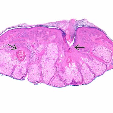
Sebaceous Hyperplasia Low-magnification view of sebaceous hyperplasia. There are normal-appearing lobules of sebocytes surrounding invaginations of epidermis that resemble the infundibula of hair follicles .
Sebaceous Hyperplasia Attached to Follicular Infundibulum Lobules of sebocytes surround an invagination of epidermis that resembles the infundibulum of a hair follicle. The lobules of sebocytes are rimmed by a single compressed layer of small basaloid cells .
Sebaceous Hyperplasia at High Magnification High-power view of sebocytes shows bubbly cytoplasm and a central round or scalloped nucleus.
Sebaceous Hyperplasia at High Magnification High-magnification view of the edge of a lobule of sebocytes shows the central sebocytes with round to scalloped nuclei and bubbly cytoplasm, rimmed by a compressed layer of small, basaloid cells .
TERMINOLOGY
Definitions
• Hyperplasia (overgrowth) of sebaceous glands
• Plump lobules of sebaceous glands arranged around central follicular structures
CLINICAL ISSUES
Site
• Commonly on face
• Rarely on trunk or other sites
Presentation
• Yellow to flesh-colored to slightly pink papule
• Often there is central dell
• Telangiectasias may be present
• Often biopsied to rule out basal cell carcinoma
Laboratory Tests
• Generally not performed
• Some have suggested that sebaceous hyperplasia on sites other than face is sufficiently rare that evaluation for Muir-Torre syndrome is warranted; however, recent study of vulvar sebaceous hyperplasia showed no association with Muir-Torre syndrome
Treatment
• Not necessary in most cases
• Biopsy (shave removal) may be curative
• Some studies suggest effective treatment with oral retinoids or lasers
Only gold members can continue reading. Log In or Register to continue


 .
.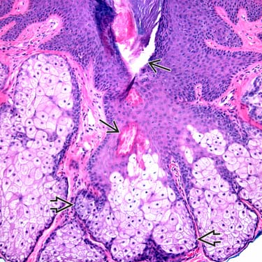
 that resembles the infundibulum of a hair follicle. The lobules of sebocytes are rimmed by a single compressed layer of small basaloid cells
  .
.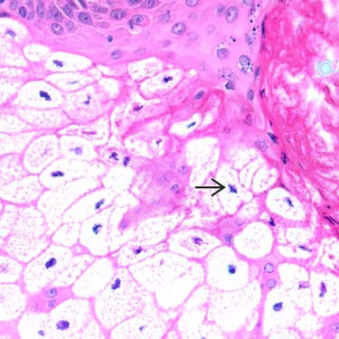
 nucleus.
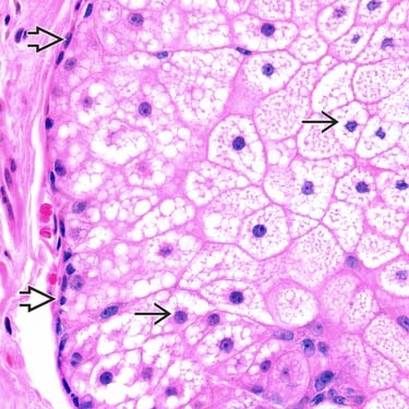
 with round to scalloped nuclei and bubbly cytoplasm, rimmed by a compressed layer of small, basaloid cells
  .
.


