Sebaceous Adenoma

 Located centrally within lobules


• Basaloid/germinative cells at periphery of lobules

• Cytologic atypia not prominent

• Presence of mitotic figure in germinative layer does not indicate malignant potential


Ancillary Tests


• Sebocytes express EMA, podoplanin (D2-40), androgen receptor, and adipophilin


Top Differential Diagnoses


• Sebaceoma
image Composed of > 50% germinative/basaloid cells

• Sebaceous hyperplasia
image Superficial sebaceous lobules surrounding dilated follicle/pore

• Clear cell basal cell carcinoma
image Lack of scalloped nuclei

image Cytoplasm not truly bubbly

• Tricholemmoma

image
Scanning Magnification of a Sebaceous Adenoma
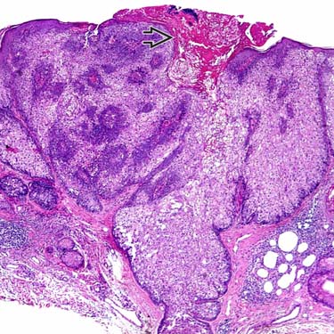
In this low magnification of a large sebaceous adenoma, the tumor is a well-circumscribed, multilobular proliferation with multiple attachments to the epidermis and superficial holocrine necrosis image.


image
High Magnification of Sebaceous Adenoma
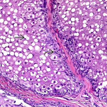
Sebocytes have a central, scalloped/crenulated nuclei with bubbly cytoplasm image. Basaloid/germinative cells rim the sebocytes image.

image
Sebaceous Adenoma With Irregular Lobules
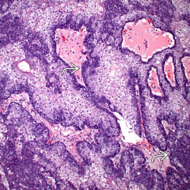
This example of sebaceous adenoma shows a proliferation of small, irregularly-shaped sebaceous lobules composed of bland-appearing sebocytes surrounded by an expanded basaloid layer image.

image
Traumatized Sebaceous Adenoma
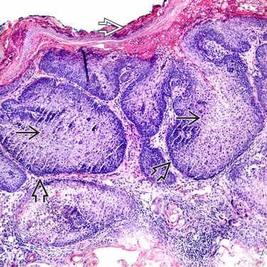
This is an example of traumatized sebaceous adenoma, with overlying hemorrhage and serum crust image. The lesion is composed of central sebocytes image and peripheral basaloid cells image. The basaloid cells form 1-3 layers at the periphery of lobules.


TERMINOLOGY


Definitions


• Benign tumor

• > 50% of tumor is composed of mature sebocytes

• > 1 layer of basaloid/germinative cells at periphery of sebocyte-filled lobules


CLINICAL ISSUES


Site


• Often on head and neck, but any site possible


Presentation


• Yellow, tan, to pink papule


Treatment


• Reexcision not necessary


Prognosis


• Benign tumor

• No malignant potential


Associated Syndromes


• Even 1 sebaceous adenoma can be associated with Muir-Torre syndrome
image Muir-Torre syndrome

Only gold members can continue reading. Log In or Register to continue

Stay updated, free articles. Join our Telegram channel

Apr 24, 2017 | Posted by in PATHOLOGY & LABORATORY MEDICINE | Comments Off on Sebaceous Adenoma

Full access? Get Clinical Tree

Get Clinical Tree app for offline access