Patient Story
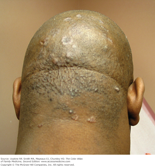
A 42-year-old man presents with “multiple bumps” that had been growing on his scalp, the back of the neck, and on preexisting scars (Figure 175-1). These lesions started developing slowly over a period of 1 year. The differential diagnosis of these lesions included cutaneous sarcoidosis, acne keloidalis nuchae, and pseudofolliculitis barbae. A punch biopsy was performed and the diagnosis of sarcoidosis was made.
Introduction
Sarcoidosis is a multisystem granulomatous disease most commonly involving the skin, lungs, lymph nodes, liver, and eyes. Patients of African descent are more commonly affected compared to white patients. Diagnosing cutaneous sarcoidosis is critical as 30% of these patients have been found to have systemic involvement. Diverse presentations of cutaneous sarcoidosis have been reported in addition to variants of specific sarcoidosis syndromes.
Synonyms
Epidemiology
- Cutaneous manifestations occur in approximately 25% of systemic sarcoidosis patients.
- The ratio between patients with only cutaneous sarcoid versus multisystem involvement is 1:3.
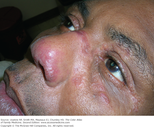
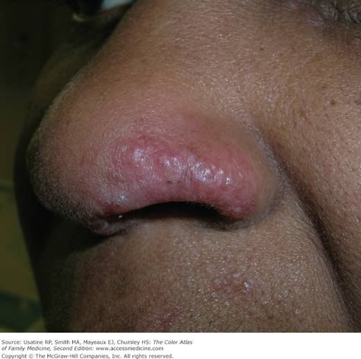
- Specific cutaneous involvement is seen most commonly in older, female patients of African descent (Figures 175-2 and 175-3).
- Common types are maculopapular, lupus pernio, cutaneous, or subcutaneous nodules, and infiltrative scars.
- Erythema nodosum (EN) occurs in 3% to 34% of patients with sarcoidosis and is the most common associated skin finding (see Chapter 178, Erythema Nodosum).
- Sarcoidosis-related EN is more prevalent in whites, especially Scandinavians. Irish and Puerto Rican females are also affected more often.
- EN occurs between the second and fourth decades of life, more commonly in women.
- Nonspecific lesions of sarcoidosis reported, besides EN, include erythema multiforme, calcinosis cutis, prurigo, and lymphedema. Nail changes can include clubbing, onycholysis, subungual keratosis, and dystrophy, with or without underlying changes in the bone (cysts).
Etiology and Pathophysiology
- Sarcoidosis is a granulomatous disease with involvement of multiple organ systems with an unknown etiology.
- The typical findings in sarcoid lesions are characterized by the presence of circumscribed granulomas of epithelioid cells with little or no caseating necrosis, although fibrinoid necrosis is not uncommon.
- Granulomas are usually in the superficial dermis but may involve the thickness of dermis and extend to the subcutaneous tissue. These granulomas are referred to as “naked” because they only have a sparse lymphocytic infiltrate at their margins.
Diagnosis
Cutaneous involvement is either specific or nonspecific.
- Specific:
- Typical noncaseating granulomas, no evidence of infection, foreign body, or other causes.
- May be disfiguring, but almost always nontender and rarely ulcerate.
- Maculopapular type is most common, red-brown or purplish, usually smaller than 1 cm, and found mostly on face, neck, upper back, and limbs (Figure 175-4).
- Lupus pernio type are most distinctive lesions and present as purplish lesions resembling frostbites with shiny skin covering them, typically affecting nose, cheeks, ears, and lips and distal extremities (Figures 175-2, 175-3, and 175-5).
- Lupus pernio may occur as a syndrome involving upper respiratory tract with pulmonary fibrosis, or be associated with chronic uveitis and bone cysts.
- Annular or circinate type appear ribbon-like, with mild scaling and yellowish red in color, with centrifugal progression and central healing and depigmentation (Figure 175-1).
- Plaque sarcoidosis is typically chronic, occurring over the forehead, extremities, and shoulders, but may heal without scarring (Figure 175-6).
- Nodular cutaneous and subcutaneous plaques that are skin-colored or violaceous without epidermal involvement are typically seen in advanced systemic sarcoidosis (Figure 175-7).
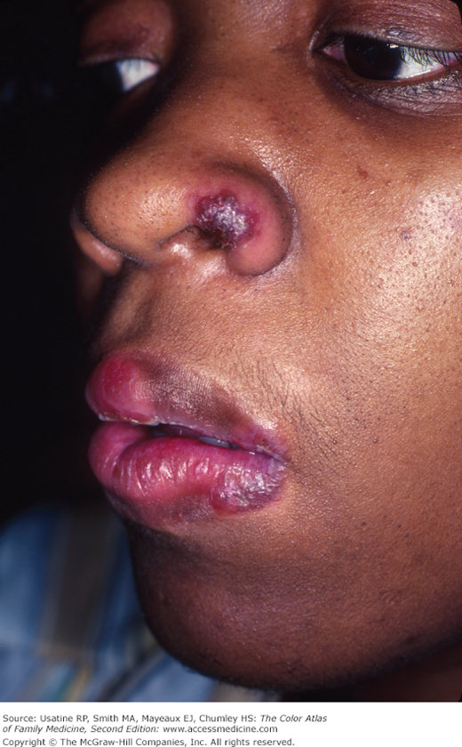
- Areas of old scars that are damaged by trauma, radiation, surgery, or tattoo may also be infiltrated with sarcoid granulomas (Figures 175-8 and 175-9). Lesions may be tender and appear indurated with red or purple discoloration.
- Typical noncaseating granulomas, no evidence of infection, foreign body, or other causes.
- Nonspecific:
- EN lesions usually are not disfiguring, but tender to touch, especially when they occur with fever, polyarthralgias, and sometimes arthritis and acute iritis.
- EN appears abruptly with warm, tender, reddish nodules on the lower extremities, most commonly the anterior tibial surfaces, ankles, and knees.
- EN nodules are 1 to 5 cm, usually bilateral, and evolve through color stages: first bright red, then purplish, and lastly a bruise-like yellow or green appearance.
- EN bouts occur with fatigue, fever, symmetrical polyarthritis, and skin eruptions that typically last 3 to 6 weeks with more than 80% of cases resolving within 2 years.4
- EN is seen in the setting of Löfgren syndrome, appearing in conjunction with hilar lymphadenopathy (bilateral most often), and occasionally anterior uveitis and/or polyarthritis.
- Löfgren syndrome is associated with right paratracheal lymph node involvement seen on x-ray.
- Ulceration is typically not observed in EN, which heal without scarring.
- Other nonspecific lesions of sarcoidosis include lymphedema, calcinosis cutis, prurigo, and erythema multiforme.
- Nail changes seen in sarcoidosis include clubbing, onycholysis, and subungual keratosis.
- EN lesions usually are not disfiguring, but tender to touch, especially when they occur with fever, polyarthralgias, and sometimes arthritis and acute iritis.