Rhabdomyoma
Cyril Fisher, MD, DSc, FRCPath
Key Facts
Terminology
Benign tumor with skeletal muscle differentiation
Extracardiac tumors can be of adult or fetal histologic type
Etiology/Pathogenesis
Some fetal rhabdomyomas associated with nevoid basal cell syndrome
Clinical Issues
Adults; mean: 6th-7th decades; 75% males
Fetal rhabdomyoma mostly childhood
Genital rhabdomyoma mostly in middle-aged women
Most often in head and neck region, especially fetal rhabdomyoma
Genital lesions mostly in vagina, occasionally vulva or cervix
Microscopic Pathology
Adult rhabdomyoma
Circumscribed
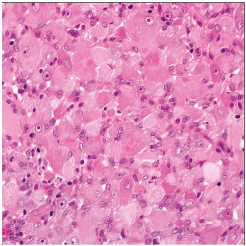
Large polygonal cells with abundant eosinophilic cytoplasm
Fetal rhabdomyoma
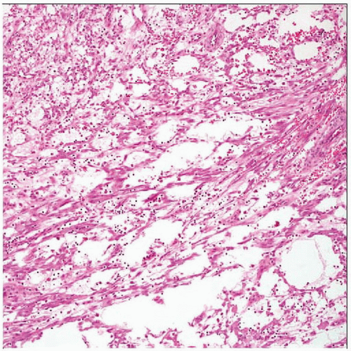
Immature (myxoid) type has long spindle cells in myxoid stroma
Intermediate (juvenile) type has spindled and round cells with variable skeletal muscle differentiation
No atypia or necrosis; mitoses usually absent
Ancillary Tests
Lesional cells are immunoreactive for desmin, myogenin, and MYOD1
 Hematoxylin & eosin shows adult rhabdomyoma composed of large polygonal cells with copious eosinophilic cytoplasm (varying in staining intensity) and small round nuclei with uniform nucleoli. |
TERMINOLOGY
Definitions
Benign tumor with skeletal muscle differentiation
Can arise in heart (cardiac rhabdomyoma) or extracardiac locations
Extracardiac tumors can be of adult or fetal histologic type
ETIOLOGY/PATHOGENESIS
Developmental Anomaly
No associations for most extracardiac lesions
Cardiac rhabdomyoma can be associated with tuberous sclerosis
Some fetal rhabdomyomas associated with nevoid basal cell syndrome
PTCH mutations
Inhibitory receptor in sonic hedgehog signaling pathway
CLINICAL ISSUES
Epidemiology
Incidence
Rare
Age
Adults; mean: 6th-7th decades
Fetal rhabdomyoma mostly in childhood; median: 4 years
About 1/2 in 1st year or congenital
Rare examples in adults up to 6th decade
Gender
75% in males
Genital rhabdomyoma mostly in middle-aged women
Rare cases in males
Site
Most often in head and neck region, especially fetal rhabdomyoma
Larynx, oropharynx, mouth, neck
Genital lesions mostly in vagina, occasionally vulva or cervix
Rare examples in males in paratesticular region or epididymis
Presentation
Incidental finding
Painless mass
Difficulty breathing
Treatment
Surgical approaches
Simple complete excision
Prognosis
Excellent after complete excision
Can recur if incompletely excised
MACROSCOPIC FEATURES


