History
Key features of the history in a patient with respiratory disease are shown in Table 4.1.
Table 4.1 Key features of the History in Respiratory Disease
| Feature | Details | Rationale |
| Basic details | Age, sex, current and previous occupations | Identify risk of occupational lung disease |
| Symptoms | Dyspnoea, chest pain, wheeze, cough, sputum and haemoptysis | Establish pattern of symptoms and their likely causes |
| Past medical history | Previous episodes or other lung disease | Suggestive of chronic or relapsing symptoms, i.e. asthma, COPD |
| Allergies | Identify known allergies in patient or family | Potential for allergic lung diseases |
| Smoking | Establish accurate smoking history | Increased risk of chronic lung disease and cancer |
Examination
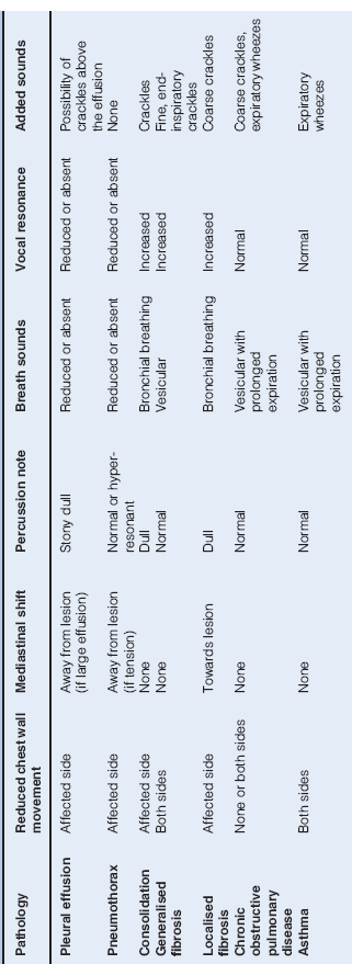
Key abnormalities detected on examination of the chest are shown in Table 4.2.
Table 4.2 Physical Findings in Common Chest Diseases
General observation: note
- dyspnoea
- cyanosis
- evidence of loss of weight
Examine the hands for
- clubbing
- tobacco staining
- coarse tremor of outstretched hands
- bounding radial pulse
Check
- the pulse rate
- the height of the jugular venous pressure
- the tongue for cyanosis
Observe
- the shape of the chest and spine
- scars
- chest movements for symmetry and expansion
- the use of accessory muscles in the neck and shoulders
- visibly enlarged cervical lymph nodes
Count
- the respiratory rate
Examine the front and back of the chest in a logical manner, usually by palpating, percussing and auscultating the front of the chest first, followed by the rear. When examining the back of the chest, ask the patient to put their hands on their hips to facilitate examination of the lung bases laterally.
Palpation
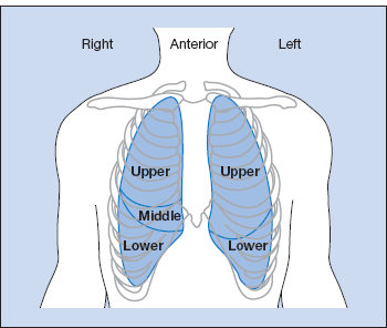
The anterior surface markings of the lungs are shown in Fig. 4.1.
Figure 4.1 Surface markings of the lungs. Oblique fissures run along the line of the fifth/sixth rib; a horizontal fissure runs from the fourth costal cartilage to the sixth rib in the mid-axillary line. Note: On auscultation posteriorly you are listening mainly to lower lobes. Anteriorly you are listening mainly to upper lobes and on the right the middle lobe.
Palpate for
- chest expansion, comparing the movements of the two sides
- the trachea in the suprasternal notch to assess mediastinal shift with the patient’s head partially extended. (Local anatomical and pathological variants may produce tracheal deviation in the absence of lung disease, e.g. a goitre or spinal asymmetry. The position of the heart apex beat is of no help in assessing lung disease except if there is marked mediastinal shift.)
- cervical lymphadenopathy
Percussion
- examine the apices by percussing the clavicles
- move down the chest alternating right and left to compare both sides
Auscultation
Listen for
- bronchial breathing
- diminished breath sounds
- added sounds
 wheezes
 crackles
 pleural rubs
- vocal resonance
Notes
Haemoptysis
Aetiology: Common
- bronchial carcinoma
- tuberculosis
- pulmonary embolism with infarction
- infection (e.g. pneumococcal pneumonia, lung abscess and Klebsiella pneumoniae)
Aetiology: Uncommon
- foreign body – history of general anaesthetic, visit to dentist or inhalation of food
- coagulation disorders
- bronchiectatic cavities
- mitral stenosis
- Wegener’s granulomatosis
- Goodpasture syndrome
- intrapulmonary vascular tumours
Investigation of Haemoptysis
The usual clinical problem is to exclude carcinoma and tuberculosis. A full history and clinical examination will usually identify pulmonary infarction, foreign body, bronchiectasis, mitral stenosis and pulmonary oedema.
Perform
- sputum microscopy and culture, including for acid-fast bacilli
- sputum cytology for malignant cells
- chest X-ray
- CT or MRI scan to define the site and nature of the lesions seen on chest X-ray
- bronchoscopy with biopsy for cytology and culture
- CT guided biopsy of mass lesions
- isotope (V/Q) lung scan +/− spiral CT if pulmonary embolism is suspected
About 40% of patients with haemoptysis have no demonstrable cause. Patients who have had a single small haemoptysis, no other symptoms and a normal chest X-ray (postero-anterior and lateral) should have a follow-up chest X-ray after 1–2 months. Patients who have more than one small haemoptysis should be referred for bronchoscopy.
Clubbing
Finger clubbing is associated with a range of respiratory diseases, but also with disease in the cardiovascular and gastrointestinal systems. Rarely, clubbing may be familial and innocent.
Respiratory Causes
- carcinoma of bronchus
- chronic suppurative lung disease: empyema, lung abscess, bronchiectasis, cystic fibrosis
- fibrosing alveolitis
- asbestosis
- mesothelioma
Cardiac Causes
- cyanotic congenital heart disease
- subacute bacterial endocarditis
Gastrointestinal Causes
- Crohn’s disease
- ulcerative colitis
- hepatic cirrhosis
Cyanosis
Cyanosis is a clinical description which refers to the blue-ish colour of a patient’s lips and tongue (central) or fingers (peripheral). Central cyanosis is always accompanied by peripheral cyanosis.
Cyanosis is an unreliable guide to the degree of hypoxaemia. Central cyanosis is usually caused by the presence of an excess of reduced haemoglobin in the capillaries. Thus, in anaemia, severe hypoxaemia may be present without cyanosis.
Examine
- the underside of the patient’s tongue and their finger nail beds (compare nail beds with your own)
If the tongue is cyanosed, the cyanosis is central in origin and secondary to:
- chronic bronchitis and emphysema, often with cor pulmonale
- congenital heart disease (cyanosis may be present only after exercise)
- polycythaemia
- massive pulmonary embolism.
If the tongue is not cyanosed but the finger nail beds are, the cyanosis is peripheral and secondary to
- physiological causes (cold)
- pathology in peripheral vascular disease (the cyanosed parts feel cold).
Left ventricular failure may produce cyanosis that is partly central (pulmonary) and partly peripheral (poor peripheral circulation).
A rare cause of cyanosis, not caused by increased circulating reduced haemoglobin, is the presence of methaemoglobin (and/or sulphaemoglobin). The patient is relatively well and not necessarily dyspnoeic. Methaemoglobinaemia is usually drug-induced, e.g. sulphonamides, primaquine or nitrites.
Investigation of the Respiratory System
Chest Radiology
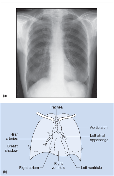
Normal chest X-rays are shown in Fig. 4.2 (postero-anterior) and Fig. 4.3 (lateral). Fig. 4.4 is a radiological chest diagram of lobar collapse. CT chest scans are shown in Fig. 4.5.
Stay updated, free articles. Join our Telegram channel

Full access? Get Clinical Tree


