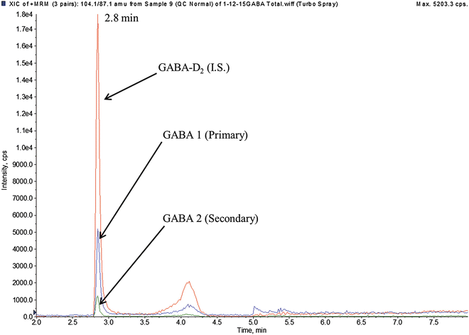
Fig. 1
HPLC-ESI-MS/MS ion chromatogram of GABA 1 (m/z 104.1 > 87.1), GABA 2 (m/z 104.1 > 69.1), GABA-D2 (m/z 106.0 > 89.1). Concentration of GABA shown is 169 nM
3.2 Sample Preparation (Total GABA )
1.
To labeled 1.5 mL microcentrifuge tubes, pipette 50 μL CSF (calibrators, controls, patient CSF).
2.
Add 50 μL of Total GABA I.S. Working Solution.
3.
Add 200 μL 6 N HCl.
4.
Cap and vortex mix tubes at maximum speed for 3 s.
5.
Place tubes in metal rack with screw-top rack.
6.
Place metal rack with tubes in electric skillet filled set at 300 °C filled with water.
7.
Boil samples for 4 h.
8.
After boiling, remove samples and allow to reach room temperature.
9.
Centrifuge for 1 min at 14,000 × g.
10.
To new labeled 1.5 mL microcentrifuge tubes, add 180 μL deproteinizing solution.
11.
Transfer 20 μL of hydrolyzed (boiled) sample to 1.5 mL tube and mix well by vortex.
12.
Centrifuge for 5 min at 14,000 × g.
13.
Transfer 160 μL supernatant into corresponding work list position in 96-well microtiter plate and cover with silicone cover.
14.
Place completed 96-well microtiter plate onto the autosampler.
15.
Inject 10 μL of sample onto HPLC-ESI-MS/MS. Representative HPLC-ESI-MS/MS ion chromatograms for free GABA and I.S. are shown in Fig. 2 (see Notes 4 and 5 ).


Fig. 2
HPLC-ESI-MS/MS ion chromatogram of GABA 1 (m/z 104.1 > 87.1), GABA 2 (m/z 104.1 > 69.1), GABA-D2 (m/z 106.0 > 89.1). Concentration of GABA shown is 8.8 μM
3.3 Data Analysis
1.
Instrumental operating parameters are given in Table 1 A, B, and C.
Table 1
HPLC-ESI-MS/MS operating conditions
A. HPLC (Free GABA) a | ||
Column temp. | 40 °C | |
Flow rate | 0.230 mL/min | |
Gradient | Time (min) | Mobile phase A (%) |
0 | 90 | |
5 | 25 | |
5.1 | 0 | |
6 | 0 | |
6.1 | 90 | |
10 | Stop | |
B. HPLC (Total GABA) a | ||
Column temp. | 40 °C | |
Flow rate | 0.230 mL/min | |
Gradient | Time (min) | Mobile phase A (%) |
0 | 90 | |
4
Stay updated, free articles. Join our Telegram channel
Full access? Get Clinical Tree
 Get Clinical Tree app for offline access

| ||