Patient Story
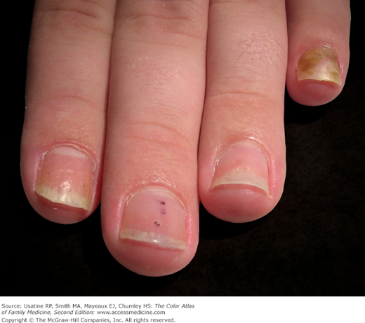
A 19-year-old man with a 4-year history of plaque psoriasis presents with nail abnormalities in several fingers (Figure 195-1). He is particularly concerned about the recently acquired greenish discoloration of his fifth digit.
Introduction
Psoriasis is a hereditary disorder of skin with numerous clinical expressions. It affects millions of people throughout the world.1 Nail involvement is common and can have a significant cosmetic impact.
Epidemiology
- Nails are involved in 30% to 50% of psoriasis patients at any given time, and up to 90% develop nail changes over their lifetime.1 In most cases, nail involvement coexists with cutaneous psoriasis, although the skin surrounding the affected nails need not be involved. Psoriatic nail disease without overt cutaneous disease occurs in 1% to 5% of psoriasis. Patients with nail involvement are thought to have a higher incidence of associated arthritis.2
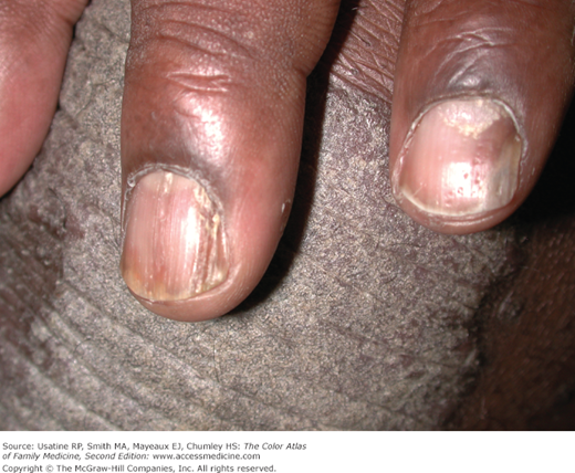
- The most common nail change seen with psoriasis is nail plate pitting (Figures 195-1 and 195-2).
Etiology and Pathophysiology
- In psoriasis, parakeratotic cells within the stratum corneum of the nail matrix alters normal keratinization.3 The proximal nail matrix forms the superficial portion of the nail plate, so that involvement in this part of the matrix results in pitting of the nail plate (Figures 195-1 and 195-2.) The pits may range in size from pinpoint depressions to large punched-out lesions. People without psoriasis can have nail pitting.
- Longitudinal matrix involvement produces longitudinal nail ridging or splitting (Figure 195-2). When transverse matrix involvement occurs, solitary or multiple “growth arrest” lines (Beau lines) may occur (see Chapter 190, Normal Nail Variants). Psoriatic involvement of the intermediate portion of the nail matrix leads to leukonychia and diminished nail plate integrity.
- Parakeratosis of the nail bed with thickening of the stratum corneum causes discoloration of the nail bed, producing the “salmon spot” or “oil drop” signs.3
- Desquamation of parakeratotic cells at the hyponychium leads to onycholysis, which may allow for bacteria and fungi infection.4
Risk Factors
Diagnosis
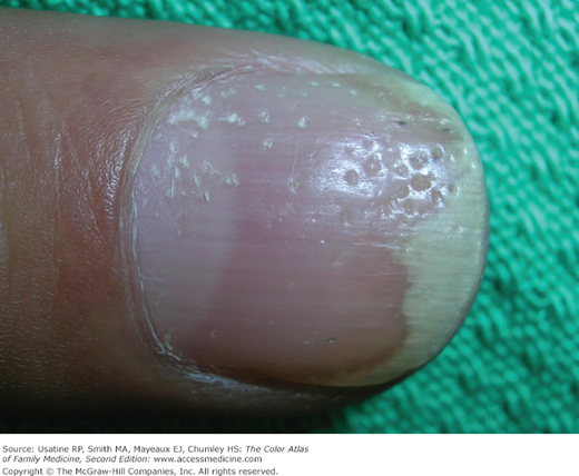
- The diagnosis of nail psoriasis is usually straightforward when characteristic nail findings coexist with cutaneous psoriasis. Nail pitting and onycholysis are the most common findings (Figure 195-3).
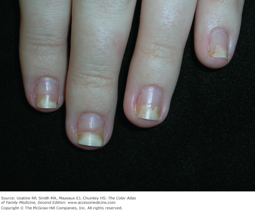
- Nail psoriasis and onychomycosis are often indistinguishable by clinical examination alone. Psoriasis at the hyponychium produces subungual hyperkeratosis and distal onycholysis (Figures 195-4 and 195-5). Trauma may accentuate this process. Secondary microbial colonization by Candida or Pseudomonas organisms may occur (Figures 195-1 and 195-5).
- Nail bed psoriasis produces localized onycholysis which often appears like a drop of oil on a piece of paper (oil drop sign) (Figures 195-2, 195-4, and 195-5). This same condition is also called the salmon patch sign.
- Extensive germinal matrix involvement may result in loss of nail integrity and transverse (horizontal) ridging (Figure 195-6).
- Psoriasis causes dermal vascular dilation and tortuosity, and in the nails is associated with splinter hemorrhages of the nail bed caused by foci of capillary bleeding. Extravasated blood becomes trapped between the longitudinal troughs of the nail bed and the overlying nail plate grows out distally along with the plate (Figure 195-7). The splinter hemorrhages of the psoriatic nail are analogous to the cutaneous Auspitz sign.
Stay updated, free articles. Join our Telegram channel

Full access? Get Clinical Tree


