Multicystic squamous neoplasm composed of mature keratinocytes lining keratin-filled spaces
Etiology/Pathogenesis
• Postulated that most cases arise in preexisting pilar (tricholemmal) cyst; may be related to chronic inflammation or trauma
Clinical Issues
• Typically occur on scalp (90% of cases) in older adults
• Much more common in female than male patients
• Most cases of PPT behave in benign fashion, but malignant PPTs are aggressive tumors that have high rate of metastasis
Complete surgical excision is recommended in order to prevent recurrence and malignant transformation
Macroscopic
• Large tumors, 6 cm or greater in most cases
• Often multicystic dermal-based tumors that may involve subcutis
Microscopic
• Cystic spaces are irregularly formed and anastomosing
• Cysts show keratinization without granular layer
• Large, multicystic, dermal-based tumor with squamous lining and spaces containing dense keratin
• Peripheral palisading of basilar layer is typically present, and there may be thickened basement membrane
• Occasional mitotic figures are present, but no high-grade atypia or increased mitotic activity should be present
Top Differential Diagnoses
• Pilar/tricholemmal cyst
Unicystic structure lined by mature squamous cells
• Proliferating epidermoid cyst
Shows overlapping features with PPT but has granular layer and laminated keratin
• Malignant PPT (squamous cell carcinoma arising in PPT)
Larger than benign PPTs, clinically often present as rapidly enlarging nodular mass lesion
Show greater cytologic atypia and mitotic activity, infiltrative features
Low Magnification of PPT Scanning magnification of proliferating pilar tumor (PPT) shows a smooth-bordered , circumscribed tumor composed of thickened cords of squamous cells surrounding a large cystic cavity containing abundant keratin material.
High Magnification of PPT High-magnification examination of a benign PPT shows bland squamous keratinocytes with abundant eosinophilic cytoplasm surrounding keratin-filled spaces. Note the lack of an intervening granular layer .
PPT With Bland Cytologic Features High-magnification examination shows the bland cytologic features of the squamous cells, which show uniform nuclei, small nucleoli, and abundant amounts of dense eosinophilic cytoplasm. Scattered dyskeratotic keratinocytes are present .
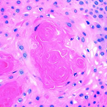
PPT With Keratinization High-magnification examination shows several small foci of dense keratin material surrounded by bland-appearing squamous cells.


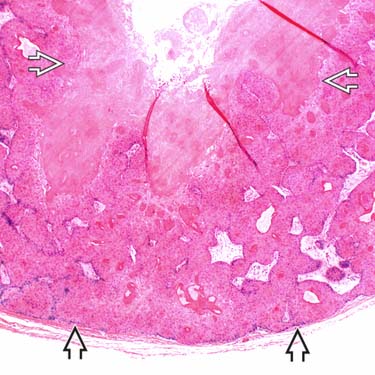
 , circumscribed tumor composed of thickened cords of squamous cells surrounding a large cystic cavity
  containing abundant keratin material.
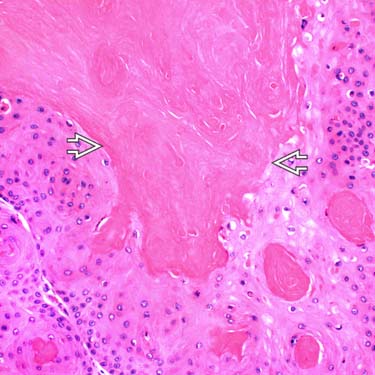
 .
.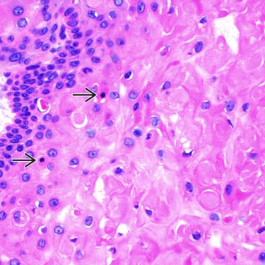
 .
.