ERCP A classic ERCP of primary sclerosing cholangitis (PSC) shows multiple segmental strictures of the biliary tree, resulting in a beaded appearance . There are also diverticular outpouchings of dilated bile ducts .
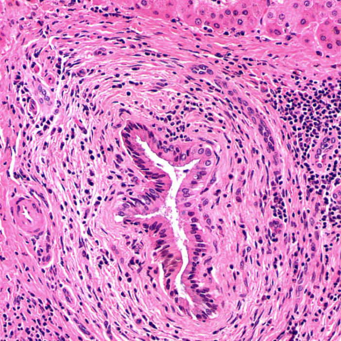
Onion Skin Lesion The classic onion skin lesion of PSC consists of concentric fibrosis around a bile duct. There is mild lymphoplasmacytic infiltration of the portal tract and lymphocytic cholangitis in the duct epithelium.
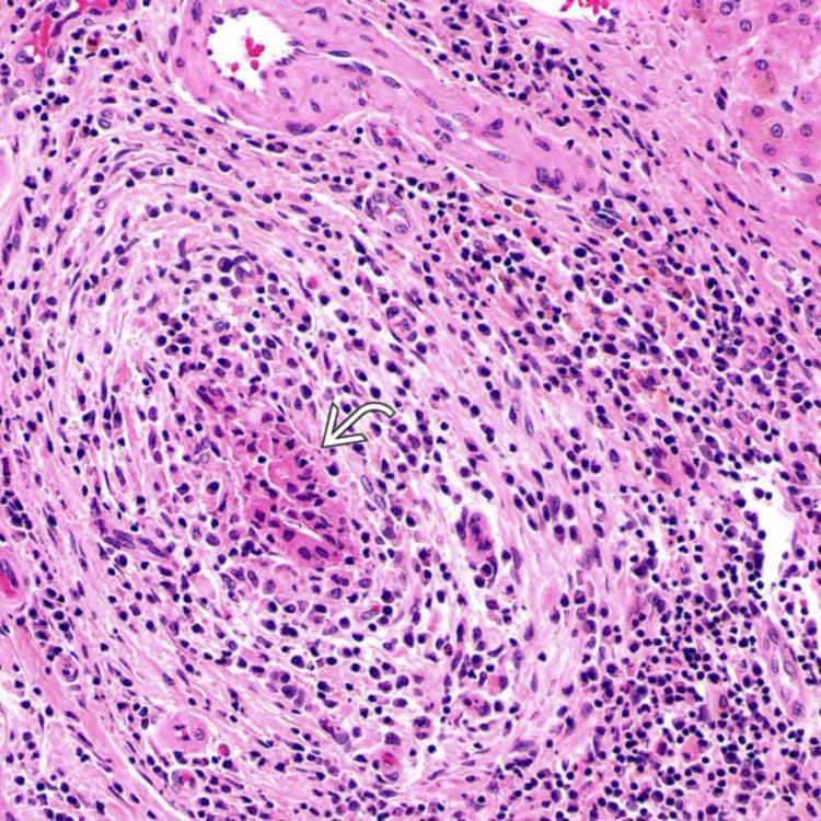
Inflammation and Duct Damage This case of PSC shows markedly damaged duct epithelium, as well as lymphoplasmacytic infiltration of the duct, periductal stroma, and portal tract. Lymphocytic cholangitis is apparent within the remaining ductal epithelium .
Duct Loss With Scar This interlobular bile duct has been entirely replaced by a round fibroobliterative scar . The hepatic artery and portal vein branches are unremarkable.
TERMINOLOGY
Abbreviations
• Primary sclerosing cholangitis (PSC)
Definitions
• Chronic cholestatic disease featuring progressive inflammation and fibrosis of intrahepatic and extrahepatic biliary tree
ETIOLOGY/PATHOGENESIS
Unknown
• Frequent association with HLA-B8 and DR3
100x increased risk of disease in 1st-degree relatives of patients with PSC


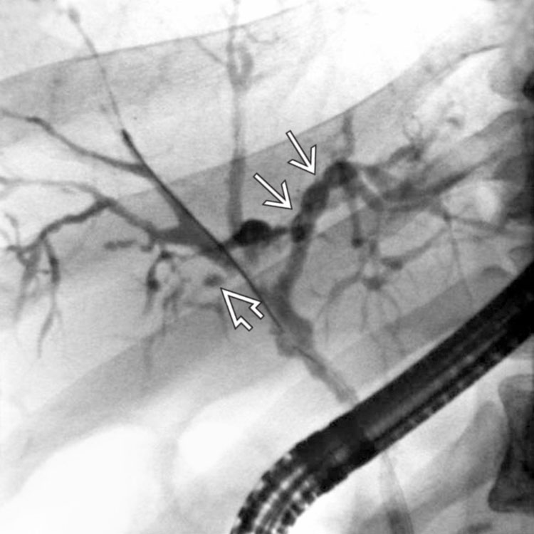
 . There are also diverticular outpouchings of dilated bile ducts
  .
.
 .
.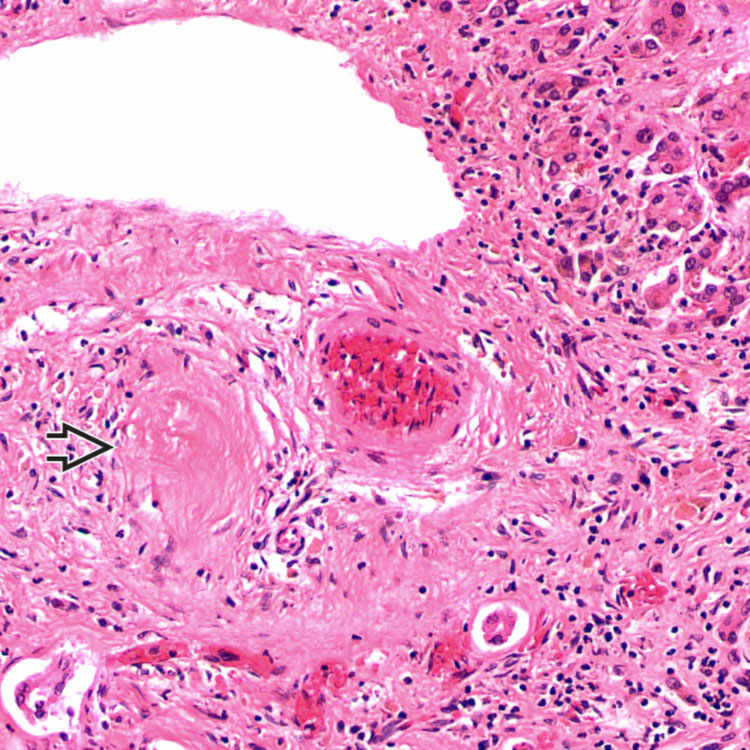
 . The hepatic artery and portal vein branches are unremarkable.


