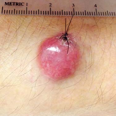
Clinical Photograph of MCC Clinical photograph of Merkel cell carcinoma (MCC) shows a well-circumscribed, erythematous dermal nodule. (Courtesy J. Wu, MD.)
MCC Involving Superficial Dermis The superficial dermal portion of this tumor shows enlarged, crowded and markedly atypical-appearing basaloid cells with several large, atypical mitotic figures easily identified.
Higher Magnification of MCC With Lymphatic Invasion High magnification of the superficial dermal portion of this tumor shows enlarged, atypical basaloid cells with several frankly atypical mitotic figures . Invasion of a superficial lymphatic vessel is also seen .
High Magnification of MCC Nuclear clearing is often seen in MCC, a feature not seen in basal cell carcinoma or most other small round blue cell tumors. Note the numerous apoptotic bodies and mitotic figures .
TERMINOLOGY
Abbreviations
• Merkel cell carcinoma (MCC)
Synonyms
• Cutaneous neuroendocrine carcinoma
• Primary small cell carcinoma of skin
• Trabecular carcinoma
Definitions
• Malignant proliferation of cutaneous neuroendocrine cells
ETIOLOGY/PATHOGENESIS
Infectious Agents
• Recent studies have shown strong link to infection with polyomavirus
Merkel cell polyoma virus infection is found in > 90% of cases by PCR studies
• Associated with immunosuppression
Organ transplant and HIV(+) patients have much higher incidence
Cell of Origin
• Postulated to represent malignant transformation of cutaneous neuroendocrine (Merkel) cells or pluripotent stem cells, but this remains speculative
CLINICAL ISSUES
Epidemiology
• Incidence
Rare
– < 500 cases/yr in USA
• Age
Typically in elderly patients (> 65 yr old)
• Sex
M > F (2.5:1)
• Ethnicity
Caucasians much more commonly affected than other races
Site
• Sun-damaged skin
• Usually head and neck or extremities
Presentation
• Dermal nodular or plaque-like mass lesion
• Rapidly enlarging dermal mass lesion
May be ulcerated &/or hemorrhagic
Natural History
• Aggressive tumors with high incidence of local recurrence, lymph node, and distant metastasis
• Clinical staging should include imaging studies, especially chest and abdominal CT scans
Treatment
• Surgical approaches
Complete and wide excision to ensure complete local removal
Consideration may be given to sentinel lymph node (SLN) biopsy
– However, SLN positivity does not seem to be very sensitive for regional lymph node involvement, as many patients progress to distant metastases
• Adjuvant therapy
Radiotherapy is generally used and may lead to remission in some cases
Chemotherapy is less effective and does not prolong overall survival
Prognosis
• High incidence of recurrence (up to 30%) and metastasis (up to 75%)
• Overall prognosis is poor
Death due to disease is high, even with treatment
Worse prognosis associated with advanced age, head and neck location, large size, and immunosuppression
MACROSCOPIC
General Features
• Nodular tumor with blue or red appearance
Only gold members can continue reading. Log In or Register to continue
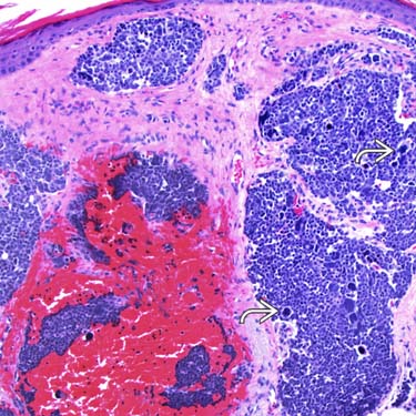
 easily identified.
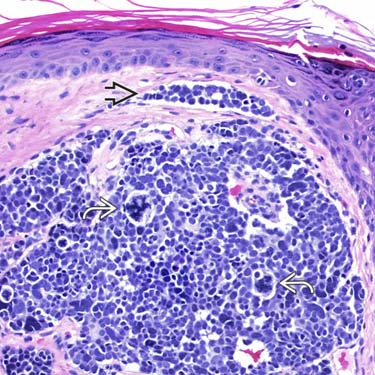
 . Invasion of a superficial lymphatic vessel is also seen
  .
.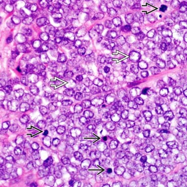
 is often seen in MCC, a feature not seen in basal cell carcinoma or most other small round blue cell tumors. Note the numerous apoptotic bodies
  and mitotic figures
  .
.


