Low Magnification of Primary Cutaneous Mucinous Carcinoma Low-power view of primary cutaneous mucinous carcinoma (PCMC) shows islands of epithelial cells floating in pools of mucin . Fibrous septa are present between some of the epithelial islands.
Intermediate Magnification of Primary Cutaneous Mucinous Carcinoma Medium-power view of PCMC shows islands of epithelial cells in pools of mucin. Fibrous septa , which characteristically divide the tumor into compartments, are present.
High Magnification of Primary Cutaneous Mucinous Carcinoma Higher magnification shows uniform cells with hyperchromatic nuclei and eosinophilic cytoplasm. An occasional mitotic figure is identified , but necrosis and marked cytologic atypia are not present.
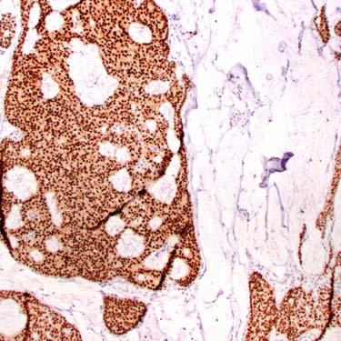
ER Immunohistochemistry in Primary Cutaneous Mucinous Carcinoma ER shows strong and diffuse nuclear staining in almost 100% of the tumor cells. This is a useful finding in excluding a metastatic mucinous colonic carcinoma, but it is not useful in excluding a metastatic breast carcinoma.


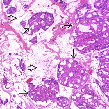
 floating in pools of mucin
  . Fibrous septa
  are present between some of the epithelial islands.
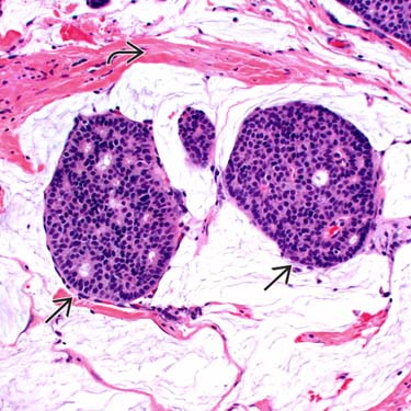
 in pools of mucin. Fibrous septa
  , which characteristically divide the tumor into compartments, are present.
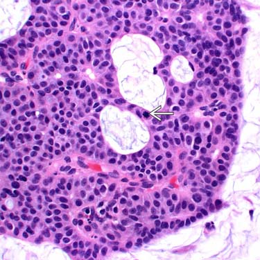
 , but necrosis and marked cytologic atypia are not present.