Poroma and Dermal Duct Tumor


Clinical Photograph of Poroma
Clinical photograph shows a traumatized poroma on the lower back of an adult male, which presented as an irritated polypoid lesion.


image
Poroma With Squamoid Features
Low magnification of a poroma with squamoid features shows a basaloid to squamoid-appearing neoplasm with multiple anastomosing connections image 
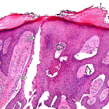
between thickened cords of tumor cells and to epidermis. Note the ectatic ductal space on the left image.

image
Poroma With Clear Cell Features
Scanning magnification of a poroma with clear cell features shows a basaloid to clear cell image 
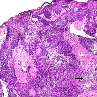
neoplasm with multiple anastomosing connections between thickened cords of tumor cells and to epidermis image.

image
Higher Magnification of Clear Cell Poroma
Higher magnification of a clear cell area of poroma shows a proliferation of small cells with abundant pale to clear-staining cytoplasm image 
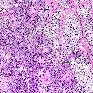
and small nuclei lacking atypia.


TERMINOLOGY


Synonyms


• Apocrine poroma

• Eccrine poroma

• Hidroacanthoma simplex (intraepidermal poroma, syringoacanthoma)

• Acrospiroma (older term, includes both poroma and hidradenoma)


Definitions


• Benign adnexal proliferation with anastomosing cords of tumor cells exhibiting ductal differentiation

• Typically has multiple epidermal attachments, except for dermal duct tumor


ETIOLOGY/PATHOGENESIS


Unknown


• Rare cases associated with radiation therapy and pregnancy
image May rarely be multiple (“poromatosis”)

• Formerly considered eccrine, but most cases likely of apocrine differentiation
image May see sebaceous &/or follicular differentiation in apocrine cases


CLINICAL ISSUES


Epidemiology


• Incidence
image Relatively common tumors

• Age
image Usually occur in middle-aged adults

• Sex
image Equal incidence in males and females


Site


• Most common on extremities, especially palmar and plantar surfaces
image May also occur on trunk, head and neck


Presentation


• Solitary pink to reddish papular or nodular lesion
image Minority of cases are pigmented

image Some cases may be very vascular and bleed easily


Natural History


• Stable lesions; do not usually regress


Treatment


• Surgical approaches
image Complete conservative excision is curative

– Typically recommended to prevent recurrence and rare transformation to porocarcinoma


Prognosis


• Excellent; most cases do not show aggressive behavior


MACROSCOPIC


General Features


• Superficial, firm papule or nodule


Size


• Generally < 1 cm but may be larger


MICROSCOPIC


Histologic Features


• Symmetric, well-circumscribed tumor with multiple epidermal attachments
image Intraepidermal component is typically sharply demarcated from adjacent normal epidermis

• Broad, anastomosing columns and thickened cords of tumor cells

• Ductal lumina small, but usually numerous
image A few small cystic structures may also be present

Only gold members can continue reading. Log In or Register to continue

Stay updated, free articles. Join our Telegram channel

Apr 24, 2017 | Posted by in PATHOLOGY & LABORATORY MEDICINE | Comments Off on Poroma and Dermal Duct Tumor

Full access? Get Clinical Tree

Get Clinical Tree app for offline access