Patient Story
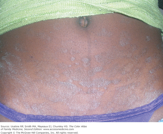
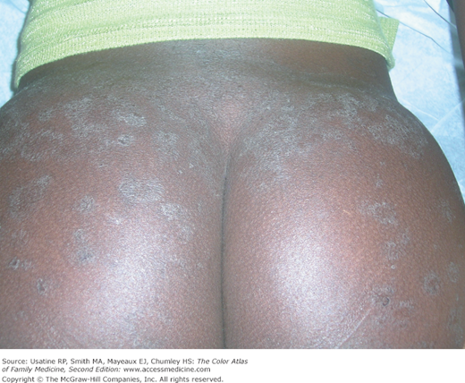
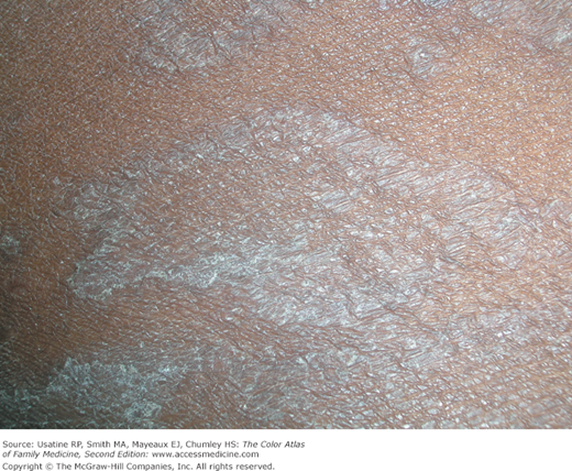
A 17-year-old young woman is brought to the office by her mom because of a rash that appeared 3 weeks ago for no apparent reason (Figures 153-1, 153-2, and 153-3). She was feeling well and the rash is only occasionally pruritic. With and without mom in the room, the young woman denied sexual activity. The diagnosis of pityriasis rosea was made by the clinical appearance even though there was no obvious herald patch. The collarette scale was visible and the distribution was consistent with pityriasis rosea. The young woman and her mom were reassured that this would resolve spontaneously. At a subsequent visit for a college physical the skin was found to be completely clear with no scarring.
Introduction
Pityriasis rosea is a common, self-limited, papulosquamous skin condition originally described in the 19th century. It is seen in children and adults. Despite the long history, its etiology remains elusive. A number of infectious etiologies have been proposed, but at present, supporting evidence is inconclusive. Pityriasis rosea has unique features including a herald patch in many cases and collarette scale that are useful in distinguishing it from other papulosquamous eruptions.
Epidemiology
- Pityriasis rosea is a papulosquamous eruption of unknown etiology.1
- It occurs throughout the life cycle. It is most commonly seen between the ages of 10 and 35 years.3
- The peak incidence is between 20 and 29 years of age.1
- The gender distribution is essentially equal.1
- The rash is most prevalent in winter months.4
Etiology and Pathophysiology
- The cause of pityriasis rosea is unknown, although numerous causes have been proposed.
- It has long been suspected that it may have a viral etiology because a viral-like prodrome often occurs prior to the onset of the rash. Human herpesviruses 6 and 7 have been proposed as causes, but numerous studies have failed to demonstrate conclusive supportive evidence.1,2
- Chlamydia pneumoniae, Mycoplasma pneumoniae, and Legionella pneumophila have been proposed as potential etiologic agents, but studies have not demonstrated any significant rise in antibody levels against any of these pathogens in patients with pityriasis rosea.1
- Pityriasis rosea has also been associated with negative pregnancy outcomes, particularly premature birth. The risk seems to be greatest when the condition occurs in the first 15 weeks of gestation.2
- Pityriasis rosea may rarely occur as the result of a drug reaction. Documented drug reactions that have produced a pityriasis rosea-like eruption include barbiturates, captopril, clonidine, interferon, bismuth, gold, and the hepatitis B vaccine.1,2
Diagnosis
- In approximately 20% to 50% of cases, the rash of pityriasis rosea is preceded by a viral-like illness consisting of upper respiratory or GI symptoms.
- This is followed by the appearance of a herald patch in 17% of cases (Figures 153-4, 153-5, and 153-6).4
- The herald patch is a solitary, oval, flesh-colored to salmon-colored lesion with scaling at the border. It often occurs on the trunk, and is generally 2 to 10 cm in diameter (Figures 153-4 and 153-5).
- One to 2 weeks after the appearance of the herald patch, other papulosquamous lesions appear on the trunk and sometimes on the extremities.
- These lesions vary from oval macules to slightly raised plaques, 0.5 to 2 cm in size. They are salmon colored (or hyperpigmented in individuals with dark skin), and typically have a collarette of scaling at the border (Figure 153-3

Stay updated, free articles. Join our Telegram channel

Full access? Get Clinical Tree


